














OnlineDPCFIFPL


Upper limb
Elbow rehabilitation: assess and empower your patients
Tessadit Aissaoui · Marion Burnier
Go further
Olecranon bursitis: telling septic from aseptic, what the clinical signs are really worth, the place of aspiration and how to manage the elbow.
Clinical examination alone cannot decide between septic bursitis and non-septic: that is exactly what justifies aspiration.
20%of septic elbow bursitis cases have fever (≥ 37.8 °C)
What follows covers each of these points in detail, with sources. It is there if you need it.
This topic is taught in a course: Elbow rehabilitation: assess and empower your patients, online with Tessadit Aissaoui.
1 / 13
12 article chapters · 52 min in total
Why the elbow gets infected more◔ 4 min
The same anatomy explains the frequency of the hygroma and its infectious risk.
Recognising a hygroma◔ 3 min
A rounded, mobile swelling that is painless through mid-range signals bursitis, not arthritis.
Signs of infection◔ 5 min
Classic percentages describe an entirely septic cohort: frequencies, not diagnostic landmarks.
What the examination alone is worth◔ 4 min
The best-performing test rests on just 11 cases, in 1989, never replicated.
The place of aspiration◔ 5 min
Aspirated fluid settles the infection question when it is plausible; nothing replaces it.
Risk factors◔ 3 min
All associated factors are weak, the strongest multiplying risk by only 1.4.
Non-septic management◔ 8 min
Compression and anti-inflammatories first line do as well as procedures, without their risks.
Septic management◔ 3 min
Medical treatment is enough most of the time: 74 % of patients are not operated on.
What is not a bursitis◔ 4 min
Test extension against resistance for any posterior swelling of the elbow, as a rule.
Case reports◔ 3 min
A missed test costs five weeks: test extension against resistance, always.
In practice◔ 3 min
Five minutes of focused examination: skin, warmth, mobility, extension and hand symptoms.
Frequently asked questions◔ 7 min
No sign confirms infection with certainty without aspiration.
Train on this
Upper limb
Tessadit Aissaoui · Marion Burnier
Next comes practice: the course that teaches this topic, with Tessadit Aissaoui.
Our courses on this topic may be covered by DPC or FIFPL, the French continuing-education funding schemes, depending on your profession and situation.
Olecranon bursitis, or elbow hygroma, is an inflammation of the bursa lying between the skin and the tip of the olecranon. This bursa has a feature that no other bursa in the body shares to the same degree: it is subcutaneous, sitting just under thin skin, over a prominent bony ridge, and it takes every load when the elbow rests on a table, an armrest or the ground. That mechanical exposure and that absence of soft-tissue cover mean the overlying skin grazes easily, and that a skin organism reaches the bursa directly.
Hence the question that governs everything else in management: is this bursitis infected? About one episode of olecranon bursitis in three is septic7, a proportion with no equivalent in the deep bursae. Getting it wrong one way means missing an infection that can reach the bone and the joint; getting it wrong the other way means aspirating and injecting a healthy bursa, a procedure whose iatrogenic risk is documented.
Three things to know from the outset, and the article sets them out in detail:
For the physiotherapist, the point is not to treat the infection but not to miss it. A posterior swelling of the elbow arriving in the clinic is frequently benign, often mechanical, sometimes inflammatory, and in a far from negligible proportion, infected. Recognising the third case and referring it the same day is the useful clinical act. The rest (protecting the elbow from pressure, adapting movements, managing load), belongs fully to rehabilitation.
This article covers the elbow. The site also covers bursitis of the knee from two angles: prepatellar bursitis and the question of the procedure, aspiration or surgery, as well as pes anserine bursitis, which is a deep bursitis and follows a different logic.
What is specific to the elbow comes down to one sentence: the olecranon bursa is subcutaneous and directly exposed to trauma and to pressure, which gives it a markedly higher infection risk than the other sites, and makes the septic-or-not distinction the central decision, whereas elsewhere it is the decision about the procedure that dominates.
A bursa is a gliding cushion. The olecranon bursa works in conditions the others never meet: under thin skin, over a prominent bone, and without a millimetre of muscle to absorb the load. That anatomy explains both how common a hygroma is and its infection risk.
The olecranon bursa is a potential space, lined with synovium, interposed between the posterior surface of the olecranon and the skin. Normally it holds a film of fluid of the order of a millilitre, clinically invisible. Its function is to let the skin glide over the bone during flexion and extension of the elbow, a movement in which the posterior skin travels several centimetres.
What sets it apart is its position: it is superficial. Between the outside world and the bursal cavity there is nothing but dermis and a very thin layer of subcutaneous tissue. Compare it with the psoas bursa, the trochanteric bursa or the pes anserine bursa: all are covered by thick, vascularised muscular or aponeurotic layers, which form a mechanical and immunological barrier. The olecranon bursa has none.
Superficial bursa or deep bursa: the same structure, two levels of exposure
Schematic section. On the left the olecranon bursa, on the right a deep bursa of the trochanteric type.
Source of the septic proportion: Stell IM, J Accid Emerg Med 1996 (PMID 8894865). The anatomical diagram is explanatory and not to scale.
The first is mechanical. The whole load of leaning on the elbow passes through this cushion. Leaning on the elbows for long stretches, working on the floor on the elbows, crawling, resting the forearm on a hard armrest: repeated microtrauma is the commonest cause of olecranon bursitis13. The old names for the condition (student's elbow, miner's elbow, plumber's elbow), all describe the same mechanism: prolonged, repeated pressure.
The second is infectious, and it is the one that structures this article. A bursa separated from the outside world by a few millimetres of thin skin, over a bony prominence that grazes at the slightest friction, offers a skin staphylococcus a short route in. That is why Staphylococcus aureus accounts for about 80 % of superficial septic bursitis15, and for 88 % of culture-positive cases in the Calgary series8: the organism that infects the bursa is the one living on the skin above it.
That short route also explains the place of trauma in the history. In the Calgary cohort, a preceding injury was found in 53 % of cases8. In other words: when you look for a point of entry, you find one every other time. When you do not find one, that rules nothing out, because a minor breach of the skin may have gone unnoticed or already healed.
Trauma is the common cause of mechanical bursitis and of septic bursitis. It therefore does not tell them apart: it precedes both.
Stell was already making this point in his 1996 review: trauma “may cause either”7, and local erythema may be present in both. A history that turns up a fall on the elbow three days earlier is not reassuring, it opens both hypotheses.
At the other sites covered on this site, the dominant question is that of the procedure: whether to aspirate, whether to operate, whether to inject. At the prepatellar bursa, also superficial, the logic is very close to that of the elbow, and the two conditions are in fact studied together in the literature9. At the pes anserinus or the greater trochanter, deep bursae, infection is a rare possibility and the debate is about the associated tendinopathy.
At the elbow, the infection question comes first, and not only in frequency. It comes first because it is the only one where the error costs dearly and quickly: a neglected mechanical bursitis remains a nuisance; a neglected septic bursitis can progress to soft-tissue spread, osteitis of the olecranon, and exceptionally to systemic infection.
The positive diagnosis of olecranon bursitis is one of the simplest in elbow pathology. It is the diagnosis of severity that is difficult. This chapter sets out the picture; the next tackles the infection question.
Olecranon bursitis presents as a rounded, well-defined swelling centred on the tip of the olecranon. Patients often describe it as having appeared quickly, sometimes overnight, and its size surprises them: a distended bursa can reach the volume of a golf ball. The consistency is tense, fluctuant when the effusion is copious.
Two clinical features carry strong orienting value, and both bear on what the bursitis is not :
This distinction is not academic. In the historical series of Ho, Tice and Kaplan, the authors explicitly noted that septic bursitis was not associated with septic arthritis and could easily be distinguished from it by the characteristic bursal swelling and the joint examination3. The two conditions rarely coexist, and a mobile elbow is a strong argument in favour of the bursa.
Beyond the mechanism, four elements of the history change what you do:
Ultrasound readily confirms the bursal effusion, measures its volume, shows synovial thickening and hyperaemia on power Doppler, and distinguishes a bursitis from a cellulitis13. It is also useful for guiding aspiration15.
What it does not say is whether the fluid is infected. This is a point on which the review by Lormeau and colleagues is explicit: ultrasound helps in diagnosing bursitis and in guiding the procedure, while telling aseptic from septic bursitis usually goes through examination of the aspirated fluid15. Thick, septated fluid with Doppler hyperaemia is compatible with infection; it is equally compatible with an advanced crystal-induced or haemorrhagic bursitis. Imaging does not replace aspiration when the question asked is that of sepsis.
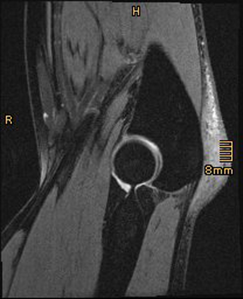
MRISagittal elbow MRI, T2 sequence: olecranon bursitis, thickened bursa with heterogeneous signal behind the olecranon.
Source : Konrads et al., SICOT-J, 2016, figure 1 · CC BY
This is the heart of the article, and it is also where the literature is most often misquoted. The percentages circulating in reviews and quick-reference guides almost all describe cohorts of patients who are already infected. A frequency within the disease is not a discriminating power.
The largest published series of septic olecranon bursitis is the Calgary one: 118 consecutive cases managed as outpatients over 21 months8. It gives the frequency of the signs with rare precision. And it is precisely that precision that misleads: every patient in that cohort is septic. There is no comparison group.
Saying “erythema is present in 92 % of septic bursitis” is accurate. Concluding that erythema allows infection to be affirmed is wrong: one would still need to know how many non septic bursitis cases show erythema. Yet Stell pointed out that local erythema can be seen in both forms7. A sign that is very sensitive but poorly specific does not help to confirm; it helps, when it is absent, to doubt.
Frequency of signs in 118 patients with confirmed septic olecranon bursitis
Caution: a cohort made up only of septic cases. These values are frequencies within the infection, not diagnostic values.
Source: Laupland KB, Davies HD, Clin Invest Med 2001 (PMID 11558851). A cohort of 118 septic olecranon bursitis cases, with no non-septic control group: these frequencies are not diagnostic sensitivities.
In that same cohort, fever defined objectively as a temperature of 37.8 °C or above was present in only 20 % of patients8. The difference with the 45 % who reported fever or chills on questioning is instructive: the symptom as declared is twice as frequent as the sign as measured.
Four patients in five with confirmed septic olecranon bursitis are afebrile at the time of examination. Taking the temperature reassures wrongly.
Local warmth, readily quoted as a cardinal sign of infection, was noted in only 36 % of cases. Here again, judging warmth by hand is a poorly reproducible test, and the figure reflects the examination method as much as the underlying biology. The next chapter shows that an instrumental measurement of the same parameter does far better.
Only two studies compare septic and non-septic bursitis from the same recruitment, and both are old and small.
Ho and Tice, 1979, on 30 olecranon and prepatellar bursitis cases of which 10 were septic: fever, tenderness, peri-bursal cellulitis and skin involvement overlying the bursa were more frequent in the septic cases; a high fluid white cell count, a low bursal-to-serum glucose ratio and a positive Gram stain distinguished septic from non-septic forms2. The authors give no sensitivity or specificity for these signs.
Smith and colleagues, 1989, on 46 consecutive olecranon bursitis cases of which 11 were septic, provide the only quantified diagnostic value available, and it does not concern a classic clinical sign but an instrumental measurement: the skin temperature differential1. That is the subject of the next chapter.
A word of caution about a widely circulated figure. One frequently reads that tenderness is present in 88 % of septic bursitis cases against 36 % of aseptic ones. We have not found this pair of values in the abstract of any verifiable primary study, and we therefore do not repeat it. What is established and citable is the direction of the difference reported by Ho and Tice2, not its size.
None of these signs taken alone proves infection, and their absence does not rule it out. Two of them together, or one alone in an at-risk patient, warrant same-day referral for aspiration.
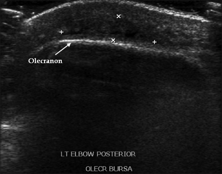
UltrasoundLongitudinal ultrasound of the posterior aspect of the elbow: effusion and echogenic debris within the olecranon bursa, arrow on the olecranon.
Source : Hirji et al., Journal of clinical imaging science, 2011, figure 29 · CC BY-NC-SA
The honest answer fits in one sentence: not enough to decide on its own, and that is precisely what justifies aspiration. But one measurable parameter does markedly better than clinical impression, and it has remained oddly little known.
In 1989, Smith and colleagues prospectively analysed 46 consecutive olecranon bursitis cases over one year, of which 11 proved septic and 35 non-septic. Alongside fluid analysis, they used a surface probe to measure skin temperature over the affected bursa and over the contralateral olecranon serving as a control1.
The results are clear-cut. The mean differential was 0.7 °C in the non-septic cases against 3.7 °C in the septic ones. In all the septic cases, the differential reached or exceeded 2.2 °C, with a range of 2.2 to 5.1 °C. Taking that 2.2 °C threshold, the measurement proved 100 % sensitive and 94 % specific. The authors conclude that it appears more useful than the bursal fluid white cell count, than the predominant cell type or than the Gram stain for telling the two forms apart early1.
Skin temperature differential between the affected and the healthy elbow
46 consecutive olecranon bursitis cases, 11 of them septic. Discriminating threshold: 2.2 °C.
Source: Smith DL, McAfee JH, Lucas LM, Kumar KL, Romney DM, Arch Intern Med 1989 (PMID 2742432). A prospective single-centre study on 46 cases.
A sensitivity of 100 % is impressive, and that is where one has to be precise rather than enthusiastic. It rests on 11 septic cases. A sensitivity of 11 out of 11 is compatible, at 95 % confidence, with a true sensitivity going down to around 72 %: the interval is wide because the numbers are small. The figure does not say that the measurement never misses an infection, it says that it missed none out of eleven.
Three further limits are worth setting out:
The temperature differential is the best documented clinical sign, and it rests on eleven infected patients in a single 1989 study. That is both far better than nothing and far less than proof.
No source supports the idea that a cluster of clinical signs would allow the sample to be dispensed with when infection is plausible. The literature's constant position, from Stell in 19967 to Lormeau in 201915, is that the distinction goes through examination of the fluid.
The reviews that propose a structured care pathway say nothing different: Blackwell and colleagues build theirs from the history and the examination, with particular emphasis on the warning signs, but it is indeed fluid analysis that governs the infective branch11.
What clinical examination does do well, on the other hand, is sort patients into three groups: those in whom infection is highly unlikely and where a procedure would be more risky than useful; those in whom it is possible and where a sample is called for; and those in whom it is probable and where the sample must not delay treatment. That is exactly the role a physiotherapist can hold on the front line, and it is already a great deal.
Aspiration is both the test that settles the question and a procedure with documented risk. That tension explains recommendations that appear contradictory. They are not: they simply do not address the same clinical situation.
The aspirated fluid should go for direct microscopy after Gram staining, cell count and differential, crystal analysis, and aerobic and anaerobic culture. Purulent fluid, a positive Gram stain or a positive culture confirm infection.
The difficulty starts with the cell count. The proposed thresholds do not agree, and the gap between them is not marginal: it runs from about 2,000 to 50,000 white cells per microlitre depending on the source. The algorithm of Baumbach and colleagues, which is the most quoted synthesis, keeps 3,000 cells per microlitre with more than 50 % polymorphs, combined with a bursal-to-serum glucose ratio below 50 %9.
The white cell thresholds proposed for confirming infection do not converge
Logarithmic scale. None of these thresholds has been prospectively validated on an independent cohort.
The threshold of 3,000 cells per microlitre with more than 50 % polymorphs is the one in the algorithm of Baumbach SF et al., Arch Orthop Trauma Surg 2014 (PMID 24305696). The other values are reported in the review literature without prospective validation. The spread is itself the message: no threshold commands consensus.
Two practical consequences follow from this spread. First, fluid below the threshold is not wholly reassuring when the clinical picture is worrying. Second, culture takes precedence over the cell count: it is culture that confirms, and cell-rich fluid with a negative Gram stain remains suspect.
This is a point that practice underestimates. In the French multicentre study by Charret and colleagues, the largest contemporary series with 272 patients managed for septic bursitis of the olecranon or the patella, 43 of the 223 bursal samples remained sterile, that is 19.3 %17. An organism was identified in only 184 patients out of 272, that is 67.6 %.
Close to one septic bursitis in five has a negative bursal culture. A sterile culture does not cancel out a clear-cut clinical picture.
The reasons are known: antibiotics already started, slow-growing organisms, a poor sample. The case of Cutibacterium acnes reported by Skedros and colleagues is instructive on that last point: the culture only turned positive on the twenty-first day of incubation, whereas a first aspiration had been reported negative after 14 days22. A standard culture stopped at five days would have concluded that there was no infection.
The opposite side is just as well documented. Bustamante and colleagues prospectively sampled 28 clinically aseptic olecranon bursitis cases and systematically sent the fluid to the laboratory. The result: zero positive cultures out of 28, zero positive Gram stains out of 28, a mean cellularity of 1,289 per cubic millimetre, and only two samples carrying calcium pyrophosphate crystals, without that changing what was done18. The authors conclude that routine analysis has limited clinical usefulness in this situation.
In other words: when the picture is frankly mechanical, analysis brings nothing back. And the procedure does cost something. Khodaee is explicit in his review: aspirating a microtraumatic bursitis is generally not recommended because of the risk of iatrogenic septic bursitis13.
An important nuance, and one often poorly reported. The systematic review by Sayegh and Strauch examined this question across 29 studies and 1,278 patients, and concludes that aspiration does not increase the risk of bursal infection in aseptic bursitis10. That finding does not cancel out Khodaee's caution: it rests mainly on level IV studies, where the procedures are performed in specialist settings and under controlled aseptic conditions. The reasonable position is that aspiration is safe when it is indicated and well performed, and that it remains useless when it is not indicated.
When aspiration is performed, it is classically done through a posterolateral approach, shifting the skin before the puncture to create a zigzag track. The logic is to avoid a short perpendicular track, which closes poorly and exposes the patient to a fistula or a chronic sinus track above the bursa. A medial approach is to be avoided because of the ulnar nerve.
This technical point explains a frequent clinical observation: an aspirated bursa that fills up again and ends up weeping through the puncture hole. It is also one of the reasons why repeated procedures are unfavourable, and why compression after aspiration is not a detail.
What to do about a posterior swelling of the elbow
An orienting tree for the front-line physiotherapist. It does not replace medical assessment, it decides how quickly to refer.
Tree adapted from Baumbach SF et al., Arch Orthop Trauma Surg 2014 (PMID 24305696) and Blackwell JR et al., Shoulder Elbow 2014 (PMID 27582935), both of which propose a pathway based on clinical triage followed by fluid analysis.
A 2024 study on nearly 55,000 subjects has renewed this chapter, and its most interesting result may be what it fails to find.
Shemesh and colleagues compared 10,301 patients with olecranon bursitis against 44,608 controls from a population database over the period 2005 to 202021. It is by far the largest published sample on the subject.
Factors associated with olecranon bursitis
Adjusted odds ratios and 95 % confidence intervals. 10,301 cases against 44,608 controls.
Source: Shemesh S, Itzikovitch R, Atzmon R, Kadar A, J Clin Med 2024;13(24):7801 (PMID 39768728). A population-based case-control study. The intervals shown are those published at 95 %.
Four associations stand out, all of small size: male sex (OR 1.406; 95 % CI 1.347 to 1.468), hyperlipidaemia (OR 1.243; 1.170 to 1.322), statin use (OR 1.130; 1.048 to 1.218) and smoking (OR 1.068; 1.020 to 1.118)21.
And diabetes is not associated (OR 0.979; p = 0.75), any more than cardiovascular disease or a history of stroke. That is a result worth pointing out, because diabetes appears in virtually every list of bursitis risk factors to be found online.
Take care not to over-read this in either direction. This study covers the occurrence of olecranon bursitis, all forms combined, not the risk of septic bursitis. Diabetes may perfectly well not favour the appearance of a hygroma while worsening the prognosis of an infected bursitis. These two questions are not the same, and the study answers only the first.
Likewise, the association with statins and hyperlipidaemia is an association, not a mechanism. An explanation by confounding (patients on statins consult more, are older, have a higher BMI) remains perfectly plausible.
None of these odds ratios comes close to the importance of the mechanical factor. The commonest cause remains chronic microtrauma from pressure13, and in the Calgary septic cohort the mean age was 44 years with 88 % men8, a profile that overlaps with manual trades loading the elbow. It is that exposure, unmeasurable in an administrative health database, that carries most of the risk.
For the non-septic forms, the soundest landmark comes from the randomised trial of Kim and colleagues: 25 patients out of 30 (83 %) treated with simple compression and anti-inflammatories had resolved at four weeks12. The natural tendency is therefore favourable.
One prognostic factor emerges from that same trial, and it is actionable: the duration of symptoms before treatment began was the only factor associated with failure at four weeks (6 weeks in case of failure against 4 weeks in case of success; p = 0.008)12. Starting management early improves the odds, whichever treatment is chosen.
For the septic forms, the prognosis under appropriate treatment is good: in Charret's series of 272 patients, the overall failure rate was 5.9 %17. Recovery can, however, be long: Stell was already noting that healing a septic olecranon bursitis can take months7.
This is the physiotherapist's ground, and the good news is that the simplest treatment is also the one the data support best. The bad news is that the overall level of evidence remains low: a single contemporary randomised trial, two old trials, and a literature dominated by retrospective series.
Kim and colleagues randomised 90 patients with non-septic olecranon bursitis into three groups of 30: compression bandaging with non-steroidal anti-inflammatory drugs, aspiration alone, or aspiration followed by a corticosteroid injection. Treatment was repeated each week if the effusion recurred, and assessment ran over four weeks12.
Resolution at 4 weeks by treatment, non-septic olecranon bursitis
Randomised trial, level II evidence. No significant difference between the three arms (p = 0.073).
Source: Kim JY, Chung SW, Kim JH, Jung JH, Sung GY, Oh KS, Lee JS, Clin Orthop Relat Res 2016 (PMID 26463567). A level II randomised trial. Seven patients lost to follow-up, hence the unequal denominators.
Three lessons, and a caveat the authors set out themselves.
First lesson: nothing is lost by starting with the simplest option. Compression bandaging with anti-inflammatories does as well as the invasive procedures at four weeks. It even does numerically better than aspiration alone, without the difference being significant.
Second lesson: the corticosteroid buys speed, not outcome. Resolution came at 2.3 weeks in the aspiration plus corticosteroid group, against 3.1 and 3.2 weeks in the other two (p = 0.015). One week gained on a condition that heals in more than eight cases out of ten.
Third lesson: the trial is small. The authors write explicitly that they were only powered to detect a 30 % difference, and offer their data as pilot data for sizing future trials12. A difference not demonstrated is not the demonstration of no difference.
Their conclusion deserves quoting in its movement: because olecranon bursitis recurs, and because aspiration and injection can cause complications, they suggest that compression bandaging combined with a short course of anti-inflammatories offers the best balance between safety and effectiveness, as long as future trials have not shown a clear advantage to the procedures12.
This is the point where the sources diverge most plainly, and it would be dishonest to smooth the disagreement over.
On one side, a double-blind randomised trial from 1989 on 42 patients split into four arms concludes that intrabursal injection of 20 mg of methylprednisolone is the most effective regimen for non-septic olecranon bursitis: the fastest reduction in swelling at one week, sustained improvement at six weeks, and fewer repeat aspirations at six months than in the naproxen or placebo groups6.
On the other, a long-term follow-up published five years earlier on 47 patients reviewed at a mean of 31 months gives the price of that effectiveness. The 25 patients treated with an injection of 20 mg of triamcinolone hexacetonide recovered quickly, generally within a week, but went on to show 3 infections, 5 skin atrophies and 7 chronic local pains. The 22 patients treated with simple aspiration recovered more slowly and with no complication at all. The authors conclude that a conservative approach is preferable since spontaneous resolution can be expected5.
Across 25 injected patients followed for a mean of 31 months: 3 infections, 5 skin atrophies, 7 chronic pains. Across 22 patients simply aspirated: no complication.
The systematic review comes down on the side of caution. Sayegh and Strauch, across 29 studies and 1,278 patients, find that corticosteroid injection in aseptic bursitis is associated with an increase in overall complications (p = 0.0458) and in skin atrophy (p = 0.0261), and conclude that it carries significant risks without improving the outcome10. The 2023 systematic review reaches the same practical position: reserve injection for refractory forms because of the higher complication rate19.
How to read this disagreement. The two camps are not measuring the same thing over the same horizon. The 1989 trial measures the speed of swelling reduction at one week and at six weeks: on that outcome, the corticosteroid wins. The 1984 follow-up and the 2014 review measure complications in the medium term: on that outcome, it loses. There is no contradiction about the facts, there is a choice of outcome measure. For a condition that resolves spontaneously in more than eight cases out of ten, favouring the “complications” outcome over the “speed” outcome is defensible, and it is the position the most recent sources take.
None of these studies assesses a physiotherapy intervention on its own: that is a gap in the literature which has to be named. What is assessed is a package of “compression and anti-inflammatories”, whose compression component falls directly to the physiotherapist. Clinical reasoning is therefore built from what is supported, while saying so.
Removing the pressure is the causal measure. As long as the elbow keeps bearing on a hard surface, the bursa keeps being irritated. That means concrete work analysing situations: workstation and armrests, sleeping position, resting on the steering wheel, occupational movements on the floor, pressure during transfers in a patient who uses their elbows. Padded elbow protection has an obvious logic here, even if its own benefit has not been isolated in a trial.
Compression is the documented active component of the best-performing arm in Kim's trial. A compression bandage worn continuously for the first few days, watching vascular and skin tolerance, is justified.
Movement should be maintained, not forced. The elbow does not stiffen spontaneously in an uncomplicated bursitis, since movement is preserved from the outset. The aim is to maintain it, in particular the terminal flexion that the mass mechanically obstructs, without trying to gain range by forcing on an inflamed bursa.
What to avoid. Firm massage over the bursa, local friction techniques and local heat have no rationale on a distended, inflamed bursa, and are strictly to be avoided as long as infection has not been ruled out. If infection is in doubt, no local technique should be applied before the medical opinion.
Levels of evidence applied to olecranon bursitis
A GRADE approach. The overall level of the literature on this subject is low: a single contemporary randomised trial.
Grading established by us from the design and the declared limits of the studies cited, following the GRADE logic. It is not taken from an official guideline: none exists on this subject, which Nchinda and Wolf point out explicitly (PMID 33840568).
| Modality | Level | What the data say |
|---|---|---|
| Removing the pressure and adapting movements | Low | A causal measure, never assessed in isolation. Microtrauma from pressure is the commonest cause13: removing it is a matter of clinical coherence more than of evidence. |
| Compression bandaging with NSAIDs | Moderate | 83 % resolution at 4 weeks, as good as the invasive procedures, without their risks12. A first-line option. |
| Aspiration alone (non-septic) | Moderate | 65 % resolution, numerically the poorest of the three arms without the difference being significant12. Does not increase the infection risk in aseptic cases10, but routine analysis of the fluid brings nothing back18. |
| Intrabursal corticosteroid injection | Low | Faster resolution (2.3 vs 3.2 weeks, p = 0.015)12, but increased complications and skin atrophy10: 3 infections, 5 atrophies, 7 chronic pains across 25 patients followed for 31 months5. To be reserved for refractory forms19. |
| Antibiotic treatment (septic form) | Moderate | The reference treatment. Overall failure 5.9 %; more failures if the duration is under 14 days (p = 0.02)17. |
| Bursectomy first line | Very low | Less effective and more risky than non-surgical treatment (resolution p = 0.0476; complications p = 0.0117)10. 29.5 % postoperative complications in a series20. Reserved for stubborn or recurrent forms. |
| Protective elbow pad | Very low | No direct assessment found. Recommended for consistency with the mechanism, cited among the usual conservative measures16. |
| Local massage, heat, frictions | Very low | No rationale on a distended, inflamed bursa. Contraindicated as long as infection has not been ruled out. |
This chapter is not the physiotherapist's direct concern, but knowing it changes how the patient is supported, how the timescales are explained, and how a treatment that goes off course is spotted.
The contemporary position is clear: medical treatment is enough in the majority of cases. In the French multicentre study on 272 patients, the success rate was equivalent between the medically managed group and the operated group, and only 26 % of patients were treated surgically17. Baumbach and colleagues had already concluded that the available data did not support the central European concept of immediate bursectomy for septic bursitis, and recommended a conservative regimen9.
Antibiotic treatment targets S. aureus first line, since it accounts for about 80 % of the organisms15. An initial intravenous route was preferred in the case of fever (p = 0.003) or extensive cellulitis (p = 0.002) in the French series, and concerned 41 % of patients17.
Treating early shortens the treatment. This is an old but robust observation: by culturing the fluid of 25 patients serially, Ho and Su showed that the time needed to sterilise the bursa correlated with the duration of symptoms before diagnosis (r = 0.68; p < 0.001). In patients treated within two weeks of symptom onset, sterility was obtained in under a week; beyond that, the response was delayed4. A delayed diagnosis therefore does not merely put healing back, it lengthens the treatment needed.
Two pieces of good-quality data point in different directions, and here again the key is that they do not describe the same situation.
On medical treatment, Charret and colleagues observe that failures were more frequent when antibiotic treatment lasted under 14 days (p = 0.02), in the surgical group as in the medical one, and conclude that a duration under 14 days deserves particular attention17.
After bursectomy, the randomised trial of Uçkay and colleagues on 164 inpatients shows that 7 days of oral antibiotics after excision of the bursa are enough, with 10 % failures in the single-stage arm14. The difference is explained: when the infected focus is removed, the bacterial load to treat is no longer the same.
That trial brings a second useful result, specific to the elbow: single-stage bursectomy with primary closure gave fewer wound dehiscences than the two-stage technique for elbow bursitis, 1 out of 66 against 9 out of 64 (p = 0.03), with a shorter hospital stay and a lower cost14.
The series of Pohl and colleagues, covering 61 open bursectomies, gives figures that invite restraint: 29.5 % postoperative complications, including 10 fluid collections, 4 ulnar nerve paraesthesias and 3 wound dehiscences, with a mean healing time of 17.4 days20.
Above all, the functional result differs by form: aseptic patients significantly improved their physical score (41.5 to 46.8; p < 0.001), septic patients did not (43.6 to 40.5; p = 0.277), a septic diagnosis being associated with a poorer result on multivariate analysis (estimate −8.40; 95 % CI −14.42 to −2.38; p = 0.008)20.
Three concrete points. First, recovery is long: Stell already pointed this out, it can take months7. Announcing that timescale saves the patient from concluding that it has failed.
Next, the posterior elbow scar is a fragile area, over which the skin tightens with every flexion. With 17.4 days of mean healing time and a documented risk of dehiscence20, progression into flexion must be conducted while watching the scar, and elbow pressure prohibited for a long time.
Lastly, ulnar nerve paraesthesia is a known postoperative complication, found in 4 patients out of 6120. Tingling along the ulnar border of the hand after bursectomy is not a trivial matter: it must be reported.
Not every posterior mass of the elbow is a hygroma, and two of the costliest errors are errors of over-confidence in the obvious diagnosis.
This is the differential diagnosis to know first, because it is functionally serious and because it is regularly taken for a bursitis. The mechanism is the same in both cases: an elbow injury followed by posterior swelling.
The sign that separates them is simple and often left out: elbow extension against resistance. Shivdasani and colleagues report the case of a 53-year-old man, a weight trainer, who felt a tearing sensation with immediate pain and swelling of the posterior aspect of the right elbow, and showed weakness on extension against resistance. The initial diagnosis was olecranon bursitis, and three aspirations were performed over several weeks before an orthopaedic opinion corrected the diagnosis, five weeks after the injury23.
The authors point precisely to the cause of the error: the extension-against-gravity test, specific to the function of the triceps tendon, is frequently left out because of the pain and the swelling23. In other words: it is precisely because the elbow is swollen and painful that the test is not done, and it is precisely there that it must be.
This is no curiosity. In the surgical series of Pohl and colleagues, 20 patients out of 61 operated on for a bursectomy, that is 32.8 %, had a triceps repair at the same time, of which 8 were complete ruptures20. An associated triceps lesion is therefore frequent in patients who end up in theatre.
For any posterior swelling of the elbow after trauma: test active extension against gravity, then against resistance. Weakness does not belong to the picture of bursitis.
Compression of the ulnar nerve at the elbow can produce a swelling or a thickening of the posteromedial region, and above all it shares with bursitis both the site and the context of prolonged elbow pressure. The difference is neurological: paraesthesias along the ulnar border of the hand, of the fourth and fifth fingers, worsened by prolonged elbow flexion and at night, with an intrinsic motor deficit at an advanced stage.
Two situations demand that it be considered explicitly. The first is a posterior swelling accompanied by distal signs: bursitis gives no symptoms in the hand. The second is the appearance of paraesthesias after a bursectomy, ulnar paraesthesia being a documented postoperative complication, found in 4 patients out of 61 in Pohl's series20.
The subject is covered in detail in our article on cubital tunnel syndrome (ulnar compression at the elbow).
Gout and chondrocalcinosis produce olecranon bursitis, sometimes as the presenting feature, and rheumatoid arthritis classically comes with nodules and bursitis in this region. Ho and Tice were already noting it: rheumatoid arthritis and gout can be accompanied by a non-septic bursitis2.
The trap is a major one, and the next chapter devotes a case to it. Finding crystals does not close the discussion, because a bursa carrying crystals can also be infected. Bustamante and colleagues did in fact find calcium pyrophosphate crystals in 2 samples out of 28 from clinically aseptic bursitis without that changing management18: crystals are frequent and rule nothing out.
| Diagnosis | What puts you on the trail | What to do |
|---|---|---|
| Triceps rupture | Injury during eccentric contraction, tearing sensation, weakness on extension against resistance, sometimes a palpable defect | Prompt surgical opinion: the result depends on the delay23 |
| Arthritis of the elbow (septic or inflammatory) | Pain throughout range, elbow held in flexion, filling of the lateral triangle | This is no longer the bursa: same-day opinion |
| Humero-ulnar osteoarthritis | Stiffness of slow onset, loss of terminal extension, crepitus, sometimes locking; no isolated fluctuant swelling | Progresses over years, not days: see our article on osteoarthritis of the elbow |
| Cubital tunnel syndrome | Paraesthesias of the 4th and 5th fingers, worse in flexion and at night | See our dedicated article; bursitis does not affect the hand |
| Gout, chondrocalcinosis | History, joint flares, tophi, a very inflamed bursa | Crystals on aspiration, but crystals do not rule out infection |
| Rheumatoid nodule | A firm, non-fluctuant, painless mass, known polyarthritis | Solid, not fluid, consistency: ultrasound settles it |
| Fracture of the olecranon | Violent direct trauma, loss of function, active extension impossible | Radiograph before any procedure |
| Infection with a slow-growing organism (mycobacteria, Cutibacterium) | A lingering bursitis, barely inflamed, recurrent, often after repeated procedures | Prolonged and mycobacterial cultures to be requested explicitly24 |
| Malignant transformation on a chronic wound | An ulcerated bursitis running for years, granulation tissue, failure to heal | Rare but described: any elbow wound that fails to heal must be seen |
Three published cases, chosen because each illustrates a different mechanism of error: the examination not done, the diagnosis that stops at the first result, and the culture stopped too soon.
A 53-year-old man, a recreational weight trainer. A tearing sensation with immediate pain and swelling of the posterior aspect of the right elbow. The general practitioner settled on olecranon bursitis and performed three aspirations over several weeks. The patient finally consulted an orthopaedic surgeon: complete rupture of the triceps tendon, repaired surgically five weeks after the injury. At one year, strength and range equal to the opposite side, a Mayo score of 100, weight training resumed without restriction at 16 weeks23.
What the case teaches. The final result is excellent, which might make the delay seem less important. That would be a misreading: repairing triceps ruptures is all the simpler for being early, and five weeks is a delay that exposes the tendon to retraction. Above all, the error was avoidable by a ten-second test. The swelling that makes testing difficult is exactly the situation where the test counts most.
A 65-year-old immunocompetent man. Swelling of the right elbow recurring two years after a first bursectomy, with no trauma, no pain, no redness, no systemic sign. Aspiration found monosodium urate crystals: a diagnosis of gout was made, and the patient received indometacin then allopurinol. Despite treatment, the fluid re-formed.
The initial tests were reassuring: a search for acid-fast bacilli negative on direct microscopy, bacteriological and mycological cultures negative. It was the mycobacterial cultures, positive at ten days, that corrected the diagnosis, confirmed on a second aspiration: Mycobacterium kansasii. Treatment with azithromycin, ethambutol and rifampicin, then bursectomy at two months, with eight months of antibiotics in total. Ethambutol had to be stopped at six months for an optic neuropathy, with persisting visual sequelae24.
What the case teaches. Three things, and all of them transfer.
A 53-year-old right-handed man, with pain and swelling at the back of the right elbow, no diabetes, no drug use, and no history whatsoever of injection into the bursa. A history, four years earlier, of septic bursitis with Staphylococcus aureus sensitive to meticillin after an abrasion, cured by debridement and a week of antibiotics.
The current episode resisted. The aspiration of 1st October 2017 stayed sterile after 14 days. A sample from 6 October eventually grew: Cutibacterium acnes, on the twenty-first day of incubation. It took five debridements in three weeks, plus curettage in the clinic, intravenous antibiotics then three months of oral amoxicillin, to eradicate an infection with probable osteitis of the olecranon. No recurrence at four and a half years22.
What the case teaches. C. acnes is usually set aside as a contaminant, and it grows slowly: a standard culture reported at five days would have missed it, and a culture reported at 14 days did miss it. When a bursitis fails to heal while the cultures are negative, the question is not only “is it infected?” but “have we looked long enough?”. This case is also a reminder that recurrence at the same site, four years after a first episode, is a risk factor in itself.
What this article changes, session by session, for a physiotherapist who sees a swollen elbow come in.
The most important instruction is not about an exercise but about a timescale. A patient leaving with a bursitis judged to be mechanical must know what should bring them back without delay: redness that spreads, pain that increases, fever or chills, and generally any worsening within 48 hours.
Two received ideas also need defusing. The first is that the size of the lump measures the severity: that is false, a large painless bursa is often less worrying than a small very painful one. The second is that the bursa has to be “emptied”: Kim's trial shows that aspiration alone does numerically worse than simple compression12, and the procedure is not neutral.
| Phase | Aim | Content |
|---|---|---|
| Days 0 to 7 | Remove the load, reduce the effusion | Strict removal of elbow pressure. Compression bandaging. Ice. Analgesia and anti-inflammatories as prescribed. Movement maintained through the free range, without forcing terminal flexion. No local massage, no heat. |
| Days 7 to 21 | Confirm the trajectory | Reassessment at day 7: the volume should be falling. Compression continued, protection of the elbow-pad type introduced for unavoidable pressure situations. Concrete analysis of the workstations and movements responsible. |
| Weeks 3 to 6 | Return to activity and prevention of recurrence | Gradual return to protected weight-bearing. Strengthening of the upper limb without direct pressure on the olecranon. Lasting change to pressure habits: that is what prevents recurrence. |
| At any time | Reopen the diagnosis | No improvement at day 7 to day 10, worsening, appearance of a red flag: re-refer. A bursitis that does not respond is a signal. |
Treating an elbow locally when infection has not been ruled out. Massage, frictions, ultrasound, heat: on a potentially infected bursa, these techniques have no rationale and may help to spread it. In case of doubt, you refer and you do not treat locally.
Being reassured by the absence of fever. This is the most predictable error, and the figure is worth remembering: only 20 % of confirmed septic bursitis cases had a temperature of 37.8 °C or above8.
Treating a recurrent bursitis as a mechanical inevitability. Recurrence after a procedure, especially in an elbow already operated on or aspirated, should raise the idea of a slow-growing infection before being put down to pressure24.
Yes, in the great majority of non-infected forms. In the only contemporary randomised trial, 25 patients out of 30 (83 %) treated with simple compression and anti-inflammatories had resolved at four weeks12. The condition is that the cause be removed: as long as the elbow keeps bearing weight, the bursa keeps being irritated.
No sign allows it to be known with certainty without aspiration, and that is precisely the message of this article. The features that should prompt a same-day consultation are: redness spreading beyond the lump, intense or night-time pain, a wound over it, fever, warmth clearly more marked than on the opposite side, or onset in under 48 hours. Beware: the absence of fever proves nothing, it is missing in 80 % of patients with confirmed septic bursitis8.
That depends entirely on the question being asked. If an infection is plausible, yes: aspiration is the test that settles it, and nothing substitutes for it. If the picture is frankly mechanical, no: routine analysis brings nothing back (no positive culture across 28 samples in a prospective series)18, and the procedure is not advised as a routine because of the risk of infecting a healthy bursa13.
It buys time and costs complications. It speeds up resolution by about a week (2.3 against 3.2 weeks)12, but the long-term follow-up of 47 patients reports, among the 25 injected, 3 infections, 5 skin atrophies and 7 chronic pains, against no complication in the 22 simply aspirated5. Recent sources reserve it for refractory forms19.
For a non-infected form managed early, reckon on three to four weeks in the majority of cases. The only prognostic factor identified is the delay before treatment starts: failures at four weeks concerned patients symptomatic for six weeks against four in those who recovered (p = 0.008)12. For an infected form, recovery is counted in weeks to months7.
Activities with no pressure or contact on the elbow can generally be continued if they do not wake the pain. Conversely, anything putting the elbow under pressure or in contact (floor sports, martial arts, bench work with elbow support, cycling on aero bars) should be suspended as long as the bursa is distended: that is the causal load. If the bursitis is infected, the stop is complete until a medical opinion.
Surgery is not a first-line treatment. The systematic review shows that it is less effective than non-surgical treatment and more productive of complications10, and a recent series reports 29.5 % postoperative complications20. It is worth discussing for genuinely stubborn or recurrent forms, after checking two things: that the causal pressure really has been removed, and that there is no unrecognised slow-growing infection.
The mechanism is close, both bursae being subcutaneous and exposed to pressure, and the literature often studies them together9. See our articles on prepatellar bursitis and on the decision to aspirate or to operate at the knee. By contrast, pes anserine bursitis is a deep bursa: its infection risk is far lower and the dominant question there is that of the associated tendinopathy.
Contrary to what one often reads, no, not for its occurrence. In the study covering 10,301 cases and 44,608 controls, diabetes was not associated (OR 0.979; p = 0.75)21. Note, however: that study covers the appearance of a bursitis, not the risk of it becoming infected nor its prognosis once infected, questions it does not answer.
What this article cannot settle. Three questions remain open in the literature, and it would be dishonest to present them as settled: the cell threshold that confirms infection (the proposed values vary by a factor of 25), the real diagnostic value of clinical signs taken as a cluster (no contemporary study has measured it), and the specific benefit of physiotherapy modalities in isolation (never assessed separately). On these three points, what is written here is clinical reasoning resting on the available data, not a recommendation founded on solid evidence.
Each reference has been checked individually: existence of the PMID and reading of the abstract to verify that the source really establishes what is attributed to it. 24 sources. Click a superscript note marker in the text: the bibliography opens and highlights the source.
Behind this article
An author who explains, a reviewer who checks.
How we write and check our content

Physiotherapist · co-founder of Physio Learning
Marked for life by his first four-hour lecture without a single image, he took a master’s in instructional design so that it would never happen to anyone again. He hunts down publication bias and unreadable slides with the same intransigence.

Head of scientific content
Physiotherapist specialising in neuro-musculoskeletal practice and holder of a master’s in public health. He checks the methodological rigour of every article: primary sources, levels of evidence, no exceptions.
