














OnlineDPCFIFPL


Upper limb
Elbow rehabilitation: assess and empower your patients
Tessadit Aissaoui · Marion Burnier
Go further
Ulnohumeral osteoarthritis: 25 % of radiographic forms after 50, 0.9 % symptomatic. Diagnosis, the ulnar nerve, physiotherapy and surgical options.
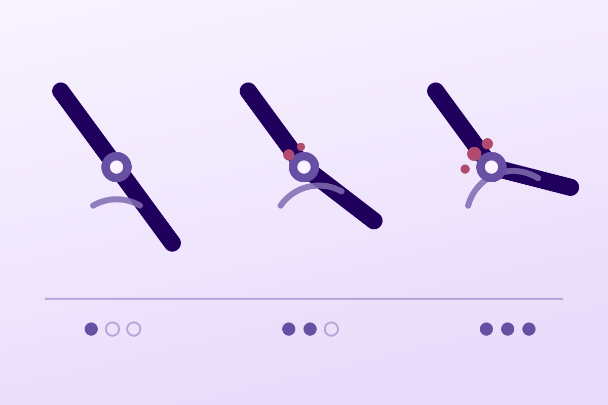
After 50, one adult in four carries the radiographic signs of elbow osteoarthritis, but fewer than one in a hundred suffers from it: imaging does not make the diagnosis.
54,5%of operated ulnar neuropathies at the elbow that rest on primary osteoarthritis
What follows covers each of these points in detail, with sources. It is there if you need it.
This topic is taught in a course: Elbow rehabilitation: assess and empower your patients, online with Tessadit Aissaoui.
1 / 10
9 article chapters · 41 min in total
Vocabulaire◔ 2 min
“Omarthrosis” comes from the Greek for shoulder; it never designates the elbow.
Définir◔ 6 min
This osteoarthritis makes bone rather than destroying cartilage, and stays silent in most who carry it.
Examiner◔ 7 min
No clinical test is validated for this osteoarthritis: neither sensitivity nor specificity published.
Imagerie◔ 3 min
Two films suffice for diagnosis; the rest prepares a procedure, not a confirmation.
Nerf ulnaire◔ 4 min
Elbow osteoarthritis is the leading identified cause of operated ulnar neuropathy, and decides the surgical timetable.
Rééduquer◔ 5 min
No controlled trial of physiotherapy exists in elbow osteoarthritis: everything is transposed.
Chirurgie◔ 5 min
Debridement and osteocapsular arthroplasty suffice in most symptomatic elbow osteoarthritis.
Cas cliniques◔ 3 min
None is reconstructed for illustration: two come from series, one is a published case with its identifier.
En pratique◔ 6 min
Rare, with no trial or guideline, this osteoarthritis needs an approach written down in advance.
Train on this
Upper limb
Tessadit Aissaoui · Marion Burnier
Next comes practice: the course that teaches this topic, with Tessadit Aissaoui.
Our courses on this topic may be covered by DPC or FIFPL, the French continuing-education funding schemes, depending on your profession and situation.
An elbow that hurts at the end of extension, that locks from time to time, in a man of fifty whose job has long forced his arms: the picture is stereotyped, the condition is little known, and its name is almost always mangled. This article covers primary osteoarthritis of the elbow, from its vocabulary to its rehabilitation, saying at each stage what the literature supports and what it does not.
A synthesis written from primary sources verified one by one on PubMed. Every figure carries its reference at the point where it is written; the full bibliography, with PMIDs, appears at the end of the article.
Elbow osteoarthritis in three figures
What three independent studies have measured, and why those three numbers tell the whole of the disease
Sources: Nakayama K et al. J Shoulder Elbow Surg 2022;31(1):123-132 (PMID 34454037) for the first two figures; Sato N et al. J Orthop Sci 2020;25(2):235-240 (PMID 31005383) for the third.
What to take away before reading the rest
An error of vocabulary is a detail only for as long as it does not change what you do. This one changes the word the patient types into their search engine, the page they open, and sometimes the joint they are told about.
“Omarthrosis” comes from the Greek ômos, the shoulder. The term means glenohumeral osteoarthritis and nothing else. The check is easy to make yourself: two German-language papers entitled literally Omarthrose and Omarthrose – Pathogenese, Klassifikation, Diagnostik und Therapie are indexed in PubMed under the English headings “Omarthrosis” and “Shoulder Osteoarthritis”12. Both deal with the glenohumeral joint; neither deals with the elbow. Using that word for an elbow is naming one joint while meaning another.
The correct name is ulnohumeral osteoarthritis, or humeroulnar, after the compartment the disease affects first. It is in fact the term used by the English-language literature, where the reference operation is called ulnohumeral arthroplasty3 and where the series speak of humeroulnar4 involvement. You also meet “cubital osteoarthritis”, vocabulary from before the revision of the anatomical nomenclature, when the ulna was called the cubitus: it is the same bone and the same joint.
Three compartments coexist at the elbow, and the disease affects them neither at the same time nor in the same way:
In the series by Doherty and Preston, the first to describe the primary form properly, radiographic involvement was present in the humeroradial compartment in 100 % of elbows, humeroulnar in 96 % and radioulnar in 85 %4. The disease is therefore pan-articular on imaging, whereas the complaint is almost always that of a hinge that jams.
Naming it correctly is not fussiness: a patient who comes in for “omarthrosis of the elbow” has already read, before seeing you, pages that were about their shoulder.
The glossary to give the patient
Elbow osteoarthritis = ulnohumeral osteoarthritis = humeroulnar osteoarthritis = cubital osteoarthritis (an old term). Omarthrosis = osteoarthritis of the shoulder, another joint, another kind of management. If the radiology report speaks of “humeroradial osteoarthritis” or “radiocapitellar osteoarthritis”, it means a compartment of the same disease.
An osteoarthritis like no other: it makes bone rather than destroying cartilage, it stays silent in most of those who carry it, and it strikes an identifiable occupational profile.
The feature that defines primary osteoarthritis of the elbow is that it is hypertrophic : the bony response dominates by far over cartilage loss. Osteophytes develop at the points of impingement, the tip of the olecranon, the coronoid process, the radial head, and the olecranon and coronoid fossae fill in, sometimes by a genuine thickening of the membrane lining them. Meanwhile, the joint space remains largely preserved5.
This feature is not incidental, it is what governs the whole strategy. Where advanced knee osteoarthritis requires the surfaces to be replaced, elbow osteoarthritis can often be treated by removing what blocks : osteophytes, loose bodies, contracted capsule. The relatively spared cartilage explains the good results of conservative procedures and also explains why prosthetic arthroplasty remains, in this indication, a last resort5.
Dalal's case-control radiographic series quantified exactly what the images show, comparing 50 osteoarthritic elbows with 50 elbows matched for age and sex. Olecranon osteophytes were present in 96 % of patients, coronoid osteophytes in 90 %, radial head osteophytes in 86 %, and osteophytes of the coronoid and radial fossae in 64 %. Thickening of the membrane of the olecranon fossa concerned 68 % of cases. Joint space narrowing, for its part, predominated in the radiocapitellar compartment (58 %) and remained rare in the humeroulnar one (16 %). In the controls, only the coronoid osteophyte was frequent (36 %); the other signs were rare6.
What the radiograph shows, and what normal ageing shows as well
Frequency of each radiographic sign: 50 osteoarthritic elbows against 50 controls matched for age and sex
Source: Dalal S, Bull M, Stanley D. J Shoulder Elbow Surg 2007;16(3):358-361 (PMID 17188910). The thin bars represent the controls; only the controls with a coronoid osteophyte reach a notable frequency.
The epidemiology of this disease remained unknown for a long time, for want of a general population study. It was established in 2022 by a Japanese survey conducted on a random draw from a municipality's resident registry: 415 residents over 50 sampled, 318 retained after excluding every cause of secondary osteoarthritis, all radiographed in two views7.
Radiographic prevalence was 25.2 % (27.3 % in men, 23.2 % in women) for a Kellgren-Lawrence grade of 2 or more. The prevalence of symptomatic forms, for its part, fell to 0.9 %. In other words, one adult over 50 in four carries radiographic osteoarthritis of the elbow, and barely one in a hundred suffers from it. The progression with age is steady and rapid7.
Radiographic prevalence by age band
318 adults aged 50 to 89, drawn at random from a municipal registry, radiographed in two views: Kellgren-Lawrence grade 2 or more
Source: Nakayama K, Kato H, Ikegami S et al. J Shoulder Elbow Surg 2022;31(1):123-132 (PMID 34454037). Japanese cohort, mean age 69.2.
Two practical lessons come out of these figures. The first: finding osteoarthritis on an elbow radiograph does not prove that it explains the pain. At 75, the probability that any given elbow carries osteoarthritic signs approaches one in three, and nine times out of ten those signs are silent. The second: this osteoarthritis is, in the authors' own words, a disease that most people “accommodate” by themselves7. It is the rarity of the symptom, and not the rarity of the lesion, that makes it seldom seen.
The surgical series draw a very consistent portrait. In Antuña, 46 operated elbows: 44 men to one woman, mean age 483. In Doherty, 16 patients: 14 men, 2 women, mean age 61, with involvement of the dominant elbow in 14 of them4. The review by Gramstad and Galatz sums up the classic background: a middle-aged man doing intense manual work5.
The Japanese survey adds factors that were measured rather than assumed. Age and body mass index were independently associated with the presence of grade 2 or higher osteoarthritis. The use of vibrating tools emerged as a factor specifically associated with the most advanced forms, those of grade 47. That last point deserves to be remembered in the clinic: jackhammer, grinder, chipping hammer, hammer drill, but also chainsaw and brushcutter in grounds maintenance workers.
One detail of Doherty's series often goes unnoticed and is nevertheless useful: these patients frequently had osteoarthritis elsewhere: the second and third metacarpophalangeal joints in 62 % of them, knees in 38 %, hips in 31 %4. Faced with isolated elbow osteoarthritis in a young person, it is therefore worth looking at the hands: associated metacarpophalangeal involvement points towards a more general arthropathy, and notably towards a search for haemochromatosis or chondrocalcinosis, two diagnoses the picture can borrow.
The same authors also established a point that tradition contradicts: the role of trauma is hard to support in the primary form4. Post-traumatic osteoarthritis exists, it is frequent and well documented, but it is another entity: it follows a radial head fracture, a complex dislocation or a fracture of the distal humerus, and it has neither the same profile nor the same prognosis.
Four background markers
Figures from the two reference series on the primary form
Sources: Antuña SA, Morrey BF, Adams RA, O'Driscoll SW. J Bone Joint Surg Am 2002;84(12):2168-2173 (PMID 12473704); Doherty M, Preston B. Ann Rheum Dis 1989;48(9):743-747 (PMID 2802795).
Key points
There is no validated clinical test for this condition: no published sensitivity, specificity or predictive value. The diagnosis therefore rests entirely on a body of arguments, and it is as well to know which ones carry weight.
Pain at terminal extension. This is the cardinal symptom, and the word “terminal” counts: the pain does not appear in the middle range but in the very last degrees, when the tip of the olecranon comes up against a filled fossa. In Antuña's series, all 46 operated elbows complained of terminal extension pain3. Patients rarely describe it in those terms; they say they can no longer “straighten the arm fully”, or that it catches when they push a heavy door or carry a shopping bag with the arm extended.
Locking. It reflects the presence of intra-articular loose bodies, found on 69 % of the radiographs in Doherty's series4 and present in 36 of the 46 elbows operated on by Antuña3. Fourteen of those 46 patients reported frank locking. It is a useful sign because it is specific and because it points straight away to a simple surgical procedure, removal of the loose body, often very rewarding.
The ulnar nerve signs. Twelve of Antuña's 46 patients complained of them before the operation3. This chapter is important enough to have its own section below; for now, remember that not looking for them means missing the commonest and most disabling complication of this osteoarthritis.
What, by contrast, is not the picture: constant pain at rest, prolonged morning stiffness, hot swelling, bilateral involvement from the outset in a young person. These features should prompt a search for something else.
Loss of range starts with extension and then progresses to flexion. The flexion contracture sets in insidiously, and the patient compensates for it so well with the shoulder that they do not notice it. Pronation and supination stay normal for a long time, which distinguishes this osteoarthritis from post-traumatic radioulnar involvement.
The classic “functional arc” marker comes from an experimental study of 1995: one hundred healthy upper limbs, a brace progressively limiting the range, twelve activities of daily living to perform at each setting. Forty-nine of the fifty subjects completed all the tasks with a range restricted to 75-120° of flexion8. That is the figure the surgical literature has used ever since to judge the need for a procedure.
That marker has aged, however, and it must be said to the patient. A three-dimensional kinematic study of 2011 measured the ranges actually required by contemporary tasks: the flexion arc needed rose to 130° ± 7 to use a telephone, with a maximum of 142° ± 3; pronation reached 65° ± 8 to type on a keyboard and supination 77° ± 13 to open a door9. The authors conclude explicitly that the functional range of the elbow is probably wider than was thought.
The functional arc of the elbow: the 1995 marker and what today's tasks demand
Flexion-extension range required by tasks, measured by three-dimensional optical capture
Sources: Vasen AP, Lacey SH, Keith MW, Shaffer JW. J Hand Surg Am 1995;20(2):288-292 (PMID 7775772) for the 75-120° arc; Sardelli M, Tashjian RZ, MacWilliams BA. J Bone Joint Surg Am 2011;93(5):471-477 (PMID 21368079) for contemporary tasks. The “table activities” band represents the order of magnitude common to both papers and is not a measured value.
| Diagnosis | What suggests it | What sets it apart from osteoarthritis |
|---|---|---|
| Lateral epicondylalgia | Pain at the lateral epicondyle, provoked by resisted wrist extension and by gripping | Ranges preserved and painless at end of range; no locking |
| Medial epicondylitis | Medial pain on resisted flexion and pronation | A tender tendon point and not a joint one; no impingement sign |
| Isolated cubital tunnel syndrome | Paraesthesiae of the 4th and 5th fingers, positive Tinel, worse with prolonged flexion | Normal radiograph; but the association is the rule rather than the exception |
| Radiocapitellar plica | Lateral pain and snapping; in Lee's series, radiocapitellar tenderness and terminal extension pain in 65 % of patients10 | A younger person, normal radiograph, thickened plica on MRI (3.7 ± 1.0 mm)10 |
| Osteochondritis dissecans of the capitellum | Adolescent or young adult, throwing sport or gymnastics, locking | Age, sporting context, focal capitellar lesion on imaging |
| Inflammatory arthritis (rheumatoid arthritis, spondyloarthritis) | Prolonged morning stiffness, synovitis, polyarticular involvement, inflammatory markers | Osteoarthritis is mechanical; the joint fluids in Doherty's series were non-inflammatory4 |
| Crystal arthropathy (gout, chondrocalcinosis) | Acute flares, effusion, calcific line, metabolic background | Its acute, recurrent character, crystals on fluid analysis |
| Olecranon bursitis | Fluctuant posterior swelling, well demarcated, superficial | Normal joint ranges, extra-articular swelling |
| Post-traumatic osteoarthritis | History of radial head fracture, dislocation or distal humeral fracture | It is another entity, with a different prognosis and different indications4 |
Red flags: stop rehabilitation and refer
Decision tree for chronic elbow pain in the adult
An approach built from the signs reported by the series cited; it is not an algorithm validated prospectively
Editorial construction from: Antuña 2002 (PMID 12473704), Doherty 1989 (PMID 2802795), Nakayama 2022 (PMID 34454037), Lee 2018 (PMID 30016689). No diagnostic algorithm has been validated prospectively in this condition.
Key points
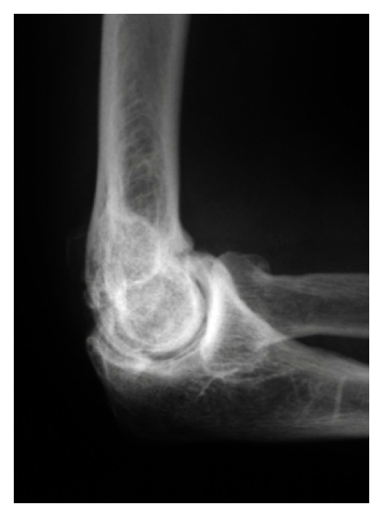
X-rayLateral elbow radiograph: osteophytes on the radial head and on the anterior and posterior aspects of the ulnohumeral joint, typical signs of elbow osteoarthritis.
Source : Biswas et al., Arthritis, 2013, figure 1 · CC BY
Two films are enough for the diagnosis. The rest of the imaging does not serve to confirm, it serves to prepare a procedure or to rule out something else, and the classifications the report uses are not all equally reliable.
Two views, AP and a strict lateral, are enough to make the diagnosis. On them you look for exactly what Dalal's series quantified: osteophytes of the olecranon, the coronoid process and the radial head, filling of the olecranon and coronoid fossae, loose bodies, and a joint space whose relative preservation is an argument in favour and not against6.
The strict lateral is the one that counts: it shows the filling of the fossae and the impinging osteophytes, that is, precisely what explains the extension pain. An AP view alone can massively underestimate the disease.
The reading trap comes down to one sentence: the coronoid osteophyte exists in 36 % of control subjects of the same age6. It is the only sign that normal ageing produces often enough to be misleading. The others (olecranon osteophyte, filling of the fossae, thickening of the membrane) are discriminating.
Three systems circulate in reports and publications. A reliability study published in Osteoarthritis and Cartilage compared them on 81 cases, having each file rated by four orthopaedic surgeons, twice11. The results deserve to be known before basing a decision on a stage.
| Classification | Medium | Inter-observer agreement (ICC) | Reading |
|---|---|---|---|
| Hastings and Rettig | Radiograph | 0.544 then 0.582 | Moderate agreement: two readers may classify the same elbow differently |
| Broberg and Morrey | Radiograph | 0.620 then 0.656 | Moderate to substantial agreement, the better of the two radiographic systems |
| CT-based classification | CT scan | 0.867 then 0.909 | Almost perfect agreement; correlated with the visual analogue scale (r = 0.754) and with the Mayo score (r = −0.614) |
The practical consequence is direct: a radiographic stage is not solid data, and the difference in severity between two successive reports may be nothing more than a difference between two readers. If a surgical decision turns on the stage, it is CT that decides, and it is also CT that, in that study, correlates best with what the patient feels.
Key points
This is the subgroup that on its own justifies a chapter: elbow osteoarthritis is the leading identified cause of surgically treated ulnar neuropathy, and it is also the variable that decides the surgical timetable.
A Japanese multicentre series reviewed the records of 413 elbows operated on for ulnar neuropathy at the elbow, diagnosed on the clinical and electrophysiological criteria of the American Association of Electrodiagnostic Medicine. An elbow lesion was present in 75.1 % of cases, and the commonest of all was primary elbow osteoarthritis, found in 54.5 % of the elbows. Next came the medial ganglion (8.5 %) and cubitus valgus (6.5 %), themselves most often associated with osteoarthritis14.
More worrying: the severity. McGowan grade III involvement (that is, with motor deficit and wasting), concerned 50.8 % of osteoarthritic elbows against 35.0 % of elbows with no associated lesion14. In other words, when osteoarthritis is the cause, the nerve is on average more damaged by the time it is operated on. The slow onset of bony compression lets the patient get used to it, and the diagnosis comes late.
Faced with an osteoarthritic elbow, the question is not “should the ulnar nerve be examined?” but “how fast is it deteriorating?”.
For a long time compression was explained by narrowing of the cubital tunnel in flexion. A dynamic CT study measured the thing in vivo, scanning 13 osteoarthritic elbows with cubital tunnel syndrome and 25 osteoarthritic elbows without, in three positions: full extension, 90° of flexion, full flexion12.
The result reverses the classic explanation. The area of the humeral osteophytes and their proportion inside the tunnel increased markedly with flexion: 24.7 mm² and 49.9 % in full flexion in symptomatic patients, against 9.0 mm² and 31.3 % in extension. And those values were significantly higher in symptomatic patients than in the others. The authors conclude that it is the effect of the medial osteophytes, humeral above all, on the nerve, and not narrowing of the tunnel, that explains the compression12.
The practical consequence is immediate and worth explaining to the patient: it is prolonged flexion that makes things worse, because it brings the osteophyte against the nerve. Sleeping with the arm bent, long phone calls, driving with the elbow bent resting on the door, working at a keyboard with the elbows tightly flexed: those are the positions to correct, and that correction is the first therapeutic step for the associated neuropathy.
There is one randomised trial, on cubital tunnel syndrome in general, not specifically on osteoarthritic forms. Seventy patients with mild or moderate symptoms were allocated to three groups: night splint, nerve gliding, or a control group. All received information on the cause of the symptoms and on the positions to avoid. At six months, 51 of the 57 patients followed up (89.5 %) had improved, and there was no difference between the three groups on any of the variables measured: not the Canadian Occupational Performance Measure, not the pain scales, not strength, not electrophysiology13.
The honest reading of that trial is not that physiotherapy is useless: it is that information and avoidance of provocative positions do most of the work, and that neither the night splint nor nerve gliding adds anything measurable in mild to moderate forms. That is good news for the patient, and it is an argument for investing the first session in explanation rather than in a technique.
What that trial does not say, on the other hand: nothing about severe forms, nothing about osteoarthritic forms specifically, and nothing about McGowan grade III, which, as we have seen, accounts for half the osteoarthritic elbows operated on. Faced with an established or progressing motor deficit, the timetable is no longer that of rehabilitation.
What should trigger a surgical opinion without waiting
Key points
This chapter begins with an admission, because leaving it out would amount to manufacturing a certainty: there is, to this day, no controlled trial of physiotherapy in primary elbow osteoarthritis. What follows is therefore reasoned transposition, and each proposal carries the level of evidence that really belongs to it.
The search was conducted for this article on PubMed, crossing elbow osteoarthritis with exercise, rehabilitation, corticosteroid injections, hyaluronic acid and platelet-rich plasma: no relevant randomised controlled trial exists at this site. The reference reviews confirm it by omission: the one by Del Core and Koehler recommends starting with “activity modification, anti-inflammatories, hand therapy and corticosteroids”15, without being able to underpin that sequence with anything other than customary consensus.
So you have to tell the patient where what you are proposing comes from. Two indirect sources are legitimate and a third is not:
Objective 1: preserve the useful arc. Loss of extension is the first to set in and the hardest to recover. Daily maintenance work, through range, without forcing against a hard stop, aims to delay the establishment of the flexion contracture. The marker to give the patient is not “you must straighten the arm completely” but the functional arc: 75 to 120° of flexion allows almost all everyday activities8, while knowing that contemporary activities, the telephone first among them, demand more9.
Objective 2: protect the ulnar nerve. Correct prolonged flexion positions, at night as at work, on the basis of the mechanism demonstrated on dynamic CT12. It is probably the measure with the best benefit-to-effort ratio in the whole arsenal.
Objective 3: reduce the impingement load. Analyse the occupational and sporting movements that take the elbow into terminal extension under load, and change them: carry close to the body rather than with the arm extended, bring loads nearer the trunk, break up the use of vibrating tools, adjust the height of the work surface.
Objective 4: maintain strength without provoking. Strengthening is done in the pain-free range, avoiding the ends of range. Triceps work in terminal extension against resistance is precisely what to avoid at the start, since it compresses the posterior impingement.
A safety principle, stated as reasoning and not as evidence
Faced with a hard, bony stop at end of range, forced passive mobilisations have no mechanical reason to work : you do not deform an osteophyte by pulling on it. They do expose the patient to post-mobilisation inflammation and to worsening pain. This position is not supported by a trial, there is none, but by the nature of the obstacle. Faced with a soft stop, by contrast, the capsular component justifies patient range-of-motion work, over months rather than weeks: in Lindenhovius's trial, the arc was still gaining between the sixth and the twelfth month17.
The ranking below applies the GRADE principles (nature of the study design, directness of the evidence, precision, risk of bias), to the precise question of primary elbow osteoarthritis. This is an editorial appraisal: no formal GRADE assessment has been published for this site, and no learned society has issued a guideline covering it.
Information and avoidance of prolonged flexion positions, when a mild to moderate ulnar neuropathy is associated. Direct evidence from a randomised trial: 89.5 % improvement at six months, with neither night splint nor nerve gliding adding anything at all13. Downgraded from “high” because the population is not exclusively osteoarthritic.
Prolonged range-of-motion work on stiffness with a capsular component, with or without a splint. Randomised trial on post-traumatic stiffness: a gain of 47° to 49° of arc at twelve months, with no difference between static progressive and dynamic splinting17. Evidence that is indirect for osteoarthritis, where the obstacle is partly bony.
Education about the disease and a structured land-based exercise programme. Core treatments for knee, hip and polyarticular osteoarthritis according to OARSI 201916, but the elbow is not covered by those guidelines. An acknowledged extrapolation.
Activity modification and management of occupational load. Recommended first line by the reference reviews155 ; consistent with the measured risk factor that vibrating tools represent7. No efficacy data of its own.
Muscle strengthening outside the ends of range. No trial at this site. Justified by functional reasoning and by preservation of the cartilage, not by a demonstration.
Intra-articular corticosteroid injection. Cited as a non-surgical option by the reviews15, with no trial at all in elbow osteoarthritis. Note: perioperative corticosteroids are among the risk factors for complications of elbow arthroscopy18.
Analgesics and anti-inflammatories. In common use, mentioned by the reviews15. OARSI does not recommend oral anti-inflammatories in the presence of cardiovascular comorbidity or frailty, and strongly advises against opioids16.
Viscosupplementation, platelet-rich plasma, shockwave therapy in elbow osteoarthritis: a systematic search conducted for this article found no trial. Offering one of these treatments here means offering an extrapolation from another joint, and the patient has a right to be told so.
Forced passive mobilisations against a hard stop. No data, and no plausible mechanism: an osteophytic block does not yield to stretching. The benefit-to-risk ratio is unfavourable by construction.
Key points
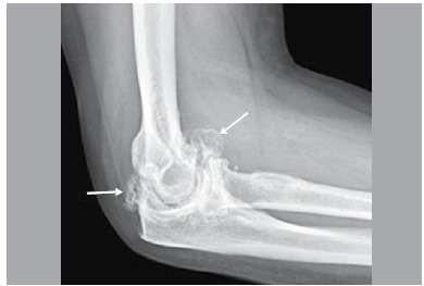
X-rayElbow radiograph at an advanced stage of osteoarthritis: destruction of the articular surface and intra-articular loose bodies, arrows, corresponding to the most severe radiographic grade.
Source : Casimiro et al., Acta ortopedica brasileira, 2016, figure 4 · CC BY
The physiotherapist does not set the indication for surgery, but is almost always the one who sees the patient long enough to spot the moment when it becomes reasonable, and the one to whom the aftermath is entrusted.
The relative preservation of the cartilage changes everything. The surgical reviews agree on a gradation that has no equivalent at the knee or the hip: debridement and osteocapsular arthroplasty are enough in most symptomatic elbow osteoarthritis, distraction arthroplasty has its place in the young person at an advanced stage, and total replacement is reserved for end-stage forms in the older, low-demand person19.
In practice, what the surgeon removes is what blocks: osteophytes of the olecranon and the coronoid, loose bodies, contracted capsule, with, in the technique known as Outerbridge-Kashiwagi, a fenestration of the olecranon fossa that restores the travel of the olecranon. Open or arthroscopic, the principle is the same.
The reference series is Antuña's: 46 elbows operated on with ulnohumeral arthroplasty between 1986 and 1996, reviewed at a mean of 80 months. The flexion-extension arc went from 79° to 101° ; at final follow-up, 35 elbows out of 46 (76 %) were painless or only slightly painful. The Mayo score was excellent for 26 elbows, good for 8, fair for 4 and poor for 83.
The British series by Phillips, with a mean follow-up of 75 months on 20 ulnohumeral arthroplasties, gives convergent results: 85 % good or excellent results on the DASH, 65 % on the Mayo score, a flexion contracture reduced by 10° on average and 20° of flexion gained. Of the 16 patients who were working at the time of the operation, 12 (75 %) returned to the same job20.
These gains in range, some twenty degrees, may seem modest. Set against the functional arc, they are not: going from 79° to 101° of arc means crossing the threshold that separates a hand that no longer reaches the face from a hand that gets back to it.
What surgical debridement gives back, and what it does not
Two independent series, mean follow-up of 80 and 75 months
Sources: Antuña 2002, J Bone Joint Surg Am (PMID 12473704): 13 patients out of 45 reported postoperative ulnar symptoms, that is 29 %, of whom 6 were reoperated on; Phillips 2003, J Bone Joint Surg Br (PMID 12729106); Tat 2022, J Shoulder Elbow Surg (PMID 35189372), survival free of conversion to total replacement at ten years, 98.5 % after open debridement and 98.0 % after the Outerbridge-Kashiwagi procedure.
A systematic review of 18 papers totalling 625 patients and 634 elbows compared the two approaches. Both improve pain, the flexion-extension arc and functional scores. The open approach gains more in flexion; arthroscopy gives more consistent results. The price differs: complications 5.1 % open against 2.0 % arthroscopic, revisions 9.5 % against 5.6 %21.
An earlier controlled, non-randomised trial pointed the same way with a useful nuance: arthroscopic debridement relieved pain better, the Outerbridge-Kashiwagi technique improved flexion better22. The choice is therefore made on the patient's dominant complaint as much as on the surgeon's habit.
The particular risk of elbow arthroscopy deserves to be known and passed on, because it is specific to this joint: a systematic review of 52 papers reports 1.5 % to 11 % total complications and 1.26 % to 7.5 % nerve injuries, with obesity, age over 65, previous surgery and perioperative corticosteroid injections as risk factors18. No level I or II study exists on the subject.
A systematic review of 49 studies and 1,995 total elbow replacements measured what the operation gives depending on the indication. For rheumatoid arthritis and fractures, long-term survival is satisfactory. For osteoarthritis, the rates reported are 50 % complications and 11 % revisions23. Conversely, survival free of conversion to a replacement after simple debridement reaches 98 % at ten years24.
The arithmetic is quickly done for a man of fifty who works with his arms: you do not put a total elbow replacement in a patient who is going to ask it to carry loads. The review by Gramstad and Galatz puts it bluntly: prosthetic arthroplasty is rarely indicated in primary osteoarthritis and must be reserved for older, low-demand people in whom the other options have failed5.
No trial has compared rehabilitation protocols after debridement of the osteoarthritic elbow. What can be tied to evidence concerns stiffness: in Lindenhovius's randomised trial of 66 patients, the flexion arc improved by 29° at three months, 40° at six months and 47° at twelve months, with no difference between dynamic splinting and static progressive splinting17. The message the authors draw from it is explicitly this: patience is justified.
Two points of vigilance specific to this surgery should guide the first weeks:
Key points
Three documented situations, taken as they were published. None is reconstructed for the sake of the example: the first two are series data, the third a case report with its identifier.
Case 1: Elbow osteoarthritis revealed by a claw hand
Spalkit S, Sinha A, Prakash M, Tripathy S. BMJ Case Reports 2021;14(5):e24277326: free full text (PMC8103949).
A 59-year-old woman, obese (body mass index 35.6), presents with weakness of the right hand developing over two years, with paraesthesiae and numbness of the fourth and fifth fingers. She describes a weak grip and difficulty cooking and cutting.
Examination finds moderate ulnar clawing of the last two fingers, weakened grip and reduced sensation in the ulnar territory. Electromyography shows a right axonal ulnar neuropathy, with unrecordable sensory and motor velocities. Ultrasound shows bowing of the nerve in the cubital tunnel with focal thickening, synovial thickening, a joint effusion and medial osteophytes arising from the olecranon. Radiographs show extensive degenerative changes with narrowing and bulky marginal humeroulnar osteophytes. MRI confirms ulnotrochlear osteophytes projecting into the cubital tunnel and displacing the nerve.
Treatment consisted of neurolysis with anterior transposition of the ulnar nerve, followed by regression of the paraesthesiae and numbness at three months.
What this case teaches. The presenting complaint was not the elbow: it was the hand. Two years went by before the diagnosis, and the involvement had become axonal. In any patient describing a weak ulnar hand, examining and radiographing the elbow costs little, and it is sometimes the only route to the cause.
Case 2: The collective picture of 46 operated elbows
Antuña SA, Morrey BF, Adams RA, O'Driscoll SW. J Bone Joint Surg Am 2002;84(12):2168-2173: PMID 12473704. This is a series, presented here as a group portrait and not as a patient.
Forty-six elbows, 45 patients, 44 men to a single woman, mean age 48. All described pain at terminal extension. Fourteen reported locking, twelve ulnar symptoms. At operation, loose bodies were removed from 36 elbows; a capsular release was needed in 19; a procedure on the ulnar nerve in 8.
At a mean follow-up of 80 months, the arc had gone from 79° to 101°, and 76 % of the elbows were painless or only slightly painful. But thirteen patients out of 45 reported postoperative ulnar symptoms, of whom six had to be reoperated on.
What this series teaches. The identikit picture is reliable: man, in his fifties, dominant elbow, impingement pain. And the ulnar nerve is the common thread: present before the operation in a quarter of patients, appearing or worsening afterwards in nearly a third. The authors draw an operational instruction from it: in a patient whose extension is limited by more than 60° or whose flexion does not reach 100°, consider preventive decompression.
Case 3: Sixteen patients followed without surgery, and a favourable course
Doherty M, Preston B. Ann Rheum Dis 1989;48(9):743-747: PMID 2802795, free full text (PMC1003867).
Sixteen patients, 14 men and 2 women, mean age 61, with no obvious trauma. Symptoms had begun on average at the age of 53 and had lasted seven years on average, with extremes ranging from one to twenty years. The joint fluids sampled in six of them were non-inflammatory; two biopsies showed non-specific synovitis.
Of the 26 affected elbows, 22 (85 %) had a favourable clinical course. Thirteen of the sixteen patients had osteoarthritis elsewhere, notably at the second and third metacarpophalangeal joints (10 of 16).
What this series teaches. It is the indispensable counterweight to the two previous cases. The disease most often runs a good course without being operated on, over years, with fluctuating symptoms. Announcing surgery straight away to a patient whose elbow is a nuisance but does not lock, and whose nerve is fine, is to ignore what this series documented more than thirty years ago.
Key points
A condition that is rare in the clinic, with no therapeutic trial and no guideline, calls for an approach written down in advance: otherwise every practitioner reinvents their own.
| What is measured | Why | Interval |
|---|---|---|
| Flexion, extension, pronation-supination in degrees | The only objective marker of the disease; the arc determines the surgical indication | Every 4 to 6 weeks |
| End-feel | A soft stop that hardens marks the progression of the bony block | At every assessment |
| Ulnar examination (sensation, pinch strength, muscle bulk) | The commonest and most disabling complication; its progression tips the balance towards surgery | At every assessment, and at every session if signs are present |
| Locking: number and circumstances | A sign of loose bodies, whose removal is the most rewarding procedure | Ask at every assessment |
| Mayo score (MEPS) or QuickDASH | Instruments used by the elbow surgical series, which makes the values comparable with the literature | Initially, then every 3 months |
| Work capacity | Return to the job is the most concrete outcome measure: 75 % after surgery20 | Initially, then at every change |
Recognition as an occupational disease: an often forgotten route
Schedule No. 69 of the French general scheme, entitled “Conditions caused by vibration and shock transmitted by certain machine tools, tools and objects and by repeated impacts of the heel of the hand on fixed elements”, explicitly mentions “osteoarthritis of the elbow with radiological signs of osteophytosis”, with a claim period of 5 years25. A minimum duration of exposure is required depending on the heading: it is for the occupational physician and the general practitioner to prepare the file. This route concerns directly the profile that epidemiology has identified, since the use of vibrating tools is associated with grade 4 forms7. The site devotes a detailed article to the process, using the shoulder as its example: acromioclavicular osteoarthritis and occupational disease.
No: the bony lesions do not regress. But the patient's question is really about the symptoms, and there the answer is more encouraging. In Doherty's series, 22 of the 26 affected elbows had a favourable clinical course without surgery4. And at population level, the disease is radiographically present in one adult over 50 in four while fewer than one in a hundred suffers from it7.
Rarely stop it, often adapt it. What provokes is end of range under load and prolonged flexion. Break up the use of vibrating tools, bring loads nearer the body, avoid pushing with the arm extended and positions with the elbow bent and supported. The use of vibrating tools is the only occupational factor formally associated with advanced forms in the general population study7.
Because sleeping with the arm bent brings the medial osteophytes against the ulnar nerve. Dynamic CT has shown it: their area inside the cubital tunnel goes from 9.0 mm² in extension to 24.7 mm² in full flexion12. Correcting the night-time position is the first treatment, and in mild to moderate forms, information alone proved as effective as a night splint in a randomised trial13.
It is cited as an option by the reference reviews15, but no trial has evaluated it in elbow osteoarthritis. It must therefore be announced for what it is: an individual therapeutic trial, with no efficacy data specific to this site. Note also that perioperative corticosteroid injections are among the risk factors for complications of elbow arthroscopy18.
Months, not weeks. The most informative randomised trial on elbow stiffness (post-traumatic, therefore with a capsular component) shows an arc gain of 29° at three months, 40° at six months and 47° at twelve months17. The authors conclude explicitly that patience is justified. In osteoarthritis, where part of the obstacle is bony, you have to be more cautious still in what you promise.
It is very unlikely. At the elbow, the surgical strategy is first to remove the osteophytes and the loose bodies, and survival free of conversion to a replacement reaches 98 % at ten years after simple debridement24. Total replacement remains reserved for end-stage forms in the older, low-demand person: put in for osteoarthritis, it shows 50 % complications and 11 % revisions in the reference systematic review23.
No. That is in fact the commonest situation: 25.2 % radiographic prevalence after 50 against 0.9 % symptomatic forms7. And an isolated coronoid process osteophyte is found in 36 % of control subjects of the same age6. An image is not a disease.
Yes, provided they are clear about what is demonstrated and what is not. Two things rest on randomised trials: information and correction of provocative positions for the ulnar nerve13, and prolonged range-of-motion work on stiffness with a capsular component17. The rest (maintaining the arc, strengthening outside the ends of range, load management) is a matter of coherent clinical reasoning, which the patient has a right to know as such.
To go further on this site
Every reference has been checked individually in PubMed: existence of the identifier, accuracy of the journal and the year, and reading of the abstract to check that the source does establish what is attributed to it.
Method and limitations
The sources were searched in PubMed via the NCBI E-utilities, crossing elbow osteoarthritis with epidemiology, diagnosis, imaging, ulnar neuropathy, exercise, injections and surgery. Every identifier was resolved and every abstract read before citation. Main limitation: the literature at this site is almost exclusively surgical and retrospective, only one randomised trial cited here concerns the elbow itself, and it deals with post-traumatic stiffness, not osteoarthritis. The levels of evidence in the table of modalities are an editorial appraisal applying the GRADE principles, and not a published GRADE assessment. Article written on 14 August 2026.
Behind this article
An author who explains, a reviewer who checks.
How we write and check our content

Physiotherapist · co-founder of Physio Learning
Marked for life by his first four-hour lecture without a single image, he took a master’s in instructional design so that it would never happen to anyone again. He hunts down publication bias and unreadable slides with the same intransigence.

Head of scientific content
Physiotherapist specialising in neuro-musculoskeletal practice and holder of a master’s in public health. He checks the methodological rigour of every article: primary sources, levels of evidence, no exceptions.
