














On-siteDPCFIFPL

Lower limb
Simplifying hip rehabilitation and empowering your patients
Mehmet Gemthe.hip.physio
Go further
In sports hernia there is no true hernia: the Doha classification, examination of the inguinal canal, imaging, rehabilitation and return to sport.

Pain and tenderness of the inguinal canal, with no palpable hernia: this is not a hernia, but an inguinal entity, and rehabilitation is the first-line treatment.
94%of patients operated on for chronic groin pain are men
What follows covers each of these points in detail, with sources. It is there if you need it.
This topic is taught in a course: Simplifying hip rehabilitation and empowering your patients, on-site with Mehmet Gem.
1 / 13
12 article chapters · 52 min in total
In brief◔ 2 min
Of 102 patients operated on for groin pain, only two had a true hernia at surgery.
Terminology◔ 6 min
The entities often add up: 44% of multiple causes in Taylor, 33% in Hölmich.
Anatomy◔ 4 min
The nerve component is massive: 96.2% in surgical series.
Epidemiology◔ 5 min
No single figure describes the frequency of the inguinal entity in the sporting population.
Examination◔ 5 min
You must examine all five entities every time, not just the one you suspect.
Differentials◔ 4 min
Femoroacetabular impingement is the leading cause in operated patients (32%): the hip is examined systematically.
Imaging◔ 4 min
No imaging criterion enters the definition: the diagnosis remains clinical.
Treating◔ 5 min
Even the surgical consensus places tailored physiotherapy as the initial treatment.
Surgery◔ 6 min
The decisive criterion is not the time elapsed, it is the quality of the conservative treatment actually given.
Case reports◔ 3 min
Two professional athletes returned to sport without surgery, in 4 and 7 weeks of rehabilitation.
In practice◔ 4 min
Correcting the word “hernia” is a clinical act, not a nuance of vocabulary.
Questions◔ 4 min
Athletic pubalgia and adductor tendinopathy are not the same thing: two distinct entities.
Train on this
Lower limb
Mehmet Gemthe.hip.physio
Next comes practice: the course that teaches this topic, with Mehmet Gem.
Our courses on this topic may be covered by DPC or FIFPL, the French continuing-education funding schemes, depending on your profession and situation.
Groin pain in the athlete · Inguinal wall · Clinical synthesis
In French, “pubalgie” names no disease. It is an umbrella, and under that umbrella the Doha agreement placed five distinct clinical entities that are not treated in the same way. This article deals with the one of the inguinal wall, what the English-language literature calls sports hernia, athletic pubalgia or inguinal disruption. An entity whose commonest name refers to a lesion that does not exist: there is no hernia.
The term is the first trap. “Pubalgie” covers five entities; “sportsman's hernia” names a hernia that does not exist. The British Hernia Society consensus (Sheen 2014, PMID 24149096) explicitly rejected the terms sportsman's hernia and sportsman's groin in favour of inguinal disruption, on the grounds that no true hernia is present. Imaging confirms it: across 102 athletes operated on for athletic pubalgia, Zoga et al. found a hernia in only two of them (PMID 18487535).
The Doha framework avoids the sorting error. Twenty-four experts from fourteen countries defined five entities on the basis of history and physical examination alone: adductor, iliopsoas, inguinal, pubic, plus hip-related groin pain (Weir 2015, PMID 26031643). The inguinal entity is defined by pain in the inguinal canal AND tenderness of that canal, with no palpable hernia.
The entities add up more than they exclude each other. Among 100 athletes assessed under Doha, 44 % had several simultaneous causes (Taylor 2018, PMID 28654441). In Hölmich's cohort of 207 patients, abdominal wall involvement was isolated in only 2 patients out of 20: the other 18 also had adductor involvement (PMID 17261557). Looking for a single entity is the second trap.
Clinical examination decides, but it is less reliable than people think. Heijboer et al. measured agreement between two independent examiners: it ranges from κ = 0.01 for the consistency of the posterior wall, that is chance, to κ = 0.72 for certain abdominal resistance tests. Their conclusion is blunt: there is no perfect clinical examination test (PMID 36643406).
Conservative treatment first, and the literature does not agree on what comes next. The Manchester consensus recommends tailored physiotherapy as initial treatment. The only randomised trial available (Paajanen 2011, PMID 21549403) gives the advantage to surgery (90 % against 27 % return to sport at 3 months), but the largest meta-analysis, 3,332 athletes, finds no difference in return-to-sport rate between surgery and rehabilitation (King 2015, PMID 26130700). That disagreement is the subject of chapter 8.
This article deals with the inguinal entity. For the adductor entity, see the dedicated article.
Both carry the word “pubalgie”, and that is precisely the problem the Doha agreement seeks to solve. Adductor tendinopathy is the commonest entity (61 % of the athletes in Taylor's cohort): pain on palpation of the adductors and on resisted adduction, with an exercise protocol validated by randomised trial. What you are reading here is the neighbouring entity: pain in the inguinal canal, examination of the wall, with no palpable hernia. The two often coexist in the same athlete: all the more reason to know how to tell them apart.
In this chapter: why an umbrella term held research back for thirty years, what the 2015 Doha agreement settled, what the 2014 Manchester consensus rejected, and the table of the five entities with their exact diagnostic criteria.
“Pubalgie” is a convenient word and that is its whole flaw. It names a painful region, not an injury mechanism; it gathers under one label an insertional tendinopathy, bony distress of the symphysis, involvement of the lower abdominal wall and sometimes a hip joint condition. The authors of the Doha agreement put it in one sentence: the heterogeneous taxonomy of groin injuries adds confusion to an already complicated field. That imprecision is not merely a problem of vocabulary: it made studies incomparable with each other for decades.
On 4 November 2014, twenty-four experts from fourteen countries met in Doha for a day of work preceded by a Delphi questionnaire and systematic reviews. The result, published in 2015 in the British Journal of Sports Medicine, is a unanimous agreement on a three-level classification (Weir 2015, PMID 26031643) :
The decisive point is methodological: these definitions rest solely on history and physical examination. None requires imaging. That is what makes them usable in the clinic on the day of the first consultation, and it is also what makes them fragile if the examination is poorly conducted: we return to that in chapter 5.
| Entity | Diagnostic criterion (Doha 2015) | What increases the probability | First line |
|---|---|---|---|
| Adductors Adductor-related |
Adductor tenderness AND pain on resisted adduction | Pain reproduced on the squeeze test; history of adductor injury | Progressive active exercise: supported by a randomised trial (Hölmich 1999) |
| Iliopsoas Iliopsoas-related |
Iliopsoas tenderness | Pain on resisted hip flexion and/or on stretching the flexors | Rehabilitation; the iliopsoas strength test is the least reliable of all (Hölmich 2004) |
| Inguinal Inguinal-related (the subject of this article) |
Pain localised to the region of the inguinal canal AND tenderness of the inguinal canal. No palpable inguinal hernia. | Pain worsened by resistance of the abdominal muscles or by Valsalva, coughing, sneezing | Tailored physiotherapy, a multidisciplinary approach (Manchester consensus 2014) |
| Pubic Pubic-related |
Local tenderness of the pubic symphysis and of the immediately adjacent bone | No specific resistance test provokes the symptoms: the Doha group notes this explicitly | Rehabilitation; faster return to sport than after surgery (10.5 against 23.1 weeks, King 2015) |
| Hip Hip-related |
No single definition: history (onset, nature, mechanical symptoms) and examination | Limited passive ranges; FADIR and FABER tests | Its own approach: see femoroacetabular impingement and labral tear |
Two years before Doha, the British Hernia Society brought together in Manchester surgeons, a musculoskeletal radiologist and a physiotherapist to settle the question of surgical vocabulary. Their conclusion, published in 2014, is explicit: the term adopted is inguinal disruption, and the terms sportsman's hernia and sportsman's groin are rejected, on the grounds that no true hernia exists (Sheen 2014, PMID 24149096).
The group described massive agreement on the existence of abnormal tension in the groin, particularly around the insertion of the inguinal ligament, with frequent findings of a detachment of the external oblique accompanied by small tears, and tissue oedema. It recommended a multidisciplinary approach with tailored physiotherapy as initial treatment, surgery consisting of releasing the tension of the inguinal canal then reinforcing it with mesh or suture.
You have to gauge what that implies in practice. A patient who is told they have a “sportsman's hernia” understands that they have a hole in their abdomen, which will have to be closed. The word makes the prognosis in the clinician's place, and it steers towards the operating theatre a condition that the consensus recommends treating first with rehabilitation. That is why this vocabulary deserves to be corrected with the patient at the first consultation.
The term athletic pubalgia remains widely used, especially in the North American literature and in imaging, where it most often refers to involvement of the insertion of rectus abdominis and of its common aponeurosis with adductor longus on the pubis. It is therefore not an exact synonym of inguinal disruption : the two terms refer to neighbouring but distinct anatomical anchor points, the first centred on the pubic insertion, the second on the inguinal canal. In French practice, both are met under the name “pubalgie du sportif”, which brings us back to the original problem.
Distribution of the entities in athletes consulting for groin pain
Two independent cohorts, two classification methods, one and the same lesson
Sources: Taylor R, et al. Clin J Sport Med. 2018;28(4):364-369 (PMID 28654441); Hölmich P. Br J Sports Med. 2007;41(4):247-252 (PMID 17261557). Both cohorts come from specialist centres: the proportions there are heavier than in a community clinic. Hölmich 2007 predates the Doha agreement and classifies into three patterns, of which “rectus abdominis” partly overlaps the inguinal entity without matching it.
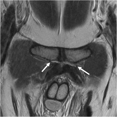
MRICoronal MRI of the pubic symphysis in a man with athletic pubalgia: bilateral clefts of fluid signal at the origin of the adductor tendons, more pronounced on the left (arrows).
Source : Li et al., Insights into Imaging, 2016, figure 13 · CC BY
In this chapter: the anatomy of the inguinal canal and of the structures that cross it, the loading mechanism that explains the footballer's pain, the share taken by the nerves, measured at 96 % in one surgical series, and why the word “weakness” is more accurate than the word “hernia”.
The inguinal canal is an oblique passage of four to six centimetres cut through the thickness of the anterior abdominal wall, just above the inguinal ligament. It transmits the spermatic cord in men, the round ligament in women, and the ilioinguinal nerve and the genital branch of the genitofemoral nerve. Its anterior wall is formed by the aponeurosis of the external oblique, its floor by the inguinal ligament, its roof by the arching fibres of the internal oblique and transversus, and its posterior wall by the transversalis fascia reinforced medially by the conjoint tendon.
It is that posterior wall that draws the attention. Farber and Wilckens described it as early as 2007 as the site of an occult hernia caused by weakness or tearing of the posterior inguinal wall, with no clinically recognisable hernia, leading to chronic groin pain (PMID 17664370). The word “occult” says a lot about the embarrassment: a hernia is postulated that no examination shows.
The pubic region is the point where two antagonistic muscle systems meet. Above, the abdominal muscles (rectus abdominis, obliques, transversus) pull the symphysis up and back. Below, the adductors, and adductor longus first among them, pull down and forward. Those two forces meet on a common aponeurosis inserted on the pubis.
The at-risk sporting movement combines hip extension, trunk rotation and violent abdominal contraction: the shot, the change of direction, the throw, the skating stride. Repeated, it imposes on that crossroads shearing forces that neither bone nor aponeurosis fully dissipates. The Manchester consensus draws from it the most cautious and probably the most accurate formulation: what is found is abnormal tension in the groin, particularly around the insertion of the inguinal ligament, with sometimes a detachment of the external oblique and small tears, and tissue oedema.
The inguinal canal: the structures involved
A schematic sagittal section of the anteroinferior abdominal wall, right side
Classical descriptive anatomy. The frequencies of nerve involvement come from the surgical series of Gerhardt M, et al. J Hip Preserv Surg. 2020;7(1):103-108 (PMID 32382436), where nerve entrapment was found intraoperatively in 96.2 % of the 51 athletes operated on. Those figures describe an operated population and do not extrapolate to patients as a whole.
The series by Gerhardt et al. covers 51 athletes operated on for groin pain between 2009 and 2015, with a mean follow-up of 4.42 years. What the surgeons found on opening is telling: nerve entrapment in 96.2 % of cases, involving the ilioinguinal nerve in 92.5 %, the iliohypogastric in 30.8 % and the genitofemoral in 13.2 %. Attenuation of the posterior inguinal wall was present and repaired in 79.3 % of cases, and scar tissue surrounding the adductor origin required debridement in 56.7 %.
A 2025 review devoted to neuropathic causes confirms that shift of attention: the anatomical variability of the nerve courses and the overlap of their sensory territories complicate diagnosis, and the clinical distinction between neuropathic pain (burning, electric shock) and nociceptive pain (dull, aching) becomes an orientation element in its own right. The authors are a reminder that conservative treatments remain first line, surgery being reserved for persistent cases (Vuckovic 2025, PMID 40032740).
In this chapter: the real weight of groin injuries in professional football over fifteen seasons, the place of the inguinal entity among the causes operated on, the profile of patients and why the published figures depend first of all on where you count.
The prospective study of UEFA elite clubs provides the most solid measure available. Across 47 European teams followed between 2001-2002 and 2015-2016, that is 268 team-seasons, hip and groin injuries account for 1,812 injuries out of 12,736, that is 14 % of the total, at a rate of 1.0 injury per 1,000 hours of exposure. Adductor-related injuries alone make up 63 % of them (Werner 2019, PMID 29691289).
The most interesting result of that study lies elsewhere. Over fifteen years, the rate of hip and groin injuries fell by 2 % a season, and that of adductor injuries by 3 %. But the burden, the number of days lost per 1,000 hours, did not move (p = 0.40). In other words: people get injured slightly less often, and stay out just as long. For a condition whose management is reputed to be difficult, the finding is worth pondering.
Fifteen seasons of European professional football
Prospective study of UEFA elite clubs, 47 teams, 268 team-seasons
Source: Werner J, Hägglund M, Ekstrand J, Waldén M. Hip and groin time-loss injuries decreased slightly but injury burden remained constant in men's professional football: the 15-year prospective UEFA Elite Club Injury Study. Br J Sports Med. 2019;53(9):539-546 (PMID 29691289). An exclusively male, professional population.
One difficulty must be stated frankly: there is no single prevalence figure for the inguinal entity, and the published proportions vary with the population recruited. Three measurements, three populations, three results:
Those figures do not contradict each other: they count different things. The last describes only the patients who made it to theatre, which by construction excludes everyone whom rehabilitation was enough to cure. That is the classic bias of surgical series, and it means never reading an operative success rate as a measure of how common the disease is.
The systematic review by Hatem et al., which brings together 47 studies and 2,737 patients operated on for chronic groin pain, gives the broadest portrait: 94 % men, mean age of 27.8 at the time of the operation (range 12 to 65), and a mean symptom duration of 13.1 months before the operation. Football dominates by far (71 %), followed by rugby (7 %), Australian rules football (5 %) and ice hockey (4 %): PMID 34541009.
That thirteen-month delay is the most telling figure in the series. It does not say that the condition takes a year to declare itself: it says that a year passes between the onset of symptoms and the procedure. Part of that delay is legitimate rehabilitation time; another part is diagnostic wandering, and it is that part the Doha framework seeks to reduce.
In this chapter: the two mandatory Doha criteria, the palpation and resistance tests named one by one, their measured inter-examiner agreement (from κ = 0.01 to κ = 0.72) and what the absence of a perfect test means for how the consultation is run.
The Doha definition comes down to one sentence, and every one of its terms counts: “Pain localised to the region of the inguinal canal AND tenderness of the inguinal canal. No palpable inguinal hernia is present.” The probability increases if the pain is worsened by a test of abdominal muscle resistance, or by a Valsalva, a cough, a sneeze.
Two practical consequences follow. First, palpation of the canal is mandatory : pain of inguinal location without tenderness on palpation does not meet the criterion. Second, looking for a palpable hernia is an exclusion criterion, not a diagnostic argument: if you feel a hernia, you are dealing with a hernia, and the patient needs a surgical opinion, not this framework.
The study by Heijboer et al., conducted at Aspetar hospital between March 2019 and October 2020, standardised the examination in 44 male athletes aged 18 to 40 with long-standing groin pain, that is 61 symptomatic sides. Two independent examiners, blind to each other, carried out the same protocol (PMID 36643406). The protocol included:
The test most often positive in athletes classified as having a defined inguinal entity is the reproduction of recognisable pain on palpation during scrotal invagination under Valsalva: 79 %. The abdominal resistance tests, for their part, were positive in only 21 to 49 % of cases depending on the test, which is enough to disqualify the idea that a negative resisted abdominal test rules out the diagnosis.
Agreement between two independent examiners
Cohen's kappa coefficient, 44 athletes, 61 symptomatic sides (Heijboer 2023)
Source: Heijboer WMP, Vuckovic Z, Weir A, et al. Clinical examination for athletes with inguinal-related groin pain: interexaminer reliability and prevalence of positive tests. BMJ Open Sport Exerc Med. 2023;9(1):e001498 (PMID 36643406). The kappa interpretation thresholds are those of Landis and Koch, by convention. A male-only population: scrotal invagination has no equivalent in women, which the study does not make up for.
A κ of 0.01 for judging the consistency of the posterior wall means that two trained clinicians, examining the same patient on the same day, agree at the level of chance. That is not a criticism of the examiners: it is the measure of a limit intrinsic to a manoeuvre that consists of judging a deep structure “firm or soft” through skin, subcutaneous tissue and scrotum.
The consequence is direct. The diagnosis of the inguinal entity is built by convergence : location of the pain, reproduction on palpation of the canal, worsening with Valsalva or abdominal resistance, absence of a palpable hernia, and above all reasoned elimination of the neighbours. Basing the decision on a single test (particularly on the perception of a bulge, whose agreement is poor) invites error.
It should be added that the reliability of groin examination is not uniformly poor. Hölmich et al. measured in 2004, in 18 athletes examined twice by two doctors and two physiotherapists, intra-observer kappas above 0.60 in 11 tests out of 14, and inter-observer kappas above 0.60 in 8 of the 10 pain tests. The only test without acceptable reliability was the iliopsoas strength test (PMID 15273182). It is therefore the deep palpation manoeuvres of the wall that are fragile, not groin examination in general.
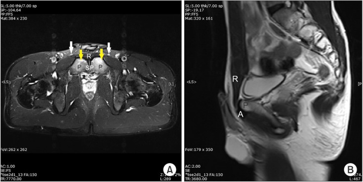
MRIMRI of the pubic symphysis in two slices, fat-saturated T2 sequences: bilateral and symmetric bone marrow oedema of both sides of the symphysis, yellow arrows, most pronounced anteroinferiorly.
Source : Yang et al., Annals of Rehabilitation Medicine, 2015, figure 4 · CC BY-NC
In this chapter: femoroacetabular impingement and the real value of the FADIR, the true inguinal hernia, the non-musculoskeletal causes the Doha agreement requires you to consider, and a sorting tree usable in the clinic.
The inguinal entity is a diagnosis of convergence, but also, in part, a diagnosis of exclusion. Three families must be ruled out: the other Doha entities, hip pathology, and what the consensus places under “other causes”.
It is the commonest and the most misleading neighbour. In the review by de Sa et al. covering 4,655 athletes operated on for groin pain, femoroacetabular impingement is the leading cause, at 32 % of indications: ahead of athletic pubalgia (24 %). Intra-articular and extra-articular causes are equal there, which makes it impossible to treat the hip as a secondary hypothesis.
The screening test is the FADIR (flexion-adduction-internal rotation). Its benchmark meta-analysis credits it with a pooled sensitivity of 0.94 - 0.99, but with diagnostic odds ratios whose confidence intervals cross unity, and the authors conclude that these tests have only screening value (Reiman 2015, PMID 25515771). In practice: a negative FADIR makes impingement unlikely and that is its whole value; a positive FADIR does not confirm it. For the full approach, see our articles on femoroacetabular impingement and hip labral tear.
The distinction is simple in principle and decisive in practice: a true hernia can be palpated. The Doha definition explicitly excludes the inguinal entity in the presence of a palpable inguinal hernia. Examination standing, at rest then on Valsalva and coughing, looks for a mass or a reducible bulge. A genuine hernia warrants a surgical opinion straight away, without going through rehabilitation.
The rarity of true hernia in this population deserves emphasising one last time: in the series by Zoga et al., of the 102 patients whose images could be compared with the operative findings, only two had a hernia. That is what justifies the consensus rejecting the term.
The Doha agreement does not stop at a list: it calls for a high level of clinical suspicion, because these causes do not reveal themselves on musculoskeletal examination. The categories retained are orthopaedic, neurological, rheumatological, urological, gastrointestinal, dermatological, oncological and surgical.
In practice, in an athlete referred for “groin pain”, you have to be able to consider testicular disease, a urinary infection, prostatitis, endometriosis in women, gastrointestinal disease in the iliac fossa, early spondyloarthritis, since sacroiliitis can refer to the groin, and a stress fracture. What these situations have in common is that they resist rehabilitation without the physiotherapist understanding why, which should in itself be a signal.
Sorting tree for groin pain in the athlete
An approach based on history and physical examination, following the structure of the Doha agreement
Built from the classification structure of Weir A, et al. Br J Sports Med. 2015;49(12):768-774 (PMID 26031643), from the verbatim definitions of each entity and from the prevalence of multiple causes reported by Taylor 2018. This tree organises an approach; it does not replace the examination and has not been prospectively validated as an algorithm.
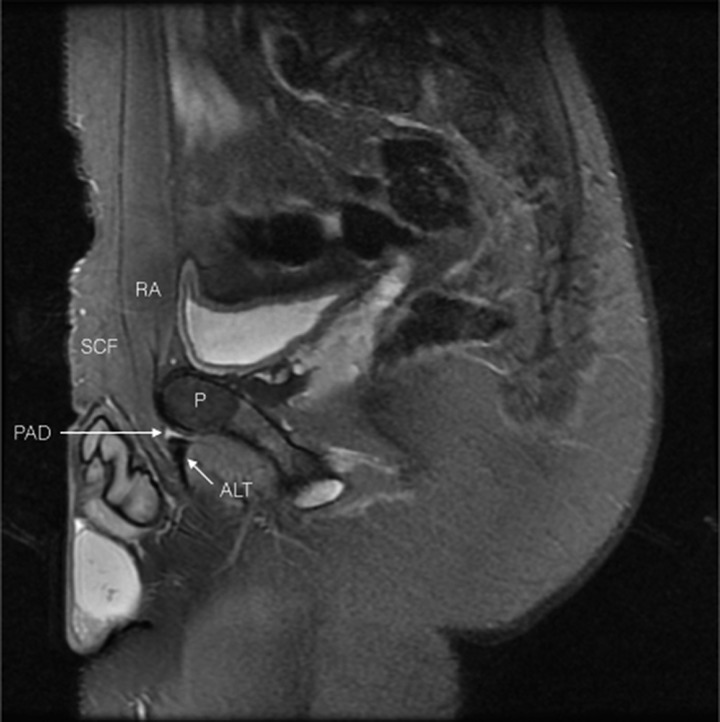
MRISagittal MRI of the groin with anatomical landmarks: pubic aponeurosis defect (PAD) at the junction of the rectus abdominis and adductor tendon, a lesion typical of athletic pubalgia.
Source : Falvey et al., British Journal of Sports Medicine, 2016, figure 3 · CC BY-NC
In this chapter: the measured performance of MRI against operative findings, what dynamic ultrasound brings and at what price, why normal imaging rules nothing out, and the rule that follows from all this.
The classic position is that of Farber and Wilckens: the role of imaging in this condition is unclear; most investigations will be normal (PMID 17664370). Twenty years later, that sentence remains largely true for the inguinal entity in the Doha sense, whose definition contains no imaging criterion. But it deserves qualifying by the quantitative data available.
The reference study is that of Zoga et al., which retrospectively compared the MRIs of 141 patients referred for groin pain with the operative findings in 102 of them, with a control group of 25 asymptomatic men (PMID 18487535). The results are mixed:
The specificity of 100 % for rectus abdominis is the usable figure: when MRI states a rectus abdominis insertion tear, it is right. The sensitivity of 68 % is the symmetrical warning: an MRI that shows nothing misses nearly a third of those lesions. Zoga also noted that patients with involvement of the rectus abdominis insertion were the most likely to go on to surgical repair of the pelvic floor.
Performance of MRI against operative findings
141 patients, of whom 102 with surgical comparison, and 25 asymptomatic controls
Source: Zoga AC, Kavanagh EC, Omar IM, et al. Radiology. 2008;247(3):797-807 (PMID 18487535). A retrospective study in a specialist centre, with a surgical reference available in 102 of the 141 patients: performance there is measured in a population with a high pre-test probability and does not transfer as it stands to the clinic.
Ultrasound has the advantage of being dynamic: it allows the wall to be loaded in real time. The Manchester consensus mentions it among the useful imaging modalities. The case reported by Yang et al. illustrates its use: a 19-year-old footballer whose abdominopelvic CT was normal, and in whom dynamic ultrasound showed a direct bulge arising from the posterior inguinal wall on abdominal tensing, with the legs raised during a full inspiration. Surgery confirmed the deficiency of the posterior wall (PMID 26798621).
That case shows the value of the method and, at the same time, its limit: it is an isolated case, operator-dependent, and it should be set against the κ of 0.29 measured by Heijboer for the clinical assessment of bulging of the posterior wall. The sign is real but hard to establish reproducibly, whatever the method used to look for it.
Three indications emerge, and one non-indication:
In this chapter: what the consensus recommends, a methodological warning about Hölmich's protocol, what the data specific to the inguinal entity actually allow you to state, and a table of modalities with their level of evidence.
The first-line recommendation is clear and comes from the most specific consensus on the question. The Manchester group, convened by the British Hernia Society, and therefore by surgeons, recommends a multidisciplinary approach with tailored physiotherapy as initial treatment, surgery coming only afterwards (PMID 24149096). That surgeons place rehabilitation in the front line deserves emphasis.
Hölmich's active exercise protocol is regularly presented as the reference treatment for groin pain. One has to be precise about what it establishes, because that is where the commonest error slips in.
The randomised trial published in the Lancet in 1999 included 68 athletes with adductor-related groin pain, as the title of the paper says explicitly: adductor-related groin pain, of a median duration of 40 weeks. The active programme, centred on strengthening and coordination of the muscles acting on the pelvis and in particular the adductors, allowed 23 patients to return to sport without pain against 4 in the passive physiotherapy group, that is an odds ratio of 12.7 (95 % CI 3.4 to 47.2) : PMID 9989713. The effect was maintained at 8-12 years of follow-up (PMID 21813441).
That distinction is not academic scruple. If the patient has both entities (a frequent case, as noted: 18 of the 20 rectus abdominis cases in Hölmich were associated with the adductors), then the protocol treats the adductor part with an excellent level of evidence, and the inguinal part by ricochet, with no guarantee. Knowing that changes how you announce the prognosis and how you interpret a failure.
The data are thin and that must be said. The systematic review by Serner et al. sifted 72 studies on the treatment of groin pain: only four were of high quality. For sportsman's hernia, the synthesis concludes to moderate level evidence in favour of surgery compared with conservative treatment (PMID 25633830).
But the same review delivers a result that must temper that conclusion: the correlation between the methodological quality of a study and the success rate it reports is moderate and inverse (r = −0.41, p < 0.001). In other words, the more rigorous a study, the less effective it finds the treatment. When the most favourable evidence comes from the weakest studies, caution is needed on both sides of the debate.
| Modality | What it targets | What the literature establishes | Level of evidence for the inguinal entity |
|---|---|---|---|
| Tailored physiotherapy first line | Reloading the abdominopelvic crossroads before considering a procedure | Explicit recommendation of the 2014 Manchester consensus, backed by multidisciplinary expert agreement | Moderate (consensus, not a trial) |
| Retraining deep abdominal control | Restoring trunk stability and force transmission | Two detailed case reports with return to sport (a professional golfer in 4 weeks, a professional ice hockey player in 7 weeks) | Low (level 4) |
| Progressive active exercise, Hölmich type | Strengthening and coordination of the pelvic muscles, adductors foremost | Randomised trial, OR 12.7, but in the adductor entity, effect maintained at 8-12 years | High for the adductors · extrapolated for the inguinal entity |
| Nerve blocks (ilioinguinal, genitofemoral) | Treating and documenting a neuropathic component | 2025 narrative review: relief in many cases, first line alongside rehabilitation | Low (narrative review) |
| Preventive adductor programme (Copenhagen) | Preventing the occurrence and recurrence of groin problems | Cluster randomised trial, 652 players: prevalence 13.5 % against 21.3 %, risk reduced by 41 % (OR 0.59) | High in preventing groin problems from all causes |
| Rest alone | Not applicable: it is not an active strategy | The “physiotherapy without active exercise” arm of Hölmich's trial achieves 4 pain-free returns to sport, against 23 in the active arm; Paajanen's conservative arm, 27 % return to sport | Contraindicated as a sole strategy |
The cluster trial by Harøy et al. deserves to be known precisely, because it is one of the few good quality randomised trials in the field. Thirty-five Norwegian semi-professional teams were randomised: 18 teams (339 players) performed an adductor strengthening programme based on a single exercise, the Copenhagen Adduction, at three levels of progression, three times a week during pre-season (6 to 8 weeks) then once a week in season (28 weeks); 17 teams (313 players) trained as usual.
Result: a mean prevalence of groin problems of 13.5 % (95 % CI 12.3-14.7) against 21.3 % (95 % CI 20.0-22.6), that is a risk reduced by 41 % (OR 0.59, 95 % CI 0.40-0.86, p = 0.008), PMID 29891614. These are groin problems from all causes, measured by self-report questionnaire, and not the inguinal entity alone: the nuance matters but takes nothing away from the value of a programme that takes five minutes.
In this chapter: the randomised trial that favours surgery, the meta-analysis that finds no difference, the techniques and their results by anatomical site, and how to hold these data together without sacrificing one of them.
This is the chapter where the literature contradicts itself, and it is better to say so at the outset than to choose the source that suits. Three good-quality pieces of work give three different answers, because they do not ask the same question.
Paajanen et al. conducted the only randomised trial directly comparing the two strategies in this indication. Sixty patients with chronic groin pain and suspected sportsman's hernia, defined by the authors as a deficiency of the posterior wall of the inguinal canal, were randomised after 3 to 6 months of symptoms into two groups of 30 (PMID 21549403) :
The results are clear-cut: 27 of the 30 operated patients (90 %) returned to sport after 3 months, against 8 of the 30 treated conservatively (27 %), p < 0.0001. Superiority in pain was significant from 1 to 12 months (p < 0.001). Seven patients in the conservative group (23 %) were eventually operated on because of persisting pain.
Two reservations, however. The first is that the conservative arm combined corticosteroid injections and anti-inflammatories with physiotherapy: that is not the progressive exercise programme current consensus recommends. The second is the sample size, 60 patients, and the absence of blinding, unavoidable when one arm involves an incision.
King et al. pooled 56 studies and 3,332 athletes to compare rates and timing of return to sport between surgery and rehabilitation, grouping diagnoses into three anatomical families: abdominal wall, adductors, pubis (PMID 26130700).
Their conclusion is that return-to-sport rates are comparable between surgery and rehabilitation in all three diagnostic groups. For pubic-related pain, rehabilitation even returns athletes to sport significantly faster: 10.5 weeks against 23.1 weeks. The “abdominal wall” group, the one that concerns us here, is the one that recovers fastest of the three, in both strategies.
The authors attach a severe warning to that result: the overall methodological quality is low, the evidence is mostly level IV, and the risk of bias is particularly high in the surgical studies.
Three pieces of work, three answers on surgery
What each measures, and why the conclusions diverge
Sources: Paajanen H, et al. Surgery. 2011;150(1):99-107 (PMID 21549403); King E, et al. Br J Sports Med. 2015;49(22):1447-1451 (PMID 26130700); Hatem M, et al. Orthop J Sports Med. 2021;9(9) (PMID 34541009). Paajanen's rates are measured at 3 months; Hatem's at the latest follow-up available, with heterogeneous follow-up durations.
The review by Hatem et al. is the most useful for informing a decision, provided you keep in mind that it compares nothing: it describes what becomes of 2,737 patients operated on across 47 studies, of which 44 are level IV and only 2 are level 1b randomised trials. Return to the previous level or higher is:
Return to the previous level is therefore significantly more likely after inguinal surgery (92 %) than after adductor surgery (75 %). The confidence intervals of the other two groups are so wide that they allow no firm conclusion: that of the symphysis runs from 47 % to 100 %.
| Technique | Principle | Published data | Level |
|---|---|---|---|
| Totally extraperitoneal (TEP) endoscopic repair with mesh | Mesh placed behind the symphysis and the painful area, endoscopically | Randomised trial: 90 % return to sport at 3 months against 27 % (Paajanen 2011) | A single randomised trial, 60 patients |
| “Minimal Repair” by open suture | The defect in the posterior wall is not enlarged; a virtually tension-free suture | Prospective cohort of 129 patients, assessment at 4 weeks; median duration of pain before the procedure: 142 days (Muschaweck 2010) | Cohort with no comparator |
| Neurolysis and wall repair according to the findings | The procedure adapted intraoperatively: neurolysis, wall repair, adductor debridement | 51 athletes, 96.1 % return to the same level at a mean of 5.9 weeks, mean follow-up 4.42 years, 2 revisions (Gerhardt 2020) | Retrospective case series |
| Tension release then reinforcement (mesh or suture) | The principle adopted by the Manchester consensus, with no stated preference of technique | Expert agreement; recommendation of a national registry of operated athletes | Consensus |
The reading that respects all three pieces of work is the following. Surgery works : return-to-sport rates after an inguinal procedure are high and consistent from one series to another. It is not for all that superior to rehabilitation in the only large-scale comparison available, and the single randomised trial that finds it superior compared surgery with a conservative arm that was not a modern progressive exercise programme.
To that is added Serner's warning: in this field, the most rigorous studies report the poorest results. A rate of 92 % coming mostly from level IV studies must be read with that implicit correction.
In practice, the elements that tilt towards a surgical opinion are: failure of a well-conducted progressive exercise programme over at least two to three months, pain that stays localised to the inguinal canal and reproducible on palpation, a clear neuropathic component, and an athlete whose season or career forces a decision. Conversely, a patient who has had only rest, anti-inflammatories and shockwave therapy has not had conservative treatment: they have had a wait.
In this chapter: three published, identified cases, two treated without surgery with return to sport, and a third that shows what dynamic ultrasound brings and where it stops. What each demonstrates, and what it does not.
The cases that follow are genuine indexed case reports , readable in full with open access on PubMed Central. They are level 4 evidence: they illustrate an approach, they demonstrate no efficacy.
Presentation. A professional golfer develops lower abdominal and groin pain after a change in his physical preparation routine. The clinical picture is consistent with a diagnosis of sports hernia.
Management. A structured programme of trunk musculature retraining, organised in progression following the neurodevelopmental sequence, targeting the neuromuscular control and stability needed for return to golf.
Result. Full return to playing golf after 13 physiotherapy sessions over 4 weeks (Becker & Bullock, Int J Sports Phys Ther 2014, PMID 25383252, full text PMC4223293).
What it brings. The authors themselves note that no case of managing this condition had been reported in a golfer, and that the available data broadly favour surgery. Their conclusion is measured: a structured rehabilitation programme may be an option prior to surgical repair, allowing some athletes to return to their sport.
Presentation. A professional ice hockey player injures his abdomen during a game; the diagnosis retained is sports hernia.
Management. Structured conservative treatment centred on trunk control and stability, with progressively increasing peripheral demands, then functional training before return to play.
Result. Return to full competition seven weeks after the injury. A rare element in a case report: the authors specify that the player was still competing in the NHL seven years later (Woodward, Parker & van der Meer, Int J Sports Phys Ther 2012, PMID 22319682, full text PMC3273884).
What it brings. The authors point to a fundamental methodological problem: the publications favourable to surgery describe poor results of conservative treatment without ever specifying what that treatment consisted of. That is exactly the criticism that can be made of the conservative arm of Paajanen's trial, and it is an argument worth knowing when a patient arrives with a “failure of rehabilitation” in their notes.
Presentation. A 19-year-old footballer presents with groin pain worsened by sporting activity. The abdominopelvic CT performed in general surgery shows no abnormality; the patient is referred to rehabilitation medicine.
Approach. History and physical examination point the way, and dynamic ultrasound reveals a direct bulge arising from the posterior inguinal wall when abdominal tension is induced by raising both legs during a full inspiration.
Result. Surgery confirmed the lesion, allowing early management (Yang et al., Ann Rehabil Med 2015, PMID 26798621, full text PMC4720758).
What it brings. A normal static investigation, here a CT, does not rule out a wall deficiency that only reveals itself under load. That is the argument for dynamic imaging when the clinical picture insists and the standard work-up stays silent.
In this chapter: the synthesis of the levels of evidence, a consultation framework, what to do in the first weeks, the referral criteria and what to tell the patient about the word “hernia”.
What is known, and how solidly
Grading of the claims in this article by the type and quality of the data
Grading established by the authors of this article from the study type and the reservations formulated by the authors themselves. It does not take up a published formal GRADE rating: no graded clinical practice guideline exists to date for the inguinal entity of the Doha agreement.
It is a consultation moment in its own right, and it is short. The patient often arrives with the phrase “sportsman's hernia”, heard at the club, from a teammate or read online. That phrase carries a precise picture: a hole in the wall, which has to be closed surgically. Yet the learned society that brings together hernia surgeons has rejected that term, on the grounds that no true hernia is present; and the only series that compared imaging with operative findings found a hernia in only two patients out of a hundred and two.
Saying that is not cheap reassurance: it is correcting a belief which, left intact, steers the patient towards the operating theatre before they have received the treatment the consensus places in the front line.
This article deals with the inguinal entity. The neighbouring entities are the subject of dedicated syntheses.
Behind this article
An author who explains, a reviewer who checks.
How we write and check our content

Physiotherapist · co-founder of Physio Learning
Marked for life by his first four-hour lecture without a single image, he took a master’s in instructional design so that it would never happen to anyone again. He hunts down publication bias and unreadable slides with the same intransigence.

Head of scientific content
Physiotherapist specialising in neuro-musculoskeletal practice and holder of a master’s in public health. He checks the methodological rigour of every article: primary sources, levels of evidence, no exceptions.
