














The patient is sitting in the waiting room and is perfectly well. He mentions, almost apologising for bringing it up, that last Thursday his right arm went heavy for twenty minutes, and that his speech was slurred. It passed on its own. He called nobody. This article deals with what is decided in those twenty minutes recounted after the event: what a transient ischaemic attack has become since its definition changed, what risk it really carries and on what timescale, what the ABCD2 score is worth and above all what it is not worth, and what the physiotherapist must do who, very often, is the first clinician to hear the story.
- Updated August 2026
- Level clinical summary
- Sources 23 verified references (PMID)
- Reading time about 40 minutes
Three figures that frame the urgency
The risk after a TIA, what a rapid pathway takes off it, and what imaging finds
Sources: Giles and Rothwell, Lancet Neurology 2007 (PMID 17993293); Rothwell et al., Lancet 2007 (PMID 17928046); Amarenco et al., New England Journal of Medicine 2016 (PMID 27096581).
In brief
- A TIA is not a small stroke, it is a stroke that has not happened yet. Since 2009 the definition is no longer chronological (under 24 hours) but tissue-based: there is a TIA when the deficit resolves and no infarction is established1.
- The risk is concentrated in the first few days. About 5.2 % stroke at 7 days across all cohorts, but 11.0 % in population studies without urgent treatment, against 0.9 % in specialist emergency pathways3.
- Speed changes the outlook, and it is the only variable we control. EXPRESS: 10.3 % then 2.1 % recurrence at 90 days, with no excess bleeding4.
- The ABCD2 score does not sort patients reliably. Prospectively validated in 2,056 patients, it proved inaccurate at every threshold8. Above all, 20 % of patients with a low score have carotid stenosis of more than 50 % or atrial fibrillation9.
- For the physiotherapist, the rule comes down to one sentence: a focal neurological deficit of sudden onset, even one that has entirely resolved, even one from a few days ago, even in a patient who feels well, calls for medical advice the same day. Resolution is not good news, it is the time you have left.
What is a TIA, and why has its definition changed?
For forty years, TIA was defined by a clock. Today it is defined by an image, and that shift is not a quarrel about nomenclature: it moved part of what used to be called TIA over to the side of established infarction, and it made imaging an urgent act rather than a confirmatory test.
From duration to tissue
The historical definition was convenient and wrong. It said: a focal neurological deficit of vascular origin that resolves completely within 24 hours. The 24-hour threshold had no biological basis; it was set by consensus in the mid-1960s, at a time when the brain could not be looked at.
Diffusion imaging showed what the threshold was hiding. In patients whose signs had all disappeared, and well before the twenty-fourth hour, an infarction was found. In 2009 the American Heart Association and the American Stroke Association therefore redefined TIA as a transient episode of neurological dysfunction caused by focal brain, spinal cord or retinal ischaemia, without acute infarction1. Duration leaves the definition; the absence of a lesion enters it.
The practical consequence is direct and often poorly understood: you cannot state that it was a TIA without imaging. Until the brain has been looked at, a resolved deficit is an ischaemic event of undetermined nature. That is exactly the reasoning that justifies the urgency, and not the other way round.
How many TIAs are actually infarctions
The figures are consistent from one cohort to another. In the SOS-TIA emergency clinic at Bichat hospital, 108 of the 643 patients with confirmed TIA, that is 17 %, had a tissue lesion5. In the international TIAregistry.org, which included 4,789 patients in 21 countries and in which 78.4 % were assessed by a specialist within 24 hours, 33.4 % had an acute cerebral infarction on imaging6.
In other words: between one patient in six and one in three referred for a deficit that has fully recovered already have a scar. The tissue has been lost; only the clinical picture has repaired itself, because the area involved was small or functionally redundant.
Key point
Complete clinical recovery says nothing about the integrity of the tissue, and nothing at all about the risk to come. A patient who is perfectly well in front of you may be carrying yesterday's infarction and a tight carotid stenosis. That is the heart of the TIA problem: the only symptom is already over by the time the patient consults.
The mechanism, in one page
A TIA is a brief interruption of blood flow in an arterial territory. Three main mechanisms account for most cases, and they have neither the same prognosis nor the same treatment.
- Large-artery atherosclerosis, first and foremost at the carotid bifurcation. A plaque ulcerates and throws platelet emboli towards the middle cerebral artery or the ophthalmic artery. It is the mechanism with the highest risk of early recurrence, and it is also the only one that calls for urgent surgery.
- Cardiac embolism, dominated by atrial fibrillation. The thrombus forms in the left atrial appendage and migrates. The treatment is anticoagulation, not antiplatelet therapy, which makes identifying the mechanism decisive.
- Small-vessel disease, or lipohyalinosis, which produces the lacunar syndromes. The risk of early recurrence is lower there, but the long-term burden of disability is not negligible.
In the TIAregistry, 23.2 % of patients had at least one extracranial or intracranial stenosis of 50 % or more, and 10.4 % atrial fibrillation6. Both of those causes can be treated, and treated well: which is precisely what makes delay culpable.
“The typical patient loses 1.9 million neurons for each minute their stroke goes untreated.” (Saver, Stroke, 2006)
That sentence concerns established stroke and not TIA20. But it describes exactly what is at stake in TIA: what is in play is not the episode that has passed, it is the infarction that threatens, and whose occurrence is counted in hours and days.
What is the real risk of stroke after a TIA, and over what period?
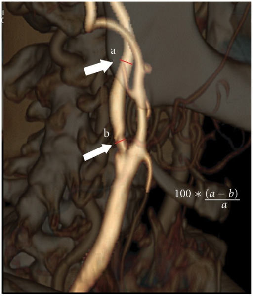
CT scan3D volume-rendered CT angiography of the carotid artery: measurement of the degree of internal carotid artery stenosis, arrows on the point of maximal stenosis and on a healthy reference segment.
Source : Stefanini et al., International journal of vascular medicine, 2012, figure 1 · CC BY
This is the question that has to be answered accurately, because it governs everything else. And the right answer is not a single figure: the risk after a TIA depends less on the patient than on what is done with them in the hours that follow.
An average figure that hides considerable heterogeneity
Giles and Rothwell's meta-analysis pooled 18 independent cohorts and 10,126 patients. The pooled risk of stroke at 7 days is 5.2 % (95 % CI 3.9 to 6.5). But the heterogeneity between studies is massive (p less than 0.0001), with risks ranging from 0 % to 12.8 %3.
The substance of the article lies in explaining that spread. The authors show that it is almost entirely explained by method, setting and treatment, and not by the patients:
The risk of stroke at 7 days depends on how care is organised, not only on the patient
Subgroups of the meta-analysis of 18 cohorts
Source: Giles MF, Rothwell PM, Lancet Neurology 2007; 18 cohorts, 10,126 patients (PMID 17993293). The extreme subgroups rest on 3 and 4 studies respectively.
A factor of twelve separates the two extremes. That is not a difference in severity between patients, it is a difference between care systems. The authors' conclusion is sober and weighty: the lowest risk is seen where treatment is given urgently in a specialist service.
The two demonstrations that changed practice
Two studies published in the same year, 2007, turned a correlation into a demonstration.
EXPRESS compared two consecutive periods in the same Oxford population. In phase 1 the patient was seen in clinic and preventive treatment was then started by the family doctor; in phase 2 the assessment and the first prescription took place the same day in clinic. The median time to assessment fell from 3 days to under 1 day, and the median time to the first prescription from 20 days to 1 day. The risk of recurrent stroke at 90 days fell from 10.3 % to 2.1 %, an adjusted hazard ratio of 0.20 (95 % CI 0.08 to 0.49; p equal to 0.0001)4. Two points deserve emphasis: the patients' baseline characteristics and the time they took to seek care were comparable between the two phases, and early treatment did not increase the risk of intracerebral haemorrhage or other bleeding.
SOS-TIA, in Paris, opened a clinic accessible round the clock and circulated its number to 15,000 doctors across the Île-de-France region. Of 1,085 patients admitted, 74 % went home the same day, and the stroke rate at 90 days was 1.24 % (95 % CI 0.72 to 2.12) when the rate predicted by the ABCD2 scores was 5.96 %5. The median symptom duration there was 15 minutes.
Key point
The risk after a TIA is not a fate written into the patient: it is a quantity that falls by 80 % when management is immediate. That is what makes delay serious. The clinician who puts things off by eight days is not making a diagnosis wait, they are letting a risk run that they could have divided by five.
The modern risk, and the risk that does not go away
Contemporary cohorts, managed in organised pathways, find figures markedly lower than those of the 2000s. TIAregistry.org gives the following Kaplan-Meier estimates after a TIA or minor stroke: 1.5 % at 2 days, 2.1 % at 7 days, 2.8 % at 30 days, 3.7 % at 90 days and 5.1 % at 1 year6.
The five-year follow-up of the same registry, covering 3,847 patients, brings the most useful information for the physiotherapist, who follows these patients for a long time. The cumulative stroke rate is 9.5 % at 5 years (95 % CI 8.5 to 10.5), and above all 43.2 % of those strokes occurred between the second and the fifth year7. The rate of cardiovascular events is 6.4 % in the first year, and another 6.4 % over the following four.
The cumulative risk of stroke after a TIA managed in a modern pathway
Kaplan-Meier estimates, international TIAregistry.org
Sources: Amarenco et al., New England Journal of Medicine 2016 (PMID 27096581) for the points from 2 days to 1 year, and 2018 (PMID 29766771) for the 5-year point, n equal to 3,847. The horizontal scale is not linear.
This result bears directly on everyday practice. The patient who had a TIA two years ago is not a cured patient: they remain, for a long time, a patient at high vascular risk. Their TIA belongs in their physiotherapy history, whatever they are consulting for today.
The trap of the recent figure
The low rates in TIAregistry are sometimes cited to play down the urgency: “the risk is only 2.1 % at 7 days”. That is a misreading. Those figures come from a cohort in which 78.4 % of patients were seen by a specialist within 24 hours and treated straight away. They describe the risk after exemplary management. They do not describe the risk for the patient sent home with an appointment in a fortnight.
How do you recognise a TIA, and what is not one?
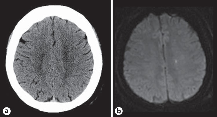
MRIBrain CT and MRI performed on the same day in a patient with a TIA: normal CT, but a very small area of restricted diffusion visible on diffusion-weighted MRI.
Source : Moreau et al., Cerebrovascular diseases extra, 2013, figure 1 · CC BY-NC
This is the difficult part, and it should be said plainly: there is no diagnostic test for TIA. The diagnosis rests entirely on a story, often reported after the event, by a patient who has nothing left to show. The rate of wrong diagnoses is consequently high, in both directions.
The clinical signature of an ischaemic event
Three features, taken together, point strongly to a vascular origin.
- The deficit is focal. It corresponds to an arterial territory, and therefore to an anatomy. A general faint turn, diffuse fatigue, tingling in all four limbs are not focal.
- Onset is sudden and the deficit is maximal from the start. Ischaemia does not spread step by step: it strikes a territory all at once. A march of symptoms over several minutes argues rather for another cause.
- The symptoms are negative. They are losses of function: loss of strength, loss of sensation, loss of speech, loss of vision. Positive phenomena (shimmering, tingling that spreads, involuntary movements) point rather to a migraine or an epileptic seizure.
The two main territories
| Territory | Typical features | What it signals |
|---|---|---|
| Carotid (anterior) | Contralateral hemiparesis or hemihypoaesthesia, aphasia if the dominant hemisphere is involved, amaurosis fugax (monocular loss of vision, described as a curtain coming down) | Prompts a search first of all for carotid stenosis on the same side. Amaurosis fugax points to the ophthalmic artery, the first branch of the internal carotid. |
| Vertebrobasilar (posterior) | Ataxia, vertigo, diplopia, dysarthria, dysphagia, bilateral visual disturbance, alternating or bilateral motor or sensory deficit, drop attack | A territory that is often missed: the signs do not fit the FAST mnemonic. Isolated vertigo is hard to tell there from a peripheral vestibular disorder. |
What the FAST mnemonic catches, and what it misses
The Face Arm Speech Test was built for prehospital alerting and it does that job well. In Harbison's study, ambulance staff trained in FAST made a correct diagnosis in 144 of the 183 stroke patients who presented to them, that is 79 %, and they brought markedly more patients in within 3 hours than general practitioners did (46 % against 12 %)12.
But FAST has a documented blind spot. Aroor and colleagues reviewed 736 consecutive records of ischaemic stroke: 14.1 % had no FAST symptom on admission. Among them, 42 % had a balance disturbance or lower limb weakness, 40 % visual symptoms, and 70 % one or the other. Adding those two items, so that the mnemonic becomes BE-FAST (Balance, Eyes, Face, Arm, Speech, Time), the proportion of unidentified strokes falls from 14.1 % to 4.4 % (p less than 0.0001)13.
What the mnemonic misses: from FAST to BE-FAST
Proportion of unidentified ischaemic strokes, consecutive series of 736 patients
Source: Aroor S, Singh R, Goldstein LB, Stroke 2017 (PMID 28082668). Single-centre series, 736 patients retained from 858 consecutive records.
For the physiotherapist, the implication is immediate: a sudden balance disturbance and a sudden visual disturbance are stroke symptoms, just as much as a drooping mouth. They are also, precisely, the complaints for which a patient may end up referred for vestibular rehabilitation or balance work rather than to the emergency department.
The mimics: what looks like a TIA without being one
The review by Nadarajan and colleagues, in Practical Neurology, sets the problem out well: the transience of the symptoms makes the differential diagnosis harder than that of established stroke, and the rate of mimics there is higher and more varied10. In Wardlaw's meta-analysis, 45 % of patients referred to a stroke prevention service were mimics9.
| Diagnosis | What points to it | What should give you pause |
|---|---|---|
| TIA | Sudden onset, maximal from the start, negative symptoms, a coherent arterial territory, vascular risk factors | No test confirms it in the clinic. The doubt is settled in the pathway, not in the treatment chair. |
| Migraine with aura | Positive symptoms (scintillating scotoma), slow progression over 5 to 30 minutes, a march of symptoms, headache afterwards, identical past episodes | Aura without headache exists, and a first aura after the age of 50 must be investigated like a TIA. |
| Focal epileptic seizure | Positive phenomena, clonic movements, automatisms, altered consciousness, a post-ictal deficit with gradual recovery | A post-ictal deficit (Todd's paralysis) mimics a TIA perfectly, and a seizure can reveal a vascular lesion. |
| Hypoglycaemia | Sweating, tremor, hunger, a context of treated diabetes, resolution once sugar is given | It can produce a genuine focal deficit. Capillary blood glucose is the first thing the emergency team does. |
| Benign paroxysmal positional vertigo or vestibular neuritis | Rotatory vertigo triggered by position, characteristic nystagmus, otherwise normal neurological examination | A cerebellar infarction sometimes presents as isolated vertigo. Headache, truncal ataxia and inability to stand unaided are warning signals. |
| Syncope, presyncope | Brief loss of consciousness, prodromes, pallor, an orthostatic context | An isolated syncope is not a TIA. But a drop attack in the posterior territory can resemble one. |
| Functional disorder | Inconsistent signs at examination, variability, distractibility | It is a diagnosis of exclusion, never a first-line diagnosis in the acute setting. |
The trap of reversed exclusion
The dangerous reasoning runs like this: “this patient has no vascular risk factor, he is young, so it is a migraine”. The correct reasoning is the opposite: the presence of risk factors raises the probability of TIA, but their absence does not eliminate it. Arterial dissections and cardiac sources of embolism strike young patients with no classic risk factor. The sorting belongs to the doctor, with imaging.
What is the ABCD2 score worth, and above all what is it not worth?
This is probably the point on which the gap between what is taught and what has been demonstrated is widest. The ABCD2 score still features in many teaching materials as the triage tool for TIA. The prospective data, however, say that it does not sort reliably, and above all that it does not see what matters.
What the score says
The score was derived and validated by Johnston, Rothwell and colleagues in 2007, by unifying two earlier scores (the California score and the ABCD score), across four independent cohorts totalling 2,893 patients2.
| Item | Criterion | Points |
|---|---|---|
| Age | 60 or over | 1 |
| Blood pressure | Blood pressure of 140/90 mmHg or more | 1 |
| Clinical features | Unilateral motor deficit | 2 |
| Speech disturbance without motor deficit | 1 | |
| Duration | 60 minutes or more | 2 |
| 10 to 59 minutes | 1 | |
| Diabetes | Diabetes present | 1 |
In the original publication, the stratification of stroke risk at 2 days was clear: 8.1 % for scores 6 and 7 (21 % of patients), 4.1 % for scores 4 and 5 (45 %), and 1.0 % for scores 0 to 3 (34 %)2.
The ABCD2 score as published: three risk strata at 2 days
Derivation and validation cohorts, 4,809 patients
Source: Johnston SC, Rothwell PM, Nguyen-Huynh MN, et al., Lancet 2007 (PMID 17258668). Risk of stroke within 2 days of the TIA.
What the score does not do
Two later pieces of work, one prospective and one meta-analytic, seriously dented the confidence that could be placed in it.
Perry's prospective validation enrolled 2,056 patients in eight Canadian emergency departments, with the score calculated by the physician before any decision about disposal. The observed stroke rate was 1.8 % at 7 days and 3.2 % at 90 days. A score above 5 had a sensitivity of 31.6 % (95 % CI 19.1 to 47.5) for stroke at 7 days: two strokes in three occurred in patients that threshold classified as low risk. A score above 2, the threshold recommended by the American Heart Association, did reach a sensitivity of 94.7 %, but with a specificity of 12.5 %, that is, at the price of classifying almost everyone as at risk. The area under the curve was 0.56 when the score was calculated by the front-line physician. The authors conclude that the score is inaccurate, whatever threshold is used8.
Wardlaw's meta-analysis, across 29 studies and 13,766 patients, adds the heaviest criticism. A score of 4 or more is sensitive (86.7 %) but not very specific (35.4 %) for recurrence at 7 days. And above all:
- 20 % of patients with a score below 4 had carotid stenosis of more than 50 % or atrial fibrillation, that is, a cause requiring urgent intervention;
- 35 to 41 % of mimics had a score of 4 or more, against 66 % of true TIAs: the score therefore does not separate TIAs from what is not one either;
- out of 1,000 patients referred, 52 % would score 4 or more, which does not reduce the clinic's workload9.
An analysis of the same TIAregistry had already shown that patients with an ABCD2 below 4 could have a 90-day stroke risk comparable to that of patients scoring 4 or more, as soon as they had large-artery atherosclerosis21.
What the ABCD2 score misses
Three independent measurements of its real performance
Sources: Perry JJ et al., CMAJ 2011 (PMID 21646462); Wardlaw JM et al., Neurology 2015 (PMID 26136519).
What predicts better than the clinical score
Imaging does what the clinical picture does not. Coutts and colleagues followed 120 patients with TIA or minor stroke, examined within 12 hours and imaged by MRI within 24 hours. The risk of stroke at 90 days, adjusted for baseline characteristics, was 4.3 % with no diffusion lesion, 10.8 % with a lesion but no arterial occlusion, and 32.6 % with both a lesion and an occlusion (p equal to 0.02)11. A factor of seven separates the extremes, in clinically similar patients.
It is that observation which led to scores incorporating imaging, such as ABCD2-I, where adding the brain and the carotids improves the identification of patients at early risk22. The general lesson does not change: what stratifies is investigation, and investigation is done urgently.
Key point
The ABCD2 score is not a reliable triage tool and it was never a licence to wait. Its only defensible use is to reinforce a decision to act urgently when it is high. It can in no way relax that decision when it is low: a score of 2 in a patient with a tight carotid stenosis describes a high-risk patient the score does not see. For the physiotherapist, the consequence is simple: there is no score to calculate in the clinic, there is a doctor to reach.
What happens in the pathway, and why do the hours count?
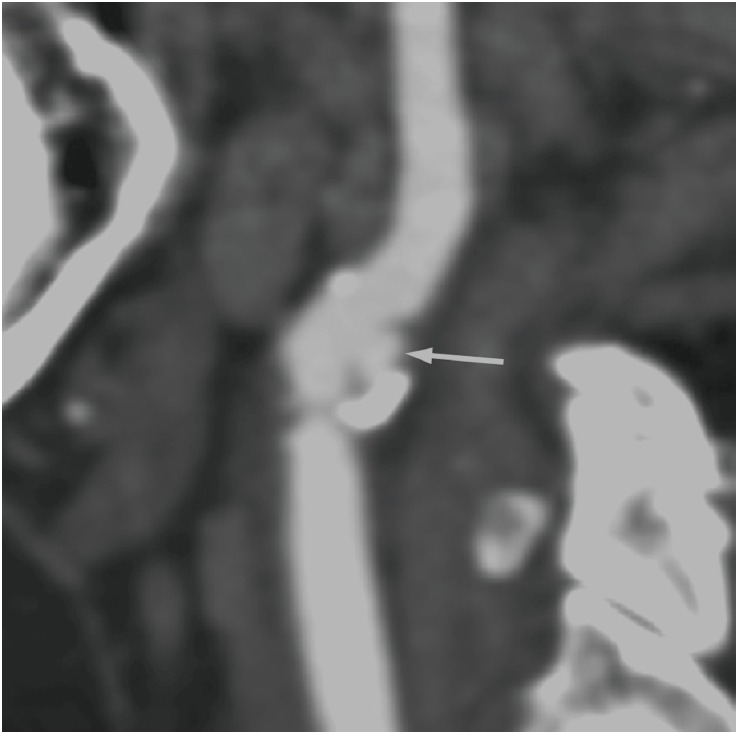
CT scanCervical CT angiography: ulcerated, calcified carotid plaque, arrow on the plaque, a frequent cause of transient ischaemic attack.
Source : Choi et al., Journal of stroke, 2015, figure 10 · CC BY-NC
Knowing what awaits the patient you refer is not idle curiosity: it is what lets you explain to them why they cannot wait until Monday, and convince them, which is often the hardest part.
The work-up, and what it looks for
Assessing a TIA aims to answer a single question: which mechanism, and therefore which treatment. It classically comprises brain imaging, investigation of the cervical and intracranial arteries, a search for a rhythm disturbance, and metabolic blood tests. The 2021 American guideline for stroke prevention sets out how it is organised16.
The search for atrial fibrillation deserves a separate mention, because it is the cause whose diagnostic yield depends most on the length of recording, and because its treatment differs radically. A patient labelled with a “cryptogenic TIA” on antiplatelet therapy may be a patient in paroxysmal atrial fibrillation who should have been anticoagulated.
Where that care takes place is not a matter of indifference. The Cochrane review of stroke units, which pooled 29 trials and 5,902 participants, shows that organised care in a dedicated unit reduces, compared with a conventional ward, the risk of a poor outcome (odds ratio 0.77; 95 % CI 0.69 to 0.87), of death (0.76; 0.66 to 0.88) and of death or dependency (0.75; 0.66 to 0.85), with moderate certainty evidence23. That is one more argument for referral to the stroke pathway where one exists, and not to a general service.
Two treatments whose benefit runs out with time
Short-course dual antiplatelet therapy. The POINT trial randomised 4,881 patients with minor ischaemic stroke or high-risk TIA between clopidogrel plus aspirin and aspirin alone. Major ischaemic events occurred in 5.0 % of patients on dual therapy against 6.5 % on aspirin alone (hazard ratio 0.75; 95 % CI 0.59 to 0.95; p equal to 0.02), most events occurring during the first week. The price is an increase in major haemorrhage, 0.9 % against 0.4 % (hazard ratio 2.32; 95 % CI 1.10 to 4.87)15. The CHANCE trial, conducted in China on the same principle, had opened this path14. The window of efficacy is narrow, which is one more argument against the deferred appointment.
Carotid endarterectomy. This is probably the most striking illustration of the cost of delay. Rothwell's pooled analysis, across 5,893 patients and 33,000 patient-years of follow-up, shows that the benefit of surgery depends not only on the degree of stenosis but on the time since the last event. For a stenosis of 50 % or more, the number of patients needing surgery to prevent one ipsilateral stroke at 5 years is 5 if the operation takes place within 2 weeks, and 125 beyond 12 weeks17.
The cost of delay: number of patients to operate on to prevent one stroke
Endarterectomy for symptomatic carotid stenosis of 50 % or more, by time since the last event
Source: Rothwell PM, Eliasziw M, Gutnikov SA, et al., Lancet 2004 (PMID 15043958). Pooled analysis of 5,893 patients, outcome: ipsilateral stroke at 5 years.
The benefit was also greater in men, and in patients aged 75 and over, which contradicts the instinct to rule older patients out.
The physiotherapist sees the patient afterwards: what should they do?
Here is the heart of the subject for our profession. The physiotherapist is not the one who makes the diagnosis of TIA nor the one who investigates it. They are very often the one who hears the story first, because they see the patient over time, regularly, and in a setting where people talk. That position gives them no additional diagnostic competence; it gives them a responsibility to refer, and that alone.
Why the physiotherapist is on the front line in spite of themselves
Three structural reasons, worth naming.
The resolution of symptoms defuses the alarm. A patient whose arm stayed heavy for twenty minutes and who has been well since has no natural reason to think emergency. So they mention it to whoever they see, which is often their physiotherapist, in passing during a session booked for something else.
Posterior circulation symptoms look like reasons for rehabilitation. A sudden balance disturbance, vertigo, unsteadiness when walking: these are common reasons for referral. The patient may arrive at the clinic with a prescription for vestibular rehabilitation or balance work for what was an ischaemic event. That is exactly the blind spot BE-FAST put figures on13.
The frequency of sessions makes the physiotherapist a sensor. Seeing a patient three times a week for six weeks is thirty chances to hear “by the way, the other day...”. No other clinician has that density of contact.
The three situations, and what to do in each
Situation 1: the episode happens during the session
This is a medical emergency, treated as such. You do not wait to see whether it passes: it is precisely the resolution that would lose the treatment window if the episode were a stroke in the making.
- Call 15 (the French emergency medical number) or 112 immediately, without waiting for the symptoms to end and without sending the patient to the emergency department under their own steam.
- Note the exact time the symptoms began, or failing that the last time the patient was seen well. That is the information that governs eligibility for thrombolysis and thrombectomy, and it is the one most often lost.
- Give nothing to drink or eat : swallowing difficulty is frequent and aspiration is a serious complication.
- Lie the patient down, head slightly raised, and monitor them without leaving them alone.
- Hand over to the call handler : time of onset, symptoms observed (and not merely reported), current medication, particularly anticoagulants and antiplatelets, and known medical history.
Situation 2: the patient describes a recent episode, now resolved
This is the most frequent and the most treacherous situation, because everything in the scene invites you to play it down: the patient is well, they minimise it, they came about their knee.
What to do is obtain medical advice the same day, with no exception based on how many days ago the episode was. In practice: reach the family doctor during the session, or refer to an urgent stroke clinic if the local pathway has one, or to the emergency department if no advice can be obtained. Do not settle for advising the patient to mention it “at some point”.
The reasoning that justifies that firmness is the one in chapter 2: the risk is concentrated in the first few days, it falls by 80 % when management is immediate4, and the benefit of any carotid surgery is divided by twenty-five once three months have passed17.
The physiotherapy session planned for that day has no reason to go ahead. Not because it would be dangerous in itself, but because the time of the session is the time that has to be given to the referral.
Situation 3: the patient had a TIA months or years ago
This patient is in secondary prevention, and will remain so. The five-year follow-up of TIAregistry is a reminder that 43.2 % of the strokes occurring within five years happen after the first year7.
The physiotherapist's role there is twofold and modest: contributing to regular physical activity, which is among the recommended secondary prevention measures16, and remaining the sensor for a new episode. A history of TIA is an assessment finding, not a decorative line of history.
Red flags: stop the session and call the emergency services
- A motor or sensory deficit of one side of the body of sudden onset, even one that has already resolved.
- Speech disturbance of sudden onset: word-finding difficulty, incoherent speech, slurred articulation.
- Facial asymmetry of recent onset.
- Loss of vision in one eye described as a curtain, or sudden loss of part of the visual field, or double vision.
- Sudden balance disturbance or unsteadiness with an inability to stand unaided, all the more so with headache: think of cerebellar infarction, which the FAST mnemonic does not see.
- Thunderclap headache, maximal from the outset, with or without a neurological sign.
- Recent neck pain or headache in a young patient, especially after even minor neck trauma, with neurological signs or a Horner's syndrome: consider arterial dissection.
In all these cases, the time the symptoms began is the most valuable information to hand over.
The four phrases that waste time
- “It's passed, that's a good sign.” The opposite is true: resolution is what defines a TIA, and a TIA is a warning.
- “We'll come back to it next session.” The next session is in three days, and half the risk is already behind us.
- “You should mention it to your doctor.” An instruction delegated to the patient is not a referral. You reach the doctor, or you refer.
- “It's surely nothing, you have no risk factors.” The absence of risk factors rules nothing out, and causes in younger patients exist.
What can physiotherapy do in secondary prevention?
Two things must be clearly distinguished here: what physical activity contributes as a modifiable risk factor, which is firmly established at population level, and what a supervised rehabilitation programme contributes after a TIA, which is far less so. Confusing the two would be selling a certainty that does not exist.
The frame: ten risk factors explain most of it
The INTERSTROKE study, across 32 countries, establishes that ten potentially modifiable risk factors are collectively associated with about 90 % of the attributable risk of stroke, in every major region of the world, in men as in women and at every age. Hypertension is more strongly associated there with intracerebral haemorrhage, whereas current smoking, diabetes, apolipoproteins and cardiac causes are more strongly associated with ischaemic stroke19.
Physical activity is one of those factors, and it is the only one the physiotherapist acts on directly in their practice. That is the rational basis for their involvement, and it is solid.
What a recent trial measured, and what it did not find
Honesty requires the following to be reported. Walton and colleagues published in 2026 in Clinical Rehabilitation a randomised controlled trial of cardiovascular rehabilitation after TIA or minor stroke: 140 participants, median age 73, randomised between a weekly six-week programme and usual care, with assessment at 6 weeks and 6 months.
The primary result is negative. No difference between groups on six-minute walk test distance in the adjusted analysis. The only significant differences concerned one dimension of quality of life: the AQoL-6D coping score at 6 weeks, in favour of rehabilitation (beta 3.6; 95 % CI 0.07 to 7.2). No other outcome differed. In subgroup analysis, the participants with a TIA proper showed a non-significant gain in walking (Cohen's d 0.40). The authors conclude that cardiovascular rehabilitation was no more effective than usual care in improving exercise capacity18.
What that negative result means, and what it does not
It does not say that physical activity is useless after a TIA: the secondary prevention argument rests on an entirely different body of evidence, epidemiological and interventional19. It says that a supervised programme of six weekly sessions, in patients of median age 73 who were mostly not very limited, did no better than usual care on walking capacity. In other words: the dose was probably too low, the outcome poorly chosen for a population with little impairment, or both. Promising the patient a measurable functional gain at the end of six sessions is not supported by this trial.
What remains defensible in practice
What do concrete clinical situations teach us?
The three situations that follow are fictional cases, built to illustrate a line of reasoning. Every figure they draw on refers back to its source; the patients themselves do not exist.
Fictional case number 1: Mr R., 68, the hand that gave way on Thursday
The scene. A rehabilitation session for an operated shoulder, sixth week. At the end of the session the patient mentions that on Thursday, while gardening, his right hand went “like cotton wool” for a quarter of an hour, and that his wife thought he was speaking oddly. It passed. It is now Monday.
The reasoning. A focal deficit, sudden onset, negative symptoms, two territories consistent with each other (hand and speech, therefore the left hemisphere). The patient has hypertension and diabetes. His ABCD2 score would be 6, but calculating it changes nothing about what to do, and chapter 4 explains why: a low score would not have licensed waiting either.
What is done. The physiotherapist reaches the family doctor during the session. Faced with their unavailability, they refer to the emergency department, explaining the reason to the patient, with the message that works best: what happened on Thursday is a warning, and the warning has an expiry date.
What the case teaches. Four days have already been lost, but not everything is lost: the benefit of investigation and treatment remains major in the first two weeks, particularly if a carotid stenosis is found17. Past delay never licenses further delay.
Fictional case number 2: Mrs T., 74, referred for balance rehabilitation
The scene. A prescription for balance rehabilitation in a patient who had, six days ago, a sudden episode of unsteadiness with double vision, lasting about an hour, now resolved. The doctor has ticked “balance disturbance in an older person”.
The reasoning. The combination of sudden unsteadiness and diplopia points to the vertebrobasilar territory. These are two items the FAST mnemonic does not contain, and precisely the ones whose addition takes the proportion of missed strokes from 14.1 % to 4.4 %13. Nothing suggests the episode was investigated.
What is done. You do not start the rehabilitation. You call the referring doctor to check whether the episode was imaged and investigated vascularly. If it was not, urgent referral takes priority over the prescription.
What the case teaches. A physiotherapy prescription is not a diagnosis, and it does not transfer responsibility for triage. The physiotherapist who starts rehabilitation without asking the question becomes, unintentionally, the link that closes the window.
Fictional case number 3: Mr B., 55, the deficit that appears during the session
The scene. During reconditioning work on a cycle ergometer, the patient stops, cannot find his words and has a right-sided facial asymmetry. It is 15.10.
What is done, in order. Call 15 immediately. Note the time: last seen well, 15.08. Patient lying down, nothing by mouth, continuous monitoring. Handover to the call handler: time, signs observed, current medication.
What the case teaches. You do not wait for resolution before calling, and that is counter-intuitive: if the symptoms resolve at 15.25, the call was not a mistake, it was the only possible course. Nobody can know, at the moment the deficit appears, whether it will turn out to be a TIA or an established infarction. The distinction is retrospective, and it is made on imaging1.
How do you apply this in the clinic?
What the physiotherapist does when faced with a transient neurological deficit
Decision tree, from the clinic to the care pathway
Built from: Easton et al., Stroke 2009 (PMID 19423857); Rothwell et al., Lancet 2007 (PMID 17928046); Wardlaw et al., Neurology 2015 (PMID 26136519); Amarenco et al., NEJM 2018 (PMID 29766771).
What to tell the patient, and how
Convincing them is the real difficulty. The patient feels well, they have things to do, and the idea of going to the emergency department for a symptom that has gone strikes them as disproportionate. Three ways of putting it work better than an instruction.
- “What happened to you is not the disease, it is the warning.” The patient grasps the idea of a fire alarm better than that of a probabilistic risk.
- “The fact that it passed on its own does not mean it has been repaired. In some patients, the scan finds a lesion.” That is true and sourced: between 17 % and 33.4 % depending on the cohort56.
- “If we find the cause today, we divide the risk by five. In a fortnight, that will no longer be true.” That is a faithful translation of EXPRESS and of Rothwell's analysis of surgical timing417.
Frequent mistakes
| Mistake | Why it is a mistake | What to do |
|---|---|---|
| Calculating an ABCD2 and being reassured by a low score | The score is inaccurate at every threshold, and 20 % of low scores carry a tight stenosis or atrial fibrillation | Refer without calculating. Triage belongs to the pathway, with imaging. |
| Sending the patient to the emergency department under their own steam during an evolving deficit | Loss of time, risk of deterioration in transit, and the call handler directs them to the right stroke unit | Call 15, which decides on the vehicle and the destination. |
| Giving a drink to a patient with a deficit | Swallowing difficulty is frequent in the acute phase and aspiration is serious | Nothing by mouth until assessment. |
| Treating sudden, uninvestigated vertigo as vestibular | Posterior circulation ischaemia presents this way, and it is FAST's documented blind spot | Check with the referring doctor that the episode has been investigated before starting. |
| Regarding an old TIA as a closed event | 43.2 % of the strokes at five years occur after the first year | Record the history in the assessment and stay alert for a new episode. |
| Promising a functional gain from a six-week exercise programme | The only recent randomised trial is negative on its primary outcome | Position physical activity as a vascular risk measure, not as performance. |
Key point
The physiotherapist's whole role faced with a TIA lies in an inversion to perform on oneself: the symptom that has disappeared is more worrying than the symptom that persists, because it sets off no spontaneous alarm in anyone. The physiotherapist does not have to decide between TIA and mimic; they have to make sure that somebody does so today.
Frequently asked questions
Is a TIA a small stroke?
No, and the distinction has practical consequences. An established stroke leaves a lesion; TIA is defined by the absence of a lesion on imaging, the clinical picture having resolved in both cases1. But TIA shares the same mechanism, the same causes and the same preventive treatment. Distinguishing them serves description, not the ranking of urgency.
How long do the symptoms of a TIA last?
Much less time than the historical 24-hour threshold suggests. In SOS-TIA, the median symptom duration was 15 minutes (interquartile range 5 to 75 minutes)5. A ten-minute episode is entirely compatible with a TIA, and its brevity is in no way reassuring.
If the symptoms have passed, do you really have to call the emergency services?
If the episode happens in front of you, yes, without waiting to see whether it will resolve. If the episode ended several hours or several days ago, calling 15 is not always the most effective route: the aim is medical advice the same day, which may come from the family doctor reached immediately or from a rapid stroke clinic where one exists. What is not acceptable is putting it off to a later date.
Can you have a TIA at 35?
Yes. The mechanisms differ: cervical artery dissection, cardiac source of embolism, patent foramen ovale, thrombophilia. The absence of classic vascular risk factors rules nothing out, and a sudden focal deficit in a young patient, especially with recent neck pain or headache, should raise the possibility of a dissection.
The patient has an ABCD2 score of 2. Is that reassuring?
No. That is the best-documented message in this file. A score below 4 comes with carotid stenosis of more than 50 % or atrial fibrillation in 20 % of cases, that is, with a cause requiring urgent treatment9, and the prospective validation concludes that the score is inaccurate whatever the threshold8.
Should physiotherapy be stopped in a patient who has had a TIA?
No, once the work-up has been done and treatment started. On the contrary, this patient is a candidate for regular physical activity, which is among the secondary prevention measures16. What must stop is the session on the day the episode is described and has not been investigated.
What is the most useful information to hand over to the emergency services?
The exact time the symptoms began, or failing that the time the patient was last seen well. That is what governs reperfusion decisions. Then come the symptoms observed, current medication (anticoagulants above all) and the medical history.
And what if I am wrong and it was a migraine?
That is not a mistake, it is how triage is supposed to work. In Wardlaw's meta-analysis, 45 % of patients referred to a specialist service were mimics9. A system that refers no mimics is a system that misses TIAs. The asymmetry of the consequences settles it: an unnecessary referral costs one consultation, a missed TIA can cost a hemiplegia.
Further reading on the site
This article deals with the transient event and its urgency. For established stroke, the site offers two complementary entries: stroke as a disease, which sets out recognition, thrombolysis, thrombectomy and secondary prevention, and rehabilitation after a stroke, which covers the recovery trajectory over months. On the differential diagnoses met in the clinic, see cerebellar ataxia for the rapid onset of a cerebellar syndrome, vestibular neuritis and benign paroxysmal positional vertigo for acute vertigo, and normal pressure hydrocephalus for gait disturbance in older people.
Bibliography: 23 verified references
- Easton JD, Saver JL, Albers GW, et al. Definition and evaluation of transient ischemic attack: a scientific statement for healthcare professionals from the American Heart Association/American Stroke Association Stroke Council. Stroke. 2009;40(6):2276-2293. PMID 19423857
- Johnston SC, Rothwell PM, Nguyen-Huynh MN, et al. Validation and refinement of scores to predict very early stroke risk after transient ischaemic attack. Lancet. 2007;369(9558):283-292. PMID 17258668
- Giles MF, Rothwell PM. Risk of stroke early after transient ischaemic attack: a systematic review and meta-analysis. Lancet Neurol. 2007;6(12):1063-1072. PMID 17993293
- Rothwell PM, Giles MF, Chandratheva A, et al. Effect of urgent treatment of transient ischaemic attack and minor stroke on early recurrent stroke (EXPRESS study): a prospective population-based sequential comparison. Lancet. 2007;370(9596):1432-1442. PMID 17928046
- Lavallée PC, Meseguer E, Abboud H, et al. A transient ischaemic attack clinic with round-the-clock access (SOS-TIA): feasibility and effects. Lancet Neurol. 2007;6(11):953-960. PMID 17928270
- Amarenco P, Lavallée PC, Labreuche J, et al. One-Year Risk of Stroke after Transient Ischemic Attack or Minor Stroke. N Engl J Med. 2016;374(16):1533-1542. PMID 27096581
- Amarenco P, Lavallée PC, Monteiro Tavares L, et al. Five-Year Risk of Stroke after TIA or Minor Ischemic Stroke. N Engl J Med. 2018;378(23):2182-2190. PMID 29766771
- Perry JJ, Sharma M, Sivilotti ML, et al. Prospective validation of the ABCD2 score for patients in the emergency department with transient ischemic attack. CMAJ. 2011;183(10):1137-1145. PMID 21646462
- Wardlaw JM, Brazzelli M, Chappell FM, et al. ABCD2 score and secondary stroke prevention: meta-analysis and effect per 1,000 patients triaged. Neurology. 2015;85(4):373-380. PMID 26136519
- Nadarajan V, Perry RJ, Johnson J, Werring DJ. Transient ischaemic attacks: mimics and chameleons. Pract Neurol. 2014;14(1):23-31. PMID 24453269
- Coutts SB, Simon JE, Eliasziw M, et al. Triaging transient ischemic attack and minor stroke patients using acute magnetic resonance imaging. Ann Neurol. 2005;57(6):848-854. PMID 15929051
- Harbison J, Hossain O, Jenkinson D, et al. Diagnostic accuracy of stroke referrals from primary care, emergency room physicians, and ambulance staff using the face arm speech test. Stroke. 2003;34(1):71-76. PMID 12511753
- Aroor S, Singh R, Goldstein LB. BE-FAST (Balance, Eyes, Face, Arm, Speech, Time): Reducing the Proportion of Strokes Missed Using the FAST Mnemonic. Stroke. 2017;48(2):479-481. PMID 28082668
- Wang Y, Wang Y, Zhao X, et al. Clopidogrel with aspirin in acute minor stroke or transient ischemic attack. N Engl J Med. 2013;369(1):11-19. PMID 23803136
- Johnston SC, Easton JD, Farrant M, et al. Clopidogrel and Aspirin in Acute Ischemic Stroke and High-Risk TIA. N Engl J Med. 2018;379(3):215-225. PMID 29766750
- Kleindorfer DO, Towfighi A, Chaturvedi S, et al. 2021 Guideline for the Prevention of Stroke in Patients With Stroke and Transient Ischemic Attack: A Guideline From the American Heart Association/American Stroke Association. Stroke. 2021;52(7):e364-e467. PMID 34024117
- Rothwell PM, Eliasziw M, Gutnikov SA, et al. Endarterectomy for symptomatic carotid stenosis in relation to clinical subgroups and timing of surgery. Lancet. 2004;363(9413):915-924. PMID 15043958
- Walton L, Preston E, Flynn A, et al. The effectiveness of cardiovascular rehabilitation for transient ischaemic attack and mild stroke: A randomised controlled trial. Clin Rehabil. 2026. PMID 42411984
- O'Donnell MJ, Chin SL, Rangarajan S, et al. Global and regional effects of potentially modifiable risk factors associated with acute stroke in 32 countries (INTERSTROKE): a case-control study. Lancet. 2016;388(10046):761-775. PMID 27431356
- Saver JL. Time is brain, quantified. Stroke. 2006;37(1):263-266. PMID 16339467
- Amarenco P, Labreuche J, Lavallée PC. Patients with transient ischemic attack with ABCD2 <4 can have similar 90-day stroke risk as patients with transient ischemic attack with ABCD2 ≥4. Stroke. 2012;43(3):863-865. PMID 22156685
- Merwick A, Albers GW, Amarenco P, et al. Addition of brain and carotid imaging to the ABCD² score to identify patients at early risk of stroke after transient ischaemic attack: a multicentre observational study. Lancet Neurol. 2010;9(11):1060-1069. PMID 20934388
- Langhorne P, Ramachandra S; Stroke Unit Trialists' Collaboration. Organised inpatient (stroke unit) care for stroke: network meta-analysis. Cochrane Database Syst Rev. 2020;4(4):CD000197. PMID 32324916
Behind this article
An author who explains, a reviewer who checks.
How we write and check our content

Physiotherapist · co-founder of Physio Learning
Marked for life by his first four-hour lecture without a single image, he took a master’s in instructional design so that it would never happen to anyone again. He hunts down publication bias and unreadable slides with the same intransigence.

Head of scientific content
Physiotherapist specialising in neuro-musculoskeletal practice and holder of a master’s in public health. He checks the methodological rigour of every article: primary sources, levels of evidence, no exceptions.