














This article deals with stroke as a disease : what it is, how it is recognised, what is decided in the first few hours and what plays out afterwards to prevent recurrence. It does not deal with rehabilitation, which is the subject of a separate and much longer article: for dose, constraint-induced therapy, body-weight-supported treadmill training, spasticity, the hemiplegic shoulder and the recovery trajectory over months, see rehabilitation after a stroke. Here we stop at the point where rehabilitation begins. For the physiotherapist, the issue is not treating acute stroke, which they will never do: it is knowing how to recognise it when it happens in front of them, and understanding why the patient who reaches them three weeks later has the recovery they have.
- Updated August 2026
- Level clinical summary
- Sources 22 verified references (PMID)
- Reading time about 40 minutes
Three figures that frame the disease
Its worldwide frequency, the speed at which it destroys, and the share of ischaemic forms
Sources: GBD 2021 Stroke Risk Factor Collaborators, Lancet Neurology 2024 (PMID 39304265); Saver JL, Stroke 2006 (PMID 16339467).
In brief
- Two diseases under one name. 65.3 % of incident strokes are ischaemic, 28.8 % are intracerebral haemorrhages and 5.8 % subarachnoid haemorrhages1. No clinical sign separates them: only imaging does, and that is why imaging comes before any treatment.
- Time is not a metaphor, it has been quantified. 1.9 million neurons a minute, that is brain ageing of 3.6 years for every untreated hour2.
- The benefit of thrombolysis falls away with delay. Within 3 hours, odds ratio 1.75; between 3 and 4.5 hours, 1.26; beyond 4.5 hours, 1.15 and not significant7.
- Thrombectomy has changed the outlook for proximal occlusions. Individual patient data meta-analysis: pooled odds ratio 2.49 and number needed to treat of 2.6 to gain one level of disability8.
- One stroke in seven shows no sign from the FAST mnemonic on admission. Adding balance and vision brings that share down from 14.1 % to 4.4 %4. That is the blind spot which concerns the physiotherapist directly.
- Recurrence can be prevented. Ten modifiable factors carry roughly 90 % of the attributable risk of stroke14.
What is a stroke, and what forms does it take?
The word stroke covers two diseases whose emergency treatment is diametrically opposed: one is treated by unblocking an artery, the other would certainly be made worse by the same act. That opposition explains the whole organisation of the acute phase, and in particular why nothing is decided before imaging.
One definition, two mechanisms
A stroke is a focal neurological deficit of sudden onset, caused by a vascular lesion of the brain. The mechanism separates two families.
- Ischaemic stroke, or cerebral infarction, where an artery blocks and deprives a territory of blood. It is by far the most common form: 65.3 % of incident strokes worldwide1. The mechanisms are the same as those of transient ischaemic attack: large-artery atherosclerosis, embolism of cardiac origin, small-artery occlusion.
- Haemorrhagic stroke, where a vessel ruptures. A distinction is drawn between intracerebral haemorrhage, within the parenchyma (28.8 % of incident strokes), and subarachnoid haemorrhage, in the meningeal spaces (5.8 %), most often through rupture of an aneurysm1.
Key point
No clinical sign at the bedside reliably distinguishes an ischaemic stroke from a haemorrhagic one. Headache, vomiting and altered consciousness are more frequent in haemorrhage, but their absence does not rule it out. That is why brain imaging comes before any treatment decision, without exception: giving a thrombolytic to a patient who is bleeding would make the haemorrhage worse.
Where transient ischaemic attack fits in
TIA shares the mechanism of ischaemic stroke, but is defined by the absence of established infarction on imaging, the clinical picture having resolved. That difference does not rank the urgency: TIA is the warning of which stroke is the realisation, and it is treated as an emergency for the same reason. The site gives it a separate article, because what it asks of the physiotherapist (knowing how to send a patient who feels fine straight to hospital) has nothing in common with what established stroke asks.
What the burden of the disease has become
The Global Burden of Disease data for 2021 set the scene. That year stroke was the third leading cause of death worldwide (7.3 million deaths, 10.7 % of all deaths), after ischaemic heart disease and COVID-19, and the fourth leading cause of disability-adjusted life years (160.5 million DALYs, 5.6 % of the total). There were 11.9 million new cases and 93.8 million people living with sequelae1.
Two trends deserve the attention of the community clinician. The first is a stalling of the fall in incidence since 2015, with rises in several regions and among people under 701. The second confirms it at French level: the Dijon registry shows that the incidence of cerebral infarction in young adults rose from 11.0 per 100,000 a year before 2003 to 22.9 afterwards, and then remained stable at that higher level15.
Four figures to place the disease
World ranking, sequelae, recent trend and the share of younger patients
Sources: GBD 2021 Stroke Risk Factor Collaborators, Lancet Neurology 2024 (PMID 39304265); Dijon Stroke Registry, Béjot et al., Neuroepidemiology 2021 (PMID 34044406).
The age trap
“He is too young to have a stroke” is still a sentence one hears, and it is wrong. In the Dijon registry, 10.5 % of the 4,451 first cerebral infarctions occurred in young adults, with a median age of 4615. In these patients, cervical artery dissection is a frequent cause, and it readily follows minor, sometimes unremarkable neck trauma. The detail of the figures is worth knowing: in the 18-45 age band, incidence rose from 5.4 per 100,000 a year before 2003 to 12.8 afterwards, and in the 45-55 band from 47 to 82 in men and from 25 to 46 in women15.
Three diseases under one word
Distribution of incident strokes worldwide, 2021
Source: GBD 2021 Stroke Risk Factor Collaborators, Lancet Neurology 2024 (PMID 39304265). Percentages of incident strokes worldwide in 2021.
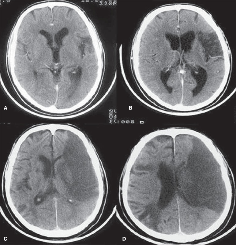
CT scanAxial brain CT: evolution of a left middle cerebral artery infarction, from partial to complete infarction with mass effect on the lateral ventricle.
Source : Garcia et al., Radiologia brasileira, 2015, figure 2 · CC BY-NC
How do you recognise a stroke, and what does the FAST mnemonic miss?
This is the part of the subject that concerns the physiotherapist most directly, and it is also the part where the most widely taught tool has a measured blind spot. A mnemonic that misses one patient in seven is not a bad tool; it is a tool whose limit you need to know.
What FAST does well
The Face Arm Speech Test was designed for prehospital alerting, and it does its job. In Harbison's study, ambulance staff trained in the test made a correct diagnosis in 144 of the 183 stroke patients who presented to them, that is 79 %. More importantly, they brought far more patients in within the useful window: 46 % within 3 hours, against 12 % for patients who went through their general practitioner first3. The proportion of non-strokes admitted was comparable whatever the referral route, at around 23 to 29 %.
What FAST misses
Aroor and colleagues reviewed 736 consecutive records of ischaemic stroke admitted over one year. 14.1 % of patients had no FAST symptom on admission. Among them, 42 % had a balance disturbance or lower-limb weakness, 40 % visual symptoms, and 70 % one or the other. Adding those two items to the mnemonic, which becomes BE-FAST, brings the proportion of unidentified strokes down to 4.4 % (p less than 0.0001)4.
BE-FAST: the six signs, and what the first two catch
Consecutive series of 736 ischaemic strokes
Source: Aroor S, Singh R, Goldstein LB, Stroke 2017 (PMID 28082668). Single-centre series, 736 patients retained from 858 consecutive records.
For a physiotherapist, this result is not an epidemiological curiosity. The two items added, balance and vision, point to the posterior circulation, and those are precisely the complaints that lead a patient to a rehabilitation clinic rather than to an emergency department. Sudden unsteadiness and double vision are stroke symptoms.
The clinical picture, territory by territory
| Territory | Typical signs | What makes the picture misleading |
|---|---|---|
| Carotid (anterior circulation) | Contralateral hemiparesis and hemihypoaesthesia, aphasia if the dominant hemisphere is affected, deviation of the head and eyes, spatial neglect, amaurosis fugax | This is the picture FAST catches well. Aphasia can be taken for confusion, and neglect for indifference. |
| Vertebrobasilar (posterior circulation) | Ataxia, vertigo, diplopia, dysarthria, dysphagia, hemianopia or cortical blindness, bilateral or alternating deficit, drop attack | This is FAST's blind spot. Vertigo with unsteadiness can be taken for a peripheral vestibular disorder, and a cerebellar infarction can be complicated by compressive oedema. |
| Lacunar (small perforating arteries) | Pure motor hemiplegia, pure sensory syndrome, ataxic hemiparesis, dysarthria and clumsy hand | The deficit is sometimes modest and the initial recovery misleading, which delays seeking care. |
What looks like a stroke without being one
Triage does not consist only in spotting stroke: it also consists in not raising the highest alarm at every neurological symptom. That said, the asymmetry of the consequences is such that doubt should always fall on the side of calling. In Harbison's study, between 23 % and 29 % of patients admitted for suspected stroke did not have one, and that proportion was comparable whatever the referral route3 : a quarter of false alarms is the normal price of a system that does not miss the real ones.
| Diagnosis | What suggests it | Why it does not spare you the call |
|---|---|---|
| Epileptic seizure and post-ictal deficit | Positive phenomena, clonic movements, tongue biting, gradual recovery of the deficit | A seizure can reveal a vascular lesion, and Todd's paralysis mimics a focal deficit perfectly. |
| Hypoglycaemia | Sweating, tremor, a context of treated diabetes, resolution once sugar is given | It produces genuine focal deficits. Capillary blood glucose is the first thing the emergency team does, not the physiotherapist. |
| Migraine with aura | Positive symptoms, progression over 5 to 30 minutes, headache afterwards, identical past episodes | A first aura after the age of 50 is investigated as a vascular event. |
| Peripheral vestibular disorder | Rotatory vertigo, characteristic nystagmus, otherwise normal neurological examination | This is FAST's measured blind spot: a cerebellar infarction can present as isolated vertigo. |
| Peripheral facial palsy | The whole half of the face is involved, forehead included, with Bell's phenomenon | In stroke, the forehead is spared. The distinction is useful but it is checked, not assumed. |
Why does every minute really count?
“Time is brain” has become a slogan, which is the surest way of ceasing to believe it. Yet the figure exists, it has been calculated, and it makes the phrase literal.
Saver modelled the tissue loss of a typical supratentorial large-vessel cerebral infarction. The mean final volume is 54 mL, the mean duration of evolution 10 hours, and the human forebrain contains about 22 billion neurons. It follows that the patient loses, for every untreated hour, 120 million neurons, 830 billion synapses and 714 kilometres of myelinated fibres. Per minute: 1.9 million neurons, 14 billion synapses, 12 kilometres of fibres. Compared with normal ageing, the ischaemic brain ages 3.6 years an hour2.
“The typical patient loses 1.9 million neurons for each minute their stroke goes untreated.” (Saver, Stroke, 2006)
This destruction is not theoretical: it can be read in the treatment trials, where the effect of the same treatment melts away as the delay lengthens. That is the subject of the next two chapters.
Key point
Three orders of magnitude are enough to hold the reasoning together during a session. A minute lost is worth 1.9 million neurons. An hour lost is worth 3.6 years of brain ageing. And moving from the 3-hour window to the 4.5-hour window drops the odds ratio of benefit from thrombolysis from 1.75 to 1.267. The time a clinician takes to decide to call is paid for on all three scales at once.
The delay is not only in hospital
It is tempting to believe that this countdown concerns the hospital. In reality it starts well before, and the prehospital part is the one a physiotherapist has leverage over. Harbison's result shows it indirectly: patients brought in by an ambulance crew trained in FAST arrived within 3 hours in 46 % of cases, against 12 % for those who went through their general practitioner first3. This is not a criticism of GPs: it is a demonstration that an intermediate link, however competent, costs hours. The physiotherapist who telephones the family doctor first, faced with an evolving deficit, adds exactly that link.
What do you do in the first few minutes, in the clinic?
A physiotherapist will never treat an acute stroke. They may, however, witness its onset, and what they do in the following five minutes weighs more on the patient's outcome than everything they will do in rehabilitation afterwards.
Immediate management of a sudden neurological deficit
- Call 15 or 112 without delay. Do not send the patient to the emergency department under their own steam: the emergency call handler decides on the vehicle and the destination, and directs the patient to a hospital with a stroke unit, whose benefit on death or dependency is proven11.
- Note the exact time the symptoms began, or failing that the last time the patient was seen well. That is the information which governs eligibility for thrombolysis and thrombectomy, and it is the one most often lost along the way.
- Give nothing by mouth, no drink and no medication: swallowing difficulty is frequent in the acute phase and aspiration is a serious complication.
- Do not try to bring the blood pressure down or to give aspirin: until the ischaemic or haemorrhagic nature is established, any treatment is a gamble.
- Lie the patient down, head slightly raised, do not leave them alone, monitor consciousness and breathing.
- Prepare the handover : time of onset, signs you observed and not merely those reported, current medication (anticoagulants above all), medical history, contact details for a relative.
What the physiotherapist does when faced with a sudden neurological deficit
Decision tree, from the clinic to the care pathway
Built from: Aroor et al., Stroke 2017 (PMID 28082668); Saver, Stroke 2006 (PMID 16339467); Emberson et al., Lancet 2014 (PMID 25106063); Langhorne and Ramachandra, Cochrane 2020 (PMID 32324916).
The mistakes that cost minutes
- Waiting to see whether it passes. Nobody can know, at onset, whether the deficit will resolve. The distinction between TIA and established infarction is retrospective and is made on imaging.
- Calling the family doctor first. Faced with an evolving deficit, the number is 15.
- Driving the patient yourself. You lose the call handling, the routing to the right unit and the preparation of the reception.
- Forgetting the time. It is the most valuable and the most fragile piece of information: it disappears as soon as the scene breaks up.
What happens in hospital, and what does the outcome depend on?
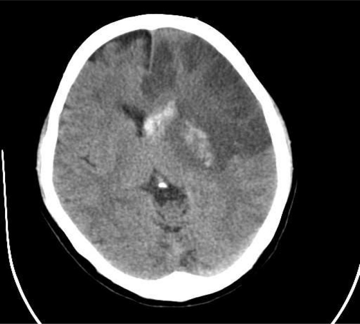
CT scanAxial brain CT, 48 hours after thrombolysis: large hypodense area of cerebral infarction with sulcal effacement and mass effect, before decompressive craniectomy.
Source : Baharvahdat et al., Iranian journal of neurology, 2014, figure 2 · CC BY
Understanding the chain of the acute phase serves two purposes: explaining to the patient and their family why things happened as they did, and understanding why two apparently similar patients arrive in rehabilitation with very different deficits.
Imaging first, always
The first decision is binary: ischaemic or haemorrhagic. It is made on CT or MRI, and nothing happens before it. That is why the delay between arrival and the start of treatment, known as door-to-needle time, is a major organisational target for emergency departments, and why the emergency call handler directs the patient to a hospital with a stroke unit rather than to the nearest hospital.
Intravenous thrombolysis, and its dependence on delay
The landmark NINDS trial, published in 1995, showed that thrombolysis with tissue plasminogen activator given within 3 hours improved outcome at three months: overall odds ratio 1.7 (95 % CI 1.2 to 2.6), treated patients being at least 30 % more likely to have minimal or no disability. The price is symptomatic intracerebral haemorrhage within 36 hours in 6.4 % of treated patients against 0.6 % on placebo, with no significant difference in mortality at three months (17 % against 21 %)5.
The ECASS III trial then extended the window. Across 821 patients treated at a median of 3 hours 59 minutes, a favourable outcome occurred in 52.4 % of patients on alteplase against 45.2 % on placebo (odds ratio 1.34; 95 % CI 1.02 to 1.76; p equal to 0.04), with 2.4 % symptomatic haemorrhage against 0.2 %, and unchanged mortality6.
Emberson's pooled analysis, on individual data from the major trials, quantifies how the benefit declines with time, and it is the most telling result in the whole file:
The benefit of thrombolysis melts away with delay
Odds ratio of a favourable outcome at 3 months, pooled analysis on individual patient data
Source: Emberson J, Lees KR, Lyden P, et al., Lancet 2014 (PMID 25106063). Bars: 95 % confidence intervals. Alteplase also increases the risk of symptomatic intracranial haemorrhage (6.8 % against 1.3 %).
A more recent development is worth knowing, because it is changing what is seen on the wards: tenecteplase, as a single bolus, has proved non-inferior and even superior on early reperfusion compared with alteplase in patients due to undergo thrombectomy10.
The stroke unit, the highest-yield intervention of all
The Cochrane review of stroke units, across 29 trials and 5,902 participants, establishes that organised care in a dedicated unit, compared with a conventional ward, reduces the risk of a poor outcome (odds ratio 0.77; 95 % CI 0.69 to 0.87), of death (0.76; 0.66 to 0.88), and of death or dependency (0.75; 0.66 to 0.85), with moderate certainty evidence11.
This benefit rests on no drug: it rests on organisation, monitoring, systematic screening for complications and teamwork. It is also what justifies the emergency service sending a patient to a more distant but better equipped hospital.
Thrombectomy: for whom, and up to when?
This is the deepest change in vascular neurology of the last twenty years, and the one that best explains why some patients arrive in rehabilitation with a deficit out of all proportion to the initial severity of their presentation.
The effect, and its size
The HERMES meta-analysis pooled individual data from 1,287 patients across five trials. Endovascular thrombectomy significantly reduces disability at 90 days, with a pooled odds ratio of 2.49 (95 % CI 1.76 to 3.53; p less than 0.0001). The number needed to treat to improve by at least one level on the modified Rankin scale is 2.68.
A number needed to treat of 2.6 is exceptional in medicine. The effect was present in every prespecified subgroup, including patients aged 80 and over (odds ratio 3.68), those randomised more than 300 minutes after symptom onset (1.76) and those not eligible for alteplase (2.43). Mortality at 90 days and the risk of haemorrhage did not differ between the groups.
The window has widened, but not for everyone
Two trials published in 2018 broke the 6-hour limit, on condition that patients were selected on imaging rather than on the clock.
- DAWN included 206 patients seen 6 to 24 hours after they were last seen well, with a mismatch between clinical severity and infarct volume. Functional independence at 90 days was 49 % in the thrombectomy group against 13 % in the control group (adjusted difference 33 points), with no excess of symptomatic haemorrhage (6 % against 3 %) or of mortality (19 % against 18 %)9.
- DEFUSE 3 included 182 patients between 6 and 16 hours, selected by perfusion imaging. Functional independence at 90 days reached 45 % against 17 % (p less than 0.001), with mortality of 14 % against 26 %12.
What the widened window does not mean
Above all it does not mean there is time. The patients in DAWN and DEFUSE 3 were selected on imaging showing tissue still salvageable: they are a favourable minority, not the rule. And Saver's analysis of the five thrombectomy trials shows that the chances of a better outcome decline as the delay between symptom onset and arterial puncture lengthens13. The widened window saves patients who would not have been treated; it does not entitle anyone to slow down.
Functional independence at 90 days in the late-window trials
Modified Rankin score of 0 to 2, thrombectomy against medical treatment alone
Sources: Nogueira RG et al., NEJM 2018 (PMID 29129157); Albers GW et al., NEJM 2018 (PMID 29364767).
What changes when the stroke is haemorrhagic?
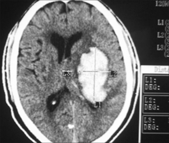
CT scanAxial brain CT: spontaneous intracerebral haematoma, a large hyperdense lesion in the left basal ganglia.
Source : Tshikwela et al., Journal of neurosciences in rural practice, 2012, figure 1 · CC BY-NC-SA
Close to a third of strokes are haemorrhagic, and their management has nothing in common with that of infarction. Physiotherapists often know this poorly, because the rehabilitation literature talks mainly about ischaemia.
Intracerebral haemorrhage accounts for 28.8 % of incident strokes, subarachnoid haemorrhage 5.8 %1. The 2022 American guideline for spontaneous intracerebral haemorrhage sets out the management16.
Three differences matter for understanding the patient who later arrives in rehabilitation.
- No reperfusion is possible. The whole logic of treatment is reversed: the aim is to limit expansion of the haematoma, to control blood pressure and to correct any anticoagulation, not to reopen a vessel.
- The dominant risk factor is high blood pressure. The INTERSTROKE study shows that hypertension is more strongly associated with intracerebral haemorrhage than with ischaemic stroke, whereas current smoking, diabetes, apolipoproteins and cardiac causes are more strongly associated with ischaemia14.
- The initial trajectory is often more severe, but recovery can be good. This is not a paradox: the haematoma compresses structures it does not always destroy, and its resorption frees functional tissue.
How is recurrence prevented?
This is the side the community physiotherapist accompanies for longest, often for years, and the one on which they have real influence even though they prescribe nothing.
Ten factors, 90 % of the risk
The INTERSTROKE study, a case-control study conducted in 32 countries, establishes that ten potentially modifiable risk factors are collectively associated with about 90 % of the attributable risk of stroke, in every major region of the world, in men as in women and at every age14. The GBD 2021 data also confirm substantial rises in DALYs attributable to high body mass index, high fasting plasma glucose, sugar-sweetened drinks, sedentary behaviour and high systolic blood pressure1.
The treatments whose benefit depends on speed
Short-course dual antiplatelet therapy. The POINT trial, in 4,881 patients with minor ischaemic stroke or high-risk TIA, showed 5.0 % major ischaemic events on clopidogrel plus aspirin against 6.5 % on aspirin alone (hazard ratio 0.75; 95 % CI 0.59 to 0.95), most events occurring in the first week, at the price of an increase in major haemorrhage (0.9 % against 0.4 %)17. The CHANCE trial had opened this path18.
Carotid endarterectomy. Rothwell's pooled analysis, across 5,893 patients, shows that for a stenosis of 50 % or more, the number of patients needing surgery to prevent one ipsilateral stroke at 5 years is 5 if surgery takes place within 2 weeks and 125 beyond 12 weeks19.
The cost of delay, once carotid stenosis has been found
Number of patients needing surgery to prevent one ipsilateral stroke at 5 years, stenosis of 50 % or more
Source: Rothwell PM, Eliasziw M, Gutnikov SA, et al., Lancet 2004 (PMID 15043958). Pooled analysis of 5,893 patients, 33,000 patient-years of follow-up.
The full set of secondary prevention measures, pharmacological and non-pharmacological, is set out in the 2021 American guideline20, which places physical activity among the recommended measures.
Key point
On preventing recurrence, the physiotherapist prescribes nothing but occupies a place no one else occupies: they see the patient every week, sometimes for months. That is where regular physical activity is played out, and that is where a new event is spotted. A history of stroke or TIA is an active assessment finding, not a line of history.
And afterwards: where does this article stop?
Handing over
This article stops at the point where rehabilitation begins, and that is not an arbitrary cut: they are two phases of care, two literatures and two different jobs. Everything to do with dose and intensity, constraint-induced movement therapy, task-oriented training, body-weight-supported treadmill training, robotics, spasticity, the painful hemiplegic shoulder, spatial neglect, post-stroke fatigue and depression, the prognosis for recovery and the question of whether recovery stops at six months is dealt with in depth in rehabilitation after a stroke.
One point of junction is worth flagging here, because it concerns the acute phase and is counter-intuitive: the AVERT trial, in 2,104 patients, showed that a very early, high-dose mobilisation protocol within 24 hours reduced the chances of a favourable outcome at 3 months (46 % against 50 %; adjusted odds ratio 0.73; 95 % CI 0.59 to 0.90)21. The prespecified dose-response analysis sharpens the message: it is the frequency of getting out of bed that improves the outcome (odds ratio 1.13 per additional daily session) whereas the amount of minutes a day worsens it (0.94)22. Shorter and more often, not longer.
What do concrete clinical situations teach us?
The two situations that follow are fictional cases, built to illustrate a line of reasoning. Every figure they draw on refers back to its source; the patients themselves do not exist.
Fictional case number 1: Mrs P., 71, the deficit that appears during a session
The scene. Rehabilitation after a hip replacement, fourth week. At 10.20 the patient stops answering correctly, her mouth deviates to the right and her right arm drops. She was normal at 10.15.
What is done. Call 15 immediately, before anything else. Time noted: last seen well 10.15. Patient lying down, nothing by mouth, continuous monitoring. Handover to the call handler: time, signs observed, any anticoagulant treatment.
What the case teaches. The five minutes gained here are not symbolic: they are worth, in order of magnitude, some ten million neurons2, and they can make the difference between treatment within 3 hours (odds ratio 1.75) and treatment between 3 and 4.5 hours (1.26)7.
Fictional case number 2: Mr K., 52, a vertigo that is not one
The scene. Referred for vestibular rehabilitation after an episode, the day before, of sudden vertigo with major unsteadiness and transient double vision. He also complains of posterior neck pain that came on after an intense training session.
The reasoning. Sudden unsteadiness and diplopia point to the posterior circulation: these are the two items whose addition to FAST brings the proportion of missed strokes down from 14.1 % to 4.4 %4. Recent neck pain in a 52-year-old man after exertion also suggests vertebral artery dissection, a classic cause of stroke in younger patients15.
What is done. You do not start vestibular rehabilitation, and above all you do not mobilise the cervical spine. You contact the referring doctor to find out whether the episode was imaged; failing that, urgent referral.
What the case teaches. A prescription is not a diagnosis. Starting cervical treatment on an undiagnosed dissection is the scenario this chapter exists to prevent.
How do you apply this in the clinic?
What the physiotherapist needs to be able to do
| Stage | What falls to them | What does not |
|---|---|---|
| Deficit evolving in front of them | Call 15, note the time of onset, nothing by mouth, monitor, hand over | Grading severity, telling ischaemia from haemorrhage, giving anything at all |
| Recent episode described | Obtain medical advice the same day, check with the referring doctor that imaging has been done | Deciding that it was peripheral vertigo or a migraine |
| Acute hospital phase | Nothing in community practice. In hospital, short and frequent mobilisation rather than long and intense | Applying a very early, high-dose mobilisation protocol, which the AVERT trial found wanting |
| Rehabilitation | The heart of the job, covered in the dedicated article | Outside the scope of this article |
| Long term | Regular physical activity, spotting a new event, supporting risk factor management | Prescribing or changing antithrombotic or antihypertensive treatment |
Key point
The physiotherapist has no treatment decision to make in the acute phase of stroke, and that is precisely why their role there is simple and entirely codifiable: recognise, call, note the time, give nothing, hand over. The only specific skill they need to add to what any trained citizen can do is knowing FAST's blind spot, because it is the patients in that blind spot who end up in their clinic rather than in the emergency department.
Frequently asked questions
What is the difference between a stroke and a TIA?
The mechanism is the same; the difference is the absence of established infarction on imaging in TIA, the clinical picture having resolved. That difference does not rank the urgency: a TIA is treated as an emergency, and the site gives it a separate article.
Can a haemorrhagic stroke be recognised without imaging?
No, not reliably. Headache, vomiting and impaired alertness are more frequent in haemorrhage, but their absence does not rule it out. That is why imaging comes before any treatment.
How long is there to treat an ischaemic stroke?
There is not one answer but two. For intravenous thrombolysis, the benefit is demonstrated up to 4.5 hours and falls sharply with delay (odds ratio 1.75 before 3 h, 1.26 between 3 h and 4.5 h, 1.15 and not significant beyond)7. For thrombectomy, patients selected on imaging have been treated with benefit up to 24 hours9. In both cases, earlier remains very clearly better.
Should aspirin be given while waiting for the emergency services?
No. Until the nature of the stroke is established, giving an antiplatelet to a patient who is bleeding is one risk, and getting a patient whose swallowing is uncertain to swallow anything at all is another.
Why not drive the patient straight to the nearest hospital?
Because the nearest hospital is not always the one with a stroke unit, or with a thrombectomy service. The benefit of the dedicated unit on death or dependency is proven (odds ratio 0.75)11, and it is the emergency call handler who knows what the network has available.
Can a young, athletic patient have a stroke?
Yes. In the Dijon registry, 10.5 % of first cerebral infarctions occurred in young adults with a median age of 46, and their incidence doubled between the period before 2003 and the one after15. Cervical artery dissection is a classic cause in that age band.
Should rehabilitation begin as early as possible?
The short answer is no, at least not in any form whatsoever. The AVERT trial showed that a very early, high-dose mobilisation protocol within 24 hours reduced the chances of a favourable outcome21, whereas the dose-response analysis shows that a high frequency of short spells out of bed improves the outcome22. The question is dealt with in detail in the rehabilitation article.
What do you say to a family who ask whether “he will recover”?
That it is too early to say in the first few days, that the recovery trajectory is judged over weeks and months, and that the outlook depends on factors which are assessed gradually. The subject is developed in the rehabilitation article, which covers prognosis and what rehabilitation really shifts.
Further reading on the site
This file is read alongside two others. Transient ischaemic attack covers the warning that comes before, and what to do when a deficit has resolved. Rehabilitation after a stroke covers everything that comes after the acute phase. On the differential diagnoses met in the clinic, see cerebellar ataxia, vestibular neuritis and benign paroxysmal positional vertigo. On gait disorders in older people, normal pressure hydrocephalus.
Bibliography: 22 verified references
- GBD 2021 Stroke Risk Factor Collaborators. Global, regional, and national burden of stroke and its risk factors, 1990-2021: a systematic analysis for the Global Burden of Disease Study 2021. Lancet Neurol. 2024;23(10):973-1003. PMID 39304265
- Saver JL. Time is brain, quantified. Stroke. 2006;37(1):263-266. PMID 16339467
- Harbison J, Hossain O, Jenkinson D, et al. Diagnostic accuracy of stroke referrals from primary care, emergency room physicians, and ambulance staff using the face arm speech test. Stroke. 2003;34(1):71-76. PMID 12511753
- Aroor S, Singh R, Goldstein LB. BE-FAST (Balance, Eyes, Face, Arm, Speech, Time): Reducing the Proportion of Strokes Missed Using the FAST Mnemonic. Stroke. 2017;48(2):479-481. PMID 28082668
- The National Institute of Neurological Disorders and Stroke rt-PA Stroke Study Group. Tissue plasminogen activator for acute ischemic stroke. N Engl J Med. 1995;333(24):1581-1587. PMID 7477192
- Hacke W, Kaste M, Bluhmki E, et al. Thrombolysis with alteplase 3 to 4.5 hours after acute ischemic stroke. N Engl J Med. 2008;359(13):1317-1329. PMID 18815396
- Emberson J, Lees KR, Lyden P, et al. Effect of treatment delay, age, and stroke severity on the effects of intravenous thrombolysis with alteplase for acute ischaemic stroke: a meta-analysis of individual patient data from randomised trials. Lancet. 2014;384(9958):1929-1935. PMID 25106063
- Goyal M, Menon BK, van Zwam WH, et al. Endovascular thrombectomy after large-vessel ischaemic stroke: a meta-analysis of individual patient data from five randomised trials. Lancet. 2016;387(10029):1723-1731. PMID 26898852
- Nogueira RG, Jadhav AP, Haussen DC, et al. Thrombectomy 6 to 24 Hours after Stroke with a Mismatch between Deficit and Infarct. N Engl J Med. 2018;378(1):11-21. PMID 29129157
- Campbell BCV, Mitchell PJ, Churilov L, et al. Tenecteplase versus Alteplase before Thrombectomy for Ischemic Stroke. N Engl J Med. 2018;378(17):1573-1582. PMID 29694815
- Langhorne P, Ramachandra S; Stroke Unit Trialists' Collaboration. Organised inpatient (stroke unit) care for stroke: network meta-analysis. Cochrane Database Syst Rev. 2020;4(4):CD000197. PMID 32324916
- Albers GW, Marks MP, Kemp S, et al. Thrombectomy for Stroke at 6 to 16 Hours with Selection by Perfusion Imaging. N Engl J Med. 2018;378(8):708-718. PMID 29364767
- Saver JL, Goyal M, van der Lugt A, et al. Time to Treatment With Endovascular Thrombectomy and Outcomes From Ischemic Stroke: A Meta-analysis. JAMA. 2016;316(12):1279-1288. PMID 27673305
- O'Donnell MJ, Chin SL, Rangarajan S, et al. Global and regional effects of potentially modifiable risk factors associated with acute stroke in 32 countries (INTERSTROKE): a case-control study. Lancet. 2016;388(10046):761-775. PMID 27431356
- Béjot Y, Duloquin G, Thomas Q, et al. Temporal Trends in the Incidence of Ischemic Stroke in Young Adults: Dijon Stroke Registry. Neuroepidemiology. 2021;55(3):239-244. PMID 34044406
- Greenberg SM, Ziai WC, Cordonnier C, et al. 2022 Guideline for the Management of Patients With Spontaneous Intracerebral Hemorrhage: A Guideline From the American Heart Association/American Stroke Association. Stroke. 2022;53(7):e282-e361. PMID 35579034
- Johnston SC, Easton JD, Farrant M, et al. Clopidogrel and Aspirin in Acute Ischemic Stroke and High-Risk TIA. N Engl J Med. 2018;379(3):215-225. PMID 29766750
- Wang Y, Wang Y, Zhao X, et al. Clopidogrel with aspirin in acute minor stroke or transient ischemic attack. N Engl J Med. 2013;369(1):11-19. PMID 23803136
- Rothwell PM, Eliasziw M, Gutnikov SA, et al. Endarterectomy for symptomatic carotid stenosis in relation to clinical subgroups and timing of surgery. Lancet. 2004;363(9413):915-924. PMID 15043958
- Kleindorfer DO, Towfighi A, Chaturvedi S, et al. 2021 Guideline for the Prevention of Stroke in Patients With Stroke and Transient Ischemic Attack: A Guideline From the American Heart Association/American Stroke Association. Stroke. 2021;52(7):e364-e467. PMID 34024117
- AVERT Trial Collaboration group. Efficacy and safety of very early mobilisation within 24 h of stroke onset (AVERT): a randomised controlled trial. Lancet. 2015;386(9988):46-55. PMID 25892679
- Bernhardt J, Churilov L, Ellery F, et al. Prespecified dose-response analysis for A Very Early Rehabilitation Trial (AVERT). Neurology. 2016;86(23):2138-2145. PMID 26888985
Behind this article
An author who explains, a reviewer who checks.
How we write and check our content

Physiotherapist · co-founder of Physio Learning
Marked for life by his first four-hour lecture without a single image, he took a master’s in instructional design so that it would never happen to anyone again. He hunts down publication bias and unreadable slides with the same intransigence.

Head of scientific content
Physiotherapist specialising in neuro-musculoskeletal practice and holder of a master’s in public health. He checks the methodological rigour of every article: primary sources, levels of evidence, no exceptions.