














On-siteDPCFIFPL

Upper limb
Hand rehabilitation and splinting: refining and deepening your practice
Jean Lichtle
Go further
Wrist osteoarthritis: SLAC and SNAC follow one and the same sequence, radioscaphoid then midcarpal, and both of them spare the radiolunate joint.
Wrist osteoarthritis is almost always the aftermath of an old injury, forgotten by the patient: a torn scapholunate ligament (SLAC) or a scaphoid that never united (SNAC).
95%of wrist osteoarthritis is periscaphoid
What follows covers each of these points in detail, with sources. It is there if you need it.
This topic is taught in a course: Hand rehabilitation and splinting: refining and deepening your practice, on-site with Jean Lichtle.
1 / 11
10 article chapters · 23 min in total
Understanding◔ 4 min
SLAC and SNAC are just two doors into the same carpal tipping, torn ligament or ununited bone.
How it progresses◔ 3 min
Wear always follows the same path and spares the radiolunate joint, which opens the door to motion-preserving surgery.
Spotting it early◔ 2 min
Once wrist osteoarthritis has set in, it cannot be treated backwards.
Testing◔ 3 min
Most wrist provocation tests have no diagnostic value on their own.
Imaging◔ 2 min
The stage decides the surgery, and capitolunate involvement is the hinge point.
Non-surgical care◔ 1 min
No trial tests exercise specifically in this osteoarthritis, the evidence comes from elsewhere.
Surgery◔ 4 min
Both salvage surgeries require an intact radiolunate joint: that is a condition, not a choice.
Rehab◔ 1 min
Grip strength remains the real marker, and it does not differ between the two techniques.
In practice◔ 1 min
The decisive question remains the old injury, often forgotten by the patient.
FAQ◔ 2 min
SLAC accounts for 57 % of degenerative wrist osteoarthritis, against 27 % for scaphotrapeziotrapezoid.
Train on this
Upper limb
Jean Lichtle
Next comes practice: the course that teaches this topic, with Jean Lichtle.
Our courses on this topic may be covered by DPC or FIFPL, the French continuing-education funding schemes, depending on your profession and situation.
Post-traumatic osteoarthritis · Wrist
A painful, stiff wrist that is losing its strength with no recent injury to explain it. The history, though, often goes back ten or twenty years: a sprain that was never investigated, or a scaphoid fracture that never united. Two mechanisms, one radiographic sequence, and one joint that is consistently spared, which is what makes the salvage procedures possible.
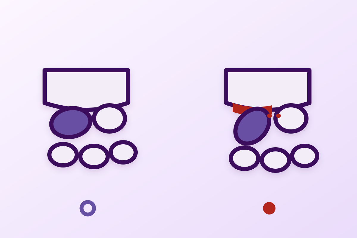
Two entities, one single mechanism. The SLAC (scapholunate advanced collapse) wrist is the sequela of a scapholunate ligament tear; the SNAC (scaphoid nonunion advanced collapse) wrist is the sequela of a scaphoid nonunion. In both cases the scaphoid loses its alignment with the radius, flexes, and the radioscaphoid joint wears out first because its surfaces no longer match1.
It is the leading cause of wrist osteoarthritis. Across 4,000 wrist radiographs reviewed and 210 cases of degenerative osteoarthritis retained after other arthritides were excluded, the SLAC pattern accounted for 57 % of cases, scaphotrapeziotrapezoid osteoarthritis 27 % and the two combined 15 %1. The 1986 series confirms it: about 95 % of wrist osteoarthritis is periscaphoid2.
The sequence is predictable, and one joint is consistently spared. Wear begins between the radius and the distal pole of the scaphoid, then reaches the capitolunate joint, and spares the radiolunate joint even at a severe stage. It is that sparing, described as early as 1987 in 64 unoperated nonunions, which makes all the salvage procedures possible: every one of them rests on a lunate that is still healthy against the radius3.
The interval is long, and it has been quantified. For a scaphoid nonunion of 4 years, 75 % of patients show radioscaphoid degenerative change; at 9 years, 60 % have midcarpal change3. What is playing out in the clinic today began one to two decades earlier.
This is not thumb base osteoarthritis. Trapeziometacarpal osteoarthritis of the thumb involves a different joint, has a different epidemiology and a different treatment. It is dealt with in its own right in a dedicated article. The confusion is common because both hurt “in the wrist on the thumb side”, but scaphotrapeziotrapezoid osteoarthritis does belong to the group described here.
And the physiotherapist has no curative treatment to offer. No trial shows that rehabilitation alters the osteoarthritic sequence. What is documented has to do with load, pain and function: the Cochrane review of exercise in hand osteoarthritis concludes that the effects are small to moderate and of low certainty on pain, function and stiffness10. Saying so does not weaken the management: it avoids promising what will not come.
The two acronyms look so alike that they get confused, and yet their origins differ radically: a torn ligament on one side, a bone that has not united on the other. This chapter sets out what separates them, and above all what makes them identical once the osteoarthritis is established.
The carpus is made of two rows, and the scaphoid bridges them. It is the only bone spanning the proximal and distal rows, which makes it both the stabiliser of the carpus and its most vulnerable piece. Its stability depends on its anchorage to the lunate through the scapholunate interosseous ligament, and on its bony integrity.
Let either give way and the mechanics fail in the same manner: the distal fragment of the scaphoid tips into flexion, while the lunate, released, extends. The review by Wessel and Wolfe, published in the Journal of Hand Surgery in 2023, notes that scapholunate injuries are the most frequent cause of carpal instability, and that left untreated they lead to collapse and to progressive deterioration of the carpus4.
Two origins, one and the same tipping
On the left the SLAC mechanism (torn scapholunate ligament), on the right the SNAC mechanism (fractured scaphoid that has not united). In both cases the distal segment of the scaphoid tips into flexion and the radioscaphoid joint loses its congruity. After Watson 1984 (PMID 6725894), Vender 1987 (PMID 3611645) and Wessel 2023 (PMID 37452815).
SNAC differs on one point that matters: the PROXIMAL fragment of the scaphoid stays attached to the lunate, and its surface facing the radius stays congruent. That is what explains the sparing of the proximal compartment.
The Swedish national registry gives the most reliable orders of magnitude: across 34,377 patients registered between 2006 and 2015, with validation of a sample of 300 records, the true adjusted incidence of scaphoid fracture is 22 per 100,000 person-years. Two figures deserve to be remembered by a rehabilitation clinician: 41 % of the fractures initially diagnosed were false positives, and the risk of nonunion was 3 % in men against 1 % in women5.
That nonunion rate looks low, and it is. But set against the incidence, it produces a steady annual stream of un-united scaphoids, some of which will only become symptomatic ten or twenty years later.
Scaphoid fracture, at the scale of a country
Swedish national registry, 34,377 patients registered between 2006 and 2015, with validation of a sample of 300 records which allowed the rates to be adjusted. After Swärd 2019 (PMID 31106681).
The false positive rate is worth remembering: a patient referred for a “scaphoid fracture” has close to a one-in-two chance of never having had one.
The hand and wrist carry several distinct forms of osteoarthritis that pain alone does not separate. Confusing them leads to treating the wrong joint, and to referring for the wrong operation.
| Entity | Joint involved | Usual origin | Where it is covered |
|---|---|---|---|
| SLAC wrist | Radioscaphoid, then capitolunate | Scapholunate ligament tear | This article |
| SNAC wrist | The same, in the same order | Scaphoid nonunion | This article |
| Scaphotrapeziotrapezoid osteoarthritis | Between the distal pole of the scaphoid, the trapezium and the trapezoid | Degenerative, sometimes associated with SLAC (15 % of Watson's cases) | This article, periscaphoid group |
| Thumb base osteoarthritis | Trapeziometacarpal, base of the thumb | Primary degenerative, markedly more common in women | Dedicated article |
| Finger osteoarthritis | Distal and proximal interphalangeal joints | Primary degenerative | Dedicated article |
This is what makes this form of osteoarthritis readable on a radiograph and operable at the right moment. The sequence is not a classification convention: it has been measured, and the joint it spares governs the whole of the surgery.
Watson and Ryu, reviewing the wear patterns, describe a progression that always starts in the same place: degenerative change begins between the radius and the distal pole of the scaphoid, because that is where joint alignment is lost first. It then reaches the capitolunate joint, secondary to carpal collapse2.
Vender and colleagues quantified this progression in 64 symptomatic scaphoid nonunions that had never been operated on. Two findings govern everything else: at four years, 75 % of patients have radioscaphoid degenerative change; at nine years, 60 % have midcarpal change. And above all: the joint between the radius and the proximal fragment of the scaphoid, along with the radiolunate joint, are consistently spared, even in the presence of severe osteoarthritis3.
The wear sequence, and the measured interval
Progression of degenerative change across 64 symptomatic, unoperated scaphoid nonunions, according to the time since injury. After Vender 1987 (PMID 3611645), completed by Watson 1986 (PMID 3955970) for the order of the compartments.
Both dates count in the clinic: the interval between injury and osteoarthritis is long, and the window for acting on the cause is well before the first symptoms.
A recent series qualifies the picture, and deserves quoting because it runs against intuition. Twenty patients with chronic scaphoid nonunions who had never had any treatment at all were reviewed: mean duration of nonunion 34 years (10 to 62 years), mean age 62. Osteoarthritis had indeed developed, but no variable (age, sex, fracture site, displacement, duration, radiographic measures) predicted the functional result, and most patients had a fair to good result despite reduced range and strength. Many wanted no intervention at all6.
The level of evidence is that of a retrospective series of 20 cases (level IV) and the selection is obvious: these are patients who have lived with it, so the least troubled by it. The usable lesson remains: the radiographic picture and functional impairment correlate poorly, and visible osteoarthritis does not mandate an operation.
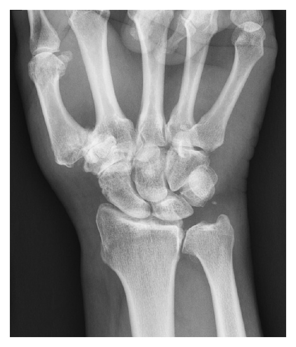
X-rayFrontal wrist radiograph: stage 3 scaphotrapeziotrapezoid joint osteoarthritis, associated with thumb base osteoarthritis, without radiocarpal involvement.
Source : Wollstein et al., Arthritis, 2012, figure 1 · CC BY
Established osteoarthritis cannot be treated backwards. But the physiotherapist regularly meets the stage before, the one where the ligament is injured without the carpus having collapsed yet. This chapter says what is established at that stage, and what is not.
Wessel and Wolfe's review describes a continuum, not a single event: rupture of the interosseous ligament first produces a desynchronisation of movement between scaphoid and lunate; it is the additional ligament injuries, or the wearing out of the secondary stabilisers, that then produce rotatory subluxation of the scaphoid and a visible scapholunate gap. The aim of any intervention at that stage is to halt the degenerative process by restoring ligament integrity and carpal kinematics4.
There is no randomised trial showing that a rehabilitation programme prevents progression to a SLAC wrist. What does exist is a neuromuscular rationale, set out by Hagert, Lluch and Rein: beyond joint congruity, static ligamentous stability and dynamic compression by muscles, a fourth factor of carpal stability has been proposed, proprioceptive and neuromuscular control. The ligaments and capsules of the carpus contain mechanoreceptors which trigger spinal reflexes for immediate stabilisation and feed higher-level loops7.
That review is narrative, and it describes a mechanism, not a clinical outcome. It justifies working on motor control of the wrist; it does not allow you to tell a patient that this work will prevent their osteoarthritis.
A figure is needed, and it is an uncomfortable one: most provocation tests of the wrist are worth almost nothing taken in isolation. Better to know that before basing management on them.
The most useful study is an Australian cross-sectional study conducted in 105 patients presenting with wrist pain and suspected ligament injury, in whom seven provocation tests were compared against arthroscopy as the reference standard, with MRI in 55 of them. The authors' verdict is blunt: most tests and MRI findings have little or no diagnostic value. The exceptions, with what they call slight usefulness, are the scaphoid shift test (positive likelihood ratio 2.88, negative 0.28), the midcarpal test (LR+ 2.67) and the distal radioulnar joint test (LR− 0.30). MRI was moderately useful for the triangular fibrocartilage complex and slightly useful for the scapholunate ligament (LR+ 4.17; LR− 0.32)8.
| Test | Positive likelihood ratio | Negative likelihood ratio | Reading |
|---|---|---|---|
| Scaphoid shift (Watson) | 2.88 | 0.28 | Slight usefulness: it points, it does not decide |
| Midcarpal test | 2.67 | not reported | Slight usefulness |
| Distal radioulnar joint test | not reported | 0.30 | Useful mainly for ruling out |
| MRI, scapholunate ligament | 4.17 | 0.32 | Slight to moderate usefulness |
| MRI, triangular fibrocartilage complex | 5.56 | 0.15 | Moderate usefulness |
| The other provocation tests | Little or no diagnostic value | Do not base management on them | |
What each examination actually shifts
Likelihood ratios measured in 105 patients with suspected wrist ligament injury, against arthroscopy as the reference. A positive ratio above 10 changes management, between 5 and 10 weighs moderately, below 2 shifts almost nothing. After Prosser 2011 (PMID 22093123).
None of these examinations reaches the threshold that changes management on its own. It is the history of the injury, and the consequence of a missed lesion, that decide on a specialist opinion.
Two practical consequences. First, a positive scaphoid shift in a young patient after a fall remains reason enough to seek an opinion, not because the test is good, but because the consequence of a missed lesion is a SLAC wrist. Second, a negative test does not entitle you to reassure: its negative likelihood ratio of 0.28 shifts the probability very little.
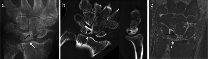
X-rayWrist radiograph and cross-sectional imaging showing stage III SLAC wrist: advanced narrowing of the radioscaphoid joint space, arrows, with extensive carpal osteoarthritis.
Source : Tischler et al., Insights into imaging, 2014, figure 10 · CC BY
The staging classification governs the surgical decision. It is also far less reproducible than is generally believed, and that is something the rehabilitation clinician needs to know before interpreting a report.
| Stage | What the radiograph shows | What remains possible |
|---|---|---|
| Stage 1 | Wear limited to the area between the radius and the distal pole of the scaphoid (radial styloid) | Procedures confined to the involved compartment; motion is largely preserved |
| Stage 2 | The whole radioscaphoid joint is involved | Proximal row carpectomy or four-corner fusion, either one |
| Stage 3 | Extension to the capitolunate (midcarpal) joint | Proximal row carpectomy becomes questionable: it depends on a healthy capitate |
| Radiolunate involvement | A rare situation, outside the usual sequence | Motion-preserving salvage no longer applies: total fusion or arthroplasty |
A German study had 38 SLAC wrists from 37 patients graded by two radiologists and two surgeons, on posteroanterior and lateral radiographs, then had the surgeons grade them again fourteen days later with the arthroscopy images in front of them. The results are severe: interobserver agreement was described as poor between radiologists and slight between surgeons, with a tendency for radiologists to overestimate severity. Above all, knowledge of the arthroscopic images changed the stage in 55 % of cases, with stages 1 and 3 then being diagnosed less often9.
What a reported stage is really worth
38 SLAC wrists from 37 patients, graded on posteroanterior and lateral radiographs by two radiologists and two surgeons, then graded again by the surgeons fourteen days later with the arthroscopy images. After Hagen 2015 (PMID 26084857).
The authors' conclusion is explicit: interpreting radiographs alone is an unreliable method for staging a SLAC wrist.
In practice: a report that gives a stage should be read as an estimate, not as a measurement. That does not invalidate the classification, which remains the common language of decisions, but it explains why two surgical opinions can diverge on the same image.
The honest question is not “how do you treat a SLAC wrist with physiotherapy” but “what is documented”. The answer is short, and it is better known than guessed at.
There is no trial of exercise specific to periscaphoid osteoarthritis. The closest data are the Cochrane review of exercise in hand osteoarthritis: seven trials included, up to five in the pooled analyses (381 participants). Compared with no exercise, exercise may improve pain (standardised mean difference −0.27; 95 % CI −0.47 to −0.07), function (−0.28; −0.58 to 0.02) and finger stiffness (−0.36; −0.58 to −0.15), with low-certainty evidence. The authors themselves write that the estimated effect sizes are small and that their clinical relevance is debatable. Adverse events were rare and mild10.
Transposing these results from finger osteoarthritis to carpal osteoarthritis is reasoning by analogy, and it should be presented as such. What remains solid: exercise does no harm, it brings a modest benefit on pain and function, and the window of tolerance can be worked on.
The physiotherapist receives these patients after the operation, and the work changes completely depending on the procedure. This chapter compares the two options on the broadest data available.
The proximal row carpectomy removes the scaphoid, the lunate and the triquetrum, and lets the capitate articulate directly in the lunate fossa of the radius.Four-corner fusion removes the scaphoid and fuses the capitate, lunate, hamate and triquetrum, preserving the radiolunate joint. Both require the same thing: an intact radiolunate joint.
Hones and colleagues pooled 61 studies and 3,174 wrists (54 % proximal row carpectomy, 46 % four-corner fusion), with a weighted mean follow-up of 61 months (12 to 216). Results: proximal row carpectomy achieved greater postoperative extension and ulnar deviation, better gains in extension, flexion and ulnar deviation, and a better pain score on a visual analogue scale. No difference in grip strength. The conversion rate to total wrist fusion was 5.2 % after carpectomy against 11 % after four-corner fusion, with a nonunion rate of 8.9 % for the latter and 2.2 % hardware removal. The level of evidence is therapeutic IV11.
Proximal row carpectomy against four-corner fusion
Meta-analysis of 61 studies, 3,174 wrists operated on for SLAC or SNAC, weighted mean follow-up 61 months. After Hones 2024 (PMID 38416092). Therapeutic level of evidence IV.
The authors conclude in favour of proximal row carpectomy. The weight of that verdict remains that of aggregated observational studies, with unmatched populations.
A series with a minimum ten-year follow-up usefully qualifies this. Among patients operated on between 1982 and 2003, fifteen wrists were reviewed at a mean follow-up of 18 years (11 to 27). Mean flexion-extension arc 68.6 degrees, mean QuickDASH 7.8 (only one score above 16), and yet 73 % of radiographs showed radiolunate degenerative change, 27 % of it severe. In other words: the patients were doing well while their images were deteriorating12.
A necessary caveat: fifteen wrists reviewed out of 489 operated on over the period. The selection bias is major, and the authors do not hide it.
When the priority is to relieve pain without sacrificing motion, selective denervation of the wrist exists. A systematic review of 12 studies out of 993 identified reports return to work of up to 94 %, with satisfaction of up to 92 %, grip strength increased by 7 to 64 %, and pain scores improved by 36 to 92 %. The authors stress the variability of the results and the need for better evidence on the technique and on nerve identification13. Those very wide ranges say more about the heterogeneity of the series than they measure an effect.
In a patient whose scaphoid has not united but whose osteoarthritis is not established, the operation still aims at union. The largest meta-analysis, 78 studies and 7,671 patients, reports a mean union rate of 88.7 % for non-vascularised grafts against 87.5 % for vascularised grafts, with no significant difference (p = 0.685). Neither the type of fixation nor the source of the graft altered the result. On the other hand, the studies that excluded proximal pole fractures and avascular necrosis reached 96.5 % union against 86.8 % for the others (p < 0.001)14. Location and vascularity dictate the outcome, not the sophistication of the graft.
No protocol is validated by a randomised trial in this indication. What can be done, on the other hand, is to derive the aims and the constraints from what the operation changed mechanically, and to say so as such.
There is no fusion to protect: the constraint is that of the soft tissues and of the new congruity between capitate and lunate fossa. Motion returns faster, and that is what the meta-analysis finds as greater postoperative range. Attention goes to the gradual return of axial compression loading, and to grip strength, which remains the weak point common to both techniques.
A fusion has to unite, and the documented risk is precisely there: 8.9 % nonunion in the meta-analysis. Progression is therefore dictated by the surgeon and by radiographic checks, not by the patient's comfort. The motion obtained will be less, which does not prevent excellent long-term functional results, as the 18-year follow-up shows.
A working summary, from first contact to referral.
The question that changes the diagnosis is the simplest one: “have you ever injured this wrist, even a very long time ago?”. A fall on the hand at twenty, six weeks in a cast, a sprain that was never investigated: that is what separates post-traumatic periscaphoid osteoarthritis from primary osteoarthritis, and it guides how the images are read.
No. Thumb base osteoarthritis is osteoarthritis of the trapeziometacarpal joint, at the base of the thumb. SLAC and SNAC involve the joints around the scaphoid, between the radius and the carpus. Two different joints, two different mechanisms and two different approaches. The subject is dealt with in its own right in the article on thumb base osteoarthritis.
That one does belong to the periscaphoid group: it accounted for 27 % of degenerative wrist osteoarthritis in Watson's series, against 57 % for SLAC. It sits between the distal pole of the scaphoid, the trapezium and the trapezoid, so just below the base of the thumb, which explains the clinical confusion with thumb base osteoarthritis.
Yes, when it involved a scapholunate ligament injury that went unnoticed. That is precisely the SLAC mechanism, and Vender's series documents an interval of several years between the injury and the first visible degenerative change.
That question is the surgeon's, and the answer depends on age, location, vascularity and how troublesome it is. What the literature contributes: union is far better when the proximal pole and avascular necrosis are out of the picture (96.5 % against 86.8 %), and a series of never-treated nonunions of several decades' standing found fair to good functional results.
On the broadest pooled data, proximal row carpectomy achieves better range and less pain, with fewer secondary conversions to total fusion. Grip strength, however, does not differ. These comparisons do not come from randomised trials, and the choice takes in age, occupation and the state of the capitate.
No, and it is better to say so. Neither technique restores the strength of the uninjured side, and the meta-analysis does not separate them on this point. The realistic aim is a usable, pain-free hand with enough strength for the intended activity.
No trial shows that it can. The proprioceptive rationale described by Hagert justifies working on motor control of the wrist, but it describes a mechanism, not a clinical outcome. What is established is that the untreated lesion leads to collapse: so it is early diagnosis and a surgical opinion that make the difference, not rehabilitation on its own.
Eighteen references, resolved one by one through the PubMed E-utilities API. For each of them the abstract was read in full and the figures cited in the article checked at source. The year kept is that of the issue, re-read on the XML record: the first “year” field of a PubMed record is sometimes a revision date, and shifts the citation by a year.
The corpus covers the immediate neighbours of this picture separately. Thumb base osteoarthritis involves the trapeziometacarpal joint, under the base of the thumb, and does not follow the same mechanics. Interphalangeal finger osteoarthritis covers the primary side of hand osteoarthritis. Upstream of the SNAC wrist, scaphoid fracture of the carpus covers the initial injury and the moment when everything is still in play: diagnosing a fracture that the radiograph does not rule out, and the immobilisation whose duration decides on union. This article picks up where that one stops, once the nonunion is established. Finally, two frequent causes of wrist pain to rule out before concluding: wrist ganglia and Kienböck's disease, which involves the very lunate on which the operations described here depend.
Behind this article
An author who explains, a reviewer who checks.
How we write and check our content

Physiotherapist · co-founder of Physio Learning
Marked for life by his first four-hour lecture without a single image, he took a master’s in instructional design so that it would never happen to anyone again. He hunts down publication bias and unreadable slides with the same intransigence.

Head of scientific content
Physiotherapist specialising in neuro-musculoskeletal practice and holder of a master’s in public health. He checks the methodological rigour of every article: primary sources, levels of evidence, no exceptions.
