














On-siteDPCFIFPL

Upper limb
Hand rehabilitation and splinting: refining and deepening your practice
Jean Lichtle
Go further
Scaphoid fracture: why the initial radiograph does not rule it out, which clinical signs count, and what too short an immobilisation ends up costing.
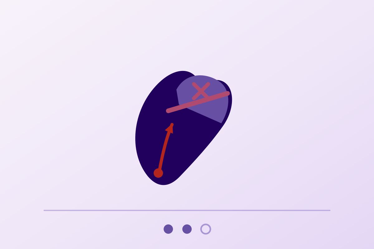
Faced with a painful wrist after a fall, with anatomical snuffbox tenderness and a normal radiograph, you treat it as a fracture until proven otherwise.
2-5%risk of scaphoid nonunion in adults
What follows covers each of these points in detail, with sources. It is there if you need it.
This topic is taught in a course: Hand rehabilitation and splinting: refining and deepening your practice, on-site with Jean Lichtle.
1 / 12
11 article chapters · 30 min in total
Understanding◔ 3 min
The scaphoid receives its blood supply against the current: a line at the waist starves the proximal pole.
Population◔ 2 min
The young man, between 20 and 29, accounts for most scaphoid fractures.
Radiograph◔ 2 min
A normal radiograph is not enough: the probability of fracture remains from 12 to 57 %.
Clinical signs◔ 3 min
The anatomical snuffbox is very sensitive, but not very specific: it does not rule anything out on its own.
Imaging◔ 4 min
In the emergency department, MRI is the most accurate investigation to confirm or rule out the occult fracture.
SWIFFT◔ 3 min
The SWIFFT trial finds no difference between early surgery and casting at one year.
Immobilisation◔ 2 min
A long then short cast at six weeks does better, except for distal third fractures.
Late diagnosis◔ 3 min
A missed fracture can still unite in a cast: 82 % at six months' delay.
Case report◔ 1 min
A 19-year-old basketball player was treated for a sprain before the fracture was seen.
In practice◔ 4 min
A "sprain" that does not heal within the expected time should prompt a return to the doctor.
Questions◔ 3 min
A normal radiograph is not enough to rule out a scaphoid fracture.
Train on this
Upper limb
Jean Lichtle
Next comes practice: the course that teaches this topic, with Jean Lichtle.
Our courses on this topic may be covered by DPC or FIFPL, the French continuing-education funding schemes, depending on your profession and situation.
It is the fracture that hides behind a wrist sprain. A young man falls on his hand, a radiograph is taken, nothing is seen, the word sprain is used and he is sent home with a soft splint. In a considerable proportion of these patients the fracture is indeed there: it simply cannot be seen yet. The scaphoid receives its blood supply against the current, from the bottom upwards, which makes its proximal pole dependent on a fracture line that has just cut off its supply. It is the only common fracture in which the main risk factor for nonunion is not the violence of the impact, but the time taken to recognise it.
Summary written from primary sources verified one by one on PubMed through the E-utilities interface: identifier resolved, journal, year and author list checked, abstract read before citation. Full bibliography at the end of the article.
Scaphoid fracture in three figures
Three results that explain why this fracture is missed, and why missing it is costly
Sources: Carpenter CR et al., Acad Emerg Med 2014, meta-analysis of 75 studies, prevalence of 12 to 57 % and a point estimate of 25 % (PMID 24673666); Jørgsholm P et al., Handchir Mikrochir Plast Chir 2020, systematic review of 42 studies (PMID 32992390); Dias JJ et al., Lancet 2020, SWIFFT randomised trial, 439 patients, adjusted difference of minus 2.1 points 95 % CI minus 5.8 to 1.6 (PMID 32771106).
What to bear in mind
The reflex that prevents nonunions
Faced with a painful wrist after a fall on the hand, with anatomical snuffbox tenderness and a normal radiograph, you do not conclude that it is a sprain. You treat it as a fracture until proven otherwise, and you organise the proof: cross-sectional imaging or a scheduled review. A physiotherapist who receives a “wrist sprain” that is not getting better, with persistent pain in the snuffbox or at the base of the thumb, must ask this question and send the patient back to the doctor rather than rehabilitate a stiffness.
Scaphoid fracture is not missed because it is rare or misleading. It is missed because a normal radiograph is falsely reassuring, and because the word “sprain” closes the file.
A bone that bridges the two rows of the carpus, and that receives its blood against the current: two anatomical peculiarities, two clinical consequences.
The scaphoid is the most lateral bone of the proximal row of the carpus. Its elongated, oblique shape carries it across the midcarpal joint line: it is the only bone that articulates with both rows, which makes it the mechanical link of the carpus. Three parts are described: the distal pole (or tubercle), the waist or middle third, and the proximal pole.
That hinge position explains its exposure. In a fall on the hand in hyperextension, the load concentrates at the junction of the two rows, precisely where the scaphoid crosses the joint line. That is why the waist accounts for most fracture lines: Jørgsholm's systematic review finds 60 to 69 % of fractures at the waist1.
The decisive peculiarity is vascular. The main arterial supply to the scaphoid enters through its distal and dorsal part, then runs inside the bone towards the proximal pole. That supply is described as retrograde : it flows against what anatomy would lead you to expect. The proximal pole has no significant arterial entry of its own; it depends entirely on that flow coming up from below.
The consequence is mechanically unavoidable. A fracture line at the waist, and all the more so at the proximal pole, interrupts the supply to the fragment lying upstream. The more proximal the line, the larger the devascularised fragment, and the slower and more uncertain union becomes. That is what sets this fracture apart from almost all others: it is not only the bone that is broken, it is also its supply line.
Buijze's review of scaphoid nonunion lists the risk factors in this order: displacement and instability first, then a delayed or missed diagnosis, an inadequate treatment, the site of the fracture line and the blood supply12. Three of those five factors depend on what the health system does, or fails to do, in the first few weeks.
The retrograde blood supply, and what a fracture line does to it
The more proximal the line, the larger the fragment deprived of blood supply
Schematic diagram illustrating the classic vascular reasoning. The distribution of fracture lines (60 to 69 % at the waist) comes from Jørgsholm P et al., Handchir Mikrochir Plast Chir 2020 (PMID 32992390). The ranking of nonunion risk factors follows Buijze GA, Ochtman L, Ring D, J Hand Surg Am 2012 (PMID 22541157).
Key point
The typical profile is useful because it guides the pre-test probability, and therefore the decision about imaging.
Duckworth's prospective Scottish study, which recorded over one year every radiographically confirmed scaphoid fracture in a defined adult population, finds 151 fractures and an annual incidence of 29 per 100,000 (95 % CI 25 to 34). The male predominance is clear (105 men out of 151, p < 0.001), and the median age of men is significantly younger than that of women (p = 0.002)2.
Jørgsholm's systematic review, covering 42 studies, sets out the age distribution: in men, the peak lies between 20 and 29 years, with reported incidences of 107 to 151 per 100,000 men; in women, the peak is earlier, between 10 and 19 years, with incidences of 14 to 46 per 100,0001. In children, the fracture mainly affects boys aged 12 and over, and remains rare before the age of 9.
The gap between Duckworth's overall incidence (29 per 100,000) and Jørgsholm's figures comes from the reference population: the first covers all adults, the second age and sex bands in which the risk concentrates. Both are consistent, and Jørgsholm in fact concludes that there is considerable heterogeneity between studies and that the epidemiological level of evidence is overall low1.
Duckworth adds a distinction useful for clinical reasoning. Low-energy falls from standing height accounted for 40.4 % of mechanisms, but men were significantly more likely to fracture in a high-energy injury (p < 0.001). The most frequent Herbert classification was type B2 (55 cases, 36.4 %), and above all, unstable fractures were more frequent in younger patients (p = 0.025) and after a high-energy injury (p = 0.042)2.
Four figures to have in mind
What the series establish about the population, the site and the outcome
Sources: Duckworth AD et al., J Trauma Acute Care Surg 2012, 151 fractures (PMID 22439232) for incidence and sex; Jørgsholm P et al., Handchir Mikrochir Plast Chir 2020 (PMID 32992390) for the site; Dias JJ et al., Bone Joint J 2026, 267 patients imaged at five years (PMID 41475366) for osteoarthritis.
This is the heart of the subject, and the reason this fracture is nicknamed the wrist sprain trap.
The widespread claim that “one scaphoid fracture in four cannot be seen on the initial radiograph” circulates a great deal, and it needs rewording to be accurate.
What Carpenter's meta-analysis establishes, from 75 studies included out of 957 citations examined, is a pre-test probability : in adult emergency department patients with clinical concern about a scaphoid injury and a non-diagnostic initial radiograph, the prevalence of fracture found on subsequent imaging ranged from 12 % to 57 %, with a point estimate of 25 %3. In other words, the figure describes the proportion of true fractures among suspected patients with a normal radiograph, and not the proportion of scaphoid fractures that escape radiography.
Two other pieces of work measure the same quantity and give lower values, which is worth saying rather than keeping the most striking figure:
These gaps are not contradictions: they reflect different populations and different thresholds of suspicion. A department that radiographs widely will have a low prevalence; one that suspects only on clear signs will have a high one. The clinical conclusion is the same in all three cases, and it is robust: a normal radiograph does not allow the fracture to be ruled out, and Carpenter states it plainly from the outset.
A methodological caveat the authors themselves make
Carpenter stresses that most of the studies included were conducted outside the emergency department, often in orthopaedic clinics, that none followed the reporting guidelines for diagnostic studies, and that the overall methodological quality assessed with QUADAS-2 was low, which increases the risk of bias in the accuracy estimates3. These figures guide the reasoning, they do not lock it down.
Three measurements of the same quantity, three different results
Proportion of true fractures among suspected patients whose initial radiograph is normal
Sources: Coventry L et al., Emerg Med J 2023 (PMID 37169546); Mallee WH et al., Cochrane Database Syst Rev 2015 (PMID 26045406); Carpenter CR et al., Acad Emerg Med 2014 (PMID 24673666). All three measure the same quantity in differently recruited populations.
Many tests, little specificity, and a single sign that genuinely helps rule the diagnosis out.
Mallee's meta-analysis identified 13 studies describing 25 clinical tests, including only studies carried out before radiographic assessment and with an acceptable reference standard. Two tests had enough data for statistical analysis:
Heterogeneity was such that the authors gave up on calculating pooled estimates. Two other tests had high sensitivity: scaphoid tubercle tenderness (sensitivity 0.82 to 1.00; specificity 0.17 to 0.57) and pain on ulnar deviation (0.67 to 1.00; 0.17 to 0.60). Above all, three studies showed that combining tests increased specificity and post-test probability while keeping sensitivity high4.
Coventry's systematic review, focused on patients with normal radiographs, assessed which signs best predict an occult fracture. Its results deserve careful reading, because they point to tests that are not much used:
| Sign | Sensitivity | Specificity | Likelihood ratio |
|---|---|---|---|
| Pain on resisted supination | 100 % | 97.9 % | LR+ 45.0 (95 % CI 6.5 to 312.5) |
| Supination strength below 10 % of the uninjured side | 84.6 % | 76.9 % | LR+ 3.7 (95 % CI 2.2 to 6.1) |
| Pain on ulnar deviation | 55.2 % | 76.4 % | LR+ 2.3 (95 % CI 1.8 to 3.0) |
| Pronation strength below 10 % of the uninjured side | 69.2 % | 64.6 % | LR+ 2.0 (95 % CI 1.2 to 3.2) |
| Absence of anatomical snuffbox tenderness | 92.1 % | 48.4 % | LR− 0.2 (95 % CI 0.0 to 0.7) |
The best-performing test, pain on resisted supination, reaches remarkable values, but its very wide confidence interval (6.5 to 312.5) betrays a limited sample. The authors say so explicitly: this sign would need external validation before being widely adopted. And their general conclusion is unambiguous: no single sign satisfactorily rules out an occult scaphoid fracture5.
That conclusion meets Carpenter's, put differently: apart from the absence of anatomical snuffbox tenderness, which significantly lowers the probability of fracture with a negative likelihood ratio of 0.15, history and physical examination alone are insufficient to confirm or rule out the diagnosis, and no validated clinical decision rule exists3.
What each sign actually contributes
Sensitivity and specificity of the main tests, in patients with normal radiographs
Single source: Coventry L, Oldrini I, Dean B, Novak A, Duckworth A, Metcalfe D, Which clinical features best predict occult scaphoid fractures? A systematic review of diagnostic test accuracy studies, Emerg Med J 2023;40(8):576-582 (PMID 37169546). Eight studies, 1,685 wrists. The resisted supination test rests on a limited sample and needs external validation, as the authors point out.
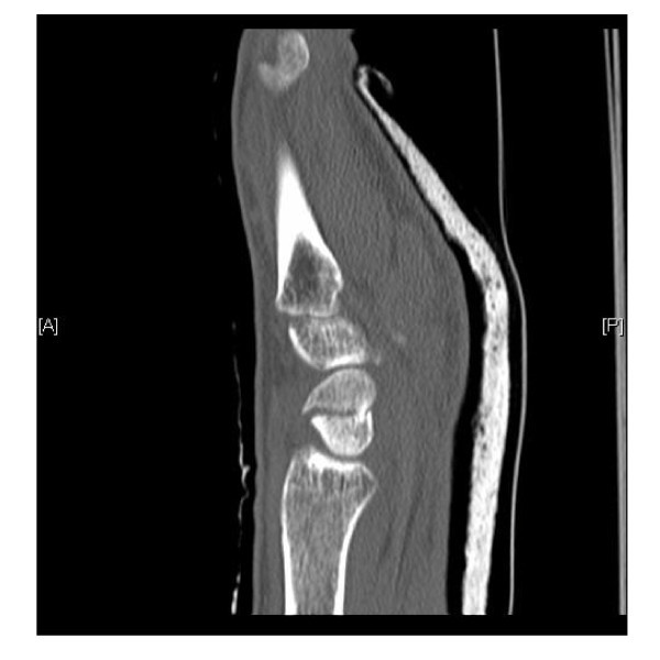
CT scanSagittal CT scan of the wrist: fracture line clearly visible at the carpal scaphoid.
Source : Rhemrev et al., International journal of emergency medicine, 2011, figure 3 · CC BY
Three possible investigations, three very different error profiles, and a trade-off that depends on what you fear most.
Mallee's Cochrane review compared CT, MRI and bone scintigraphy in suspected patients with normal radiographs. Eleven studies were included: 4 for CT (277 suspected cases), 5 for MRI (221) and 6 for scintigraphy (543). The pooled estimates are as follows6 :
| Investigation | Sensitivity | Specificity | Per 1,000 patients |
|---|---|---|---|
| Bone scintigraphy | 0.99 (95 % CI 0.69 to 1.00) | 0.86 (95 % CI 0.73 to 0.94) | 112 patients overtreated, 2 fractures missed |
| MRI | 0.88 (95 % CI 0.64 to 0.97) | 1.00 (95 % CI 0.38 to 1.00) | Not calculated in the review |
| CT | 0.72 (95 % CI 0.36 to 0.92) | 0.99 (95 % CI 0.71 to 1.00) | 8 patients overtreated, 56 fractures missed |
Reading this table is subtler than it looks, and the authors do it explicitly. Statistically, scintigraphy is the best investigation for establishing a definitive diagnosis : its sensitivity is the highest, and it is the one that misses fewest fractures. But its lower specificity becomes a problem when prevalence is low: across 1,000 patients it would lead to 112 people being overtreated, against 8 with CT. Conversely, CT overtreats almost nobody but would let 56 fractures through where scintigraphy would miss only 26.
The authors add three important caveats: scintigraphy is more invasive, involves radiation and imposes a diagnostic delay of at least 72 hours ; the confidence intervals are wide for all three investigations; and the comparisons are mostly indirect, which exposes them to confounding6.
Across 1,000 suspected patients, the errors each investigation produces
Two opposite profiles: scintigraphy misses almost nothing but overtreats a great deal, CT does the reverse
Source of the figures: Mallee WH, Wang J, Poolman RW et al., Cochrane Database Syst Rev 2015;2015(6):CD010023 (PMID 26045406), the authors' calculation on a median prevalence of 20 %. The confidence intervals of all three investigations are wide, as the review stresses. The preference for MRI in the emergency setting comes from Carpenter CR et al., Acad Emerg Med 2014 (PMID 24673666).
Carpenter reaches a more clear-cut recommendation, in the specific context of the emergency department: in patients whose initial radiograph shows no fracture but in whom concern persists,MRI is the most accurate investigation, superior to scintigraphy, CT and ultrasound, both for confirming and for ruling out the diagnosis. If MRI is not available, CT is enough to confirm a fracture but remains insufficient to rule one out. The author also notes that MRI and CT share the advantage of identifying other causes of post-traumatic wrist pain3.
The apparent divergence between these two sources can be explained. The Cochrane review reasons on pure statistical accuracy, Carpenter's meta-analysis on clinical usefulness in the emergency setting, taking in availability, delay and invasiveness. For a physiotherapist, the practical conclusion is a single one: when doubt persists, cross-sectional imaging settles it, and passive waiting is not a diagnostic strategy.
An old practice found wanting
The traditional strategy of immobilising and then repeating a radiograph later has been specifically evaluated, and it does not hold up. A diagnostic performance study had 81 orthopaedic surgeons read the initial and six-week radiographs of 34 suspected patients, taking the CT and MRI performed on admission as reference standards. Inter-observer agreement was poor (kappa of 0.15 and 0.14 depending on the display mode), sensitivity ranged from 42 to 79 %, specificity from 53 to 59 %, accuracy from 53 to 58 % and positive predictive value from 14 to 26 %. The authors conclude that six-week radiographs are unsuitable for assessing a suspected scaphoid fracture15. The Cochrane review notes moreover that this kind of radiographic follow-up served as the reference standard in several included studies, which weakens their estimates accordingly6.
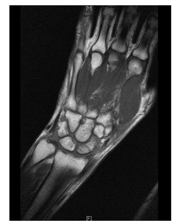
MRICoronal MRI of the wrist: fracture of the waist of the carpal scaphoid.
Source : Rhemrev et al., International journal of emergency medicine, 2011, figure 5 · CC BY
A large pragmatic trial, five years of follow-up, and a conclusion that reversed a surgical trend.
SWIFFT is a pragmatic, multicentre, open-label randomised superiority trial conducted in 31 hospitals in England and Wales. It included adults aged 16 and over with a bicortical fracture of the scaphoid waist displaced by 2 mm or less. Of 1,047 patients assessed, 439 were randomised (mean age 33, 363 men or 83 %) between early surgical fixation (n = 219) and below-elbow cast immobilisation, followed by immediate fixation if nonunion was confirmed (n = 220)7.
The primary outcome was the PRWE score at 52 weeks. The results:
The authors' recommendation is explicit: adults with a waist fracture displaced by 2 mm or less should receive initial cast immobilisation, with any suspected nonunion confirmed and then fixed immediately. That strategy avoids the risks of surgery and reserves it for the fractures that do not unite7.
The five-year follow-up, published in 2026, reinforces this result. Across 344 patients who provided a valid PRWE score (78.4 % of the initial cohort), no significant difference appeared between the groups: a mean difference of 0.6 points (95 % CI 2.4 to 3.6), p = 0.709. Neither the pain and function subscores, nor the global estimate of treatment effect differed. Grip strength and range of motion were comparable. The nonunion rate among imaged patients remained very low: 3 out of 146 (2.1 %) in the fixation group against 4 out of 121 (3.3 %) in the immobilisation group8.
The radiological arm of the same follow-up brings two pieces of information a physiotherapist needs to know9 :
That last point deserves emphasis: early surgery does not protect against the development of osteoarthritis, and that development is not silent.
SWIFFT: what early surgery brings, and what it costs
439 patients randomised, functional outcome at one and five years, complication profile
Sources: Dias JJ et al., Lancet 2020;396(10248):390-401 (PMID 32771106) for the one-year result and the complications; Dias JJ et al., Bone Joint J 2026;108-B(1):70-78 (PMID 41475372) for the five-year clinical follow-up and the nonunion rates.
This is the question where the temptation to immobilise less is most costly, and where the evidence is old but clear.
The benchmark randomised trial on this question dates from 1989 and is still cited for the simple reason that no more recent trial has produced as clear a result on this precise point. Fifty-one patients with an undisplaced scaphoid fracture were allocated to two forms of immobilisation, with follow-up at least until union and a mean duration of twelve months:
The results are as follows10 :
| Form | Mean time to union | Nonunions | Delayed unions |
|---|---|---|---|
| Long then short cast (n = 28) | 9.5 weeks | 0 | 2 |
| Short cast only (n = 23) | 12.7 weeks | 2 | 6 |
Three lessons follow. First, more restrictive initial immobilisation shortens the time to union by more than three weeks. Second, it clearly reduces the number of nonunions and delayed unions. Third, and this is the most useful point in practice, the advantage depends on the site of the fracture line : it was significant for fractures of the proximal third and the waist, whereas distal third fractures did well whatever the form of immobilisation10.
The trial has to be placed for what it is: 51 patients, a 1989 publication, immobilisation practices that have moved on since. It does not set a protocol, but it clearly documents the direction of the effect, and that direction is consistent with the vascular physiology described above.
The mistake not to make
Shortening immobilisation because the patient is finding it a nuisance, or because the pain has gone, exposes them to exactly the mechanism Gellman documented: more delayed unions and nonunions, especially if the line sits at the waist or proximally10. The disappearance of pain does not mean union in a bone whose proximal fragment may be devascularised. It is imaging, not comfort, that allows a return to activity.
The good news is real, and it does not remove the need to make the diagnosis early.
Grewal's study assessed the outcome of so-called subacute fractures, that is, diagnosed between six weeks and six months after the injury, and treated with a cast alone. Twenty-eight patients were included (20 men, 8 women, mean age 30), with 20 waist fractures, 7 proximal pole and 1 distal pole, with union assessed by CT.
The results are encouraging11 :
The authors conclude that subacute scaphoid fractures, up to six months from injury, can unite with a cast alone, even if the initial diagnosis was delayed, and that the expected time to union is shorter than previously reported. It is a level IV study, retrospective and without a control group: it describes an outcome, it does not compare strategies.
When union does not occur, the classically described course is degenerative osteoarthritis of the wrist following a stereotyped sequence, scaphoid nonunion advanced collapse or SNAC wrist12. That established osteoarthritis, its stages and its salvage procedures are the subject of a separate article on this site: carpal osteoarthritis of the wrist. This article stops where that one begins, at the fracture and its union.
But Buijze's review introduces here an intellectual caveat that deserves to be reported as it stands, because it is rarely mentioned: the correlation between symptoms and disease is poor, and the true natural history is debatable, since only symptomatic patients who present are assessed. The authors go further: it is not established that surgery alters the natural history, even when union is achieved12.
That finding is not an invitation to neglect the diagnosis, it is an invitation to measured language. Clinical reasoning remains unchanged: early diagnosis avoids heavier treatment, there is no benefit in delay, and caution is called for in any wrist pain that drags on. But telling a patient that osteoarthritis is inevitable goes beyond what the data allow.
A series to read with caution
A review of 64 patients published in 2024 reports a nonunion rate of 82.8 %, far higher than the rest of the literature. The authors give the explanation themselves: their unit is a referral centre for established nonunions, which produces a major recruitment bias. That figure therefore does not describe the risk for an unselected patient. The practical recommendation they draw from it does remain useful: clinical follow-up and CT at three months for conservative treatment, and early referral of fractures that have not united14.
Solid evidence
Solid evidence
Weak evidence
More restrictive initial immobilisation for proximal and waist fracture lines. A single randomised trial, 51 patients, 1989, but a clear direction of effect consistent with the vascular physiology10.
Weak evidence
Union is possible in a fracture diagnosed late. A retrospective series of 28 patients, level IV, without a control group11.
Very weak
The comparative accuracy of cross-sectional investigations. Eleven studies, wide confidence intervals for all three modalities, mostly indirect comparisons6.
Not recommended
Repeat radiography at six weeks as a means of diagnosis. Judged unsuitable by a level II diagnostic performance study: poor inter-observer agreement and accuracy of 53 to 58 %15.
A published case, with its identifier, that reproduces exactly the trap described in this article.
A 19-year-old basketball player, diagnosed with a “wrist sprain”
Braun S, Yelinek A, Cureus 2020;12(8):e9793 (PMID 32953308). Case report.
History. A 19-year-old patient, a college basketball player, falls on her outstretched hand during a game. She presents to an urgent care centre where the diagnosis made is a wrist sprain. Faced with persistent pain, she then consults in sports medicine, where the diagnosis made is a displaced scaphoid fracture.
Management and course. Treatment was conservative, with thumb-spica cast immobilisation, supplemented by low-intensity pulsed ultrasound as an adjunct13.
What this case teaches, and what it does not establish. It illustrates the exact sequence this article describes: a typical mechanism, a first diagnosis of sprain, and a fracture revealed by persistent symptoms. The authors also note that the diagnosis is often missed because of the particular anatomy and position of this bone. On the other hand, a single case demonstrates the efficacy of no treatment, and the low-intensity pulsed ultrasound used here cannot on any account be recommended on that basis. The value of this report is diagnostic and educational, not therapeutic.
A discrepancy in the figures worth flagging
This case reports that scaphoid fractures account for 60 to 70 % of fractures of the bones of the wrist13, whereas Carpenter's meta-analysis gives 70 % of carpal fractures3 and the SWIFFT trial 90 %7. These gaps come from the denominator used, carpal bones or wrist bones, and from the populations studied. We report them rather than choose one: the robust datum is that the scaphoid is the most frequently fractured carpal bone1, not the exact value of the percentage.
What these data change in the assessment, the referral and the conduct of rehabilitation.
The physiotherapist is not the one who requests imaging, but they are very often the one who sees these patients repeatedly, and therefore the one who can catch a missed diagnosis. Three situations should prompt a return to the doctor:
Conversely, the absence of anatomical snuffbox tenderness is the only element that substantially lowers the probability of fracture, with a negative likelihood ratio of 0.153 - 0.25 depending on the source. That does not rule it out, but it is the most useful clinical information available.
Faced with a painful wrist after a fall on the hand
A reasoning framework, and at each branch the datum that justifies it
Reasoning framework assembled from: Carpenter CR et al., Acad Emerg Med 2014, LR− of the absence of snuffbox tenderness and superiority of MRI (PMID 24673666); Coventry L et al., Emerg Med J 2023, LR− of 0.2 and the absence of any single sign allowing exclusion (PMID 37169546); Mallee WH et al., Arch Orthop Trauma Surg 2016, unsuitability of six-week radiographs (PMID 27026536). Assembling them into a single tree is a proposed line of clinical reasoning: no validated decision rule exists on this subject, as Carpenter explicitly points out.
Also on the site
No, and that is the central message of this article. In emergency department patients with clinical suspicion and a non-diagnostic initial radiograph, the pre-test probability of fracture is estimated at 25 %, with a spread from 12 to 57 % across studies3. Other work measuring the same quantity finds 9.0 %5 or a median of 20 %6. Whatever figure is used, the conclusion is identical: a normal radiograph does not rule the diagnosis out, and the strategy of a repeat radiograph at six weeks has been judged unsuitable15.
In the emergency setting, Carpenter's meta-analysis concludes that MRI is the most accurate investigation, superior to scintigraphy, CT and ultrasound, both for confirming and for ruling out the diagnosis; failing that, CT can confirm but remains insufficient to rule out3. The Cochrane review, reasoning on pure statistical accuracy, names scintigraphy as the best performing, while stressing that it is more invasive, involves radiation, imposes a delay of at least 72 hours and would lead to 112 patients out of 1,000 being overtreated against 8 with CT6. The decision rests with the prescriber.
For a bicortical waist fracture displaced by 2 mm or less, no. The SWIFFT trial found no difference in functional score at one year between early surgery and casting, whereas potentially serious complications affected 14 % of those operated on against 1 % of those in a cast7. The equivalence still held at five years, with nonunion rates of 2.1 % and 3.3 %8. The recommended strategy is cast first, with immediate fixation if nonunion is confirmed. Fractures displaced by more than 2 mm and those of the proximal pole do not fall within that frame.
Because the fracture line often cuts off the blood supply to the proximal fragment. The main artery enters through the distal pole and runs up towards the proximal pole: the more proximal the line, the more dependent the fragment deprived of supply. That is why the time to union depends on the site, as Grewal's study shows, with 11 weeks of immobilisation on average for the waist against 14 weeks for the proximal pole11.
That is precisely the decision that exposes you to nonunion. In Gellman's randomised trial, more restrictive initial immobilisation achieved union in 9.5 weeks against 12.7 weeks, with no nonunion against two in the short immobilisation group, the advantage being significant for fractures of the proximal third and the waist10. The disappearance of pain does not mean union in a bone whose proximal fragment may be devascularised: it is imaging that allows the cast to come off.
No, and that is reassuring information to give. In Grewal's series covering fractures diagnosed between six weeks and six months from injury and treated with a cast alone, the union rate reached 82 %, and 96 % after excluding patients with diabetes, comminuted fractures and humpback deformities11. It is a retrospective series of 28 patients, level IV evidence: it describes a favourable outcome, it does not guarantee an individual result.
The classically described course is the SNAC wrist, a progressive degenerative osteoarthritis. But Buijze's review adds a rarely cited caveat: the correlation between symptoms and disease is poor, the true natural history is debatable since only symptomatic patients who present are assessed, and it is not established that surgery alters that natural history, even when union is achieved12. Note also that in the SWIFFT trial, 52.4 % of patients imaged at five years had osteoarthritis in at least one joint, with a similar prevalence after surgery and after casting9.
None of the sources used for this article provides a validated return-to-sport criterion specific to this fracture, and it is better to say so than to offer a convenient timescale. The reasoning rests on union confirmed by imaging, and not on the disappearance of pain alone. Two useful markers: the immobilisation times observed range from 9.5 to 14 weeks depending on the form and the site1011, and the recommended conservative follow-up includes clinical and CT assessment at three months14.
Fifteen references, each verified individually on PubMed through the E-utilities interface: identifier resolved, title, journal, year, pagination and author list checked on the record itself, abstract read before citation. The links point to the PubMed record.
Note on method
The bibliographic base was built before writing, subject by subject, by querying PubMed through the E-utilities interface: every identifier was resolved on its XML record, every reference checked on its authors, journal, year and pagination, and every abstract read before being cited. Several displayed years differ from those commonly cited elsewhere, the record taking precedence: Mallee's meta-analysis on clinical tests is dated 2014, the Cochrane imaging review 2015, and the five-year follow-up of SWIFFT 2026. Three points are explicitly treated as uncertain rather than settled: the exact proportion of carpal fractures attributable to the scaphoid, which varies from 60 to 90 % depending on the denominator used; the prevalence of fracture among suspected cases with a normal radiograph, measured between 9 and 25 % depending on the threshold of suspicion; and the natural history of nonunion, which Buijze's review itself calls debatable. The absence of a validated return-to-sport criterion is flagged rather than filled in. The classification codes in the structured data block were checked against the official tables: ICD-11 NC53.0 on the WHO MMS linearisation, ICD-10 S62.0 on the official ICD-10 browser. Article written on 16 August 2026.
Behind this article
An author who explains, a reviewer who checks.
How we write and check our content

Physiotherapist · co-founder of Physio Learning
Marked for life by his first four-hour lecture without a single image, he took a master’s in instructional design so that it would never happen to anyone again. He hunts down publication bias and unreadable slides with the same intransigence.

Head of scientific content
Physiotherapist specialising in neuro-musculoskeletal practice and holder of a master’s in public health. He checks the methodological rigour of every article: primary sources, levels of evidence, no exceptions.
