














On-siteDPCFIFPL

Upper limb
Hand rehabilitation and splinting: refining and deepening your practice
Jean Lichtle
Go further
Distal radius fracture: what the tilt really predicts, why the distal radioulnar joint decides rotation, and when rehabilitation should begin.

An isolated radiographic angle should not alarm the patient: it is the combination of several defects, in a young, demanding patient, that matters.
96%of range and strength regained on the healthy side at eleven years
What follows covers each of these points in detail, with sources. It is there if you need it.
This topic is taught in a course: Hand rehabilitation and splinting: refining and deepening your practice, on-site with Jean Lichtle.
1 / 12
11 article chapters · 38 min in total
Understanding◔ 4 min
The radius carries most of the load of the wrist, the ulna a far smaller fraction.
Population◔ 2 min
Two very different populations hide behind the same diagnosis: the postmenopausal woman and the young patient.
Radiograph◔ 4 min
Of 84 correlations tested, only 2 linked radiographic anatomy to function.
Rotation◔ 4 min
Supination is the movement recovered least well, because of the distal radioulnar joint.
Surgery◔ 3 min
In patients aged 60 and over, no surgical technique wins out, nor does plaster.
Immobilisation◔ 3 min
Immobilising the wrist does not immobilise the limb: fingers, elbow and shoulder remain to be worked.
After removal◔ 6 min
Supervision does not always change the outcome; a randomised trial shows the opposite.
Complications◔ 3 min
The physiotherapist sees these patients more often than the surgeon, and spots the complication as it sets in.
Case report◔ 1 min
Untreated osteoporosis preceded a complex regional pain syndrome at three months.
In practice◔ 4 min
Do not dramatise an isolated angle: rotation and the functional score matter more.
Questions◔ 4 min
Recovery takes far longer than the bone takes to heal, sometimes years.
Train on this
Upper limb
Jean Lichtle
Next comes practice: the course that teaches this topic, with Jean Lichtle.
Our courses on this topic may be covered by DPC or FIFPL, the French continuing-education funding schemes, depending on your profession and situation.
This is the fracture everyone thinks they know: a fall on the hand, a dinner-fork deformity, six weeks in plaster. The detail that changes rehabilitation lies elsewhere. Dorsal tilt, the parameter everyone measures, predicts far less than its radiographic obviousness suggests, and it only becomes frankly troublesome when combined with shortening. What decides the rotation lost is the distal radioulnar joint, at the other end of the same fracture line. And the physiotherapist's work does not begin on the day the cast comes off: it begins during immobilisation, on segments the cast does not immobilise.
A synthesis written from primary sources verified one by one on PubMed through the E-utilities interface: identifier resolved, journal, year and author list checked, abstract read before citation. Full bibliography at the end of the article.
Distal radius fracture in three figures
Three independent results that shift the question from “how do we straighten this wrist?” to “what, here, really changes the patient's life?”
Sources: Soerensen S et al., Hand (N Y) 2024, population study, 5,426 fractures in 522,607 inhabitants (PMID 35856325); Chung KC et al., JAMA Netw Open 2021, WRIST randomised trial, 182 patients assessed at 24 months (PMID 34137830); Reid SA et al., J Physiother 2020, multicentre randomised trial, 67 patients, difference 12° 95 % CI 5 to 20 (PMID 32291223).
What to have in mind
Four situations that call for a medical opinion without waiting for the next session
A fractured wrist is judged on what the patient can do, not on the angle it forms. Eleven years later, range and strength come back to 96 % of the healthy side, whatever the radiograph looks like.
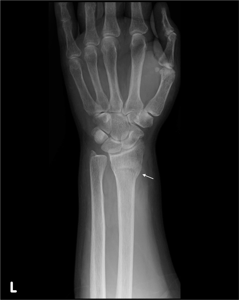
X-rayAnteroposterior wrist X-ray: distal radius fracture, arrow on the metaphyseal fracture line.
Source : Ozaki et al., Medicine, 2016, figure 3 · CC BY
Understanding the tilt means understanding why it recurs in plaster, and why it is paid for in rotation more than in flexion.
The distal radius is the widened end of the radius, the one that articulates with the carpus. It carries three articular surfaces: the scaphoid fossa and the lunate fossa, which receive the first row of the carpus, and the sigmoid notch, on the medial border, which receives the head of the ulna and forms the radial side of the distal radioulnar joint. That third surface is the one that gets forgotten, and it is the one that decides pronation and supination.
The radius carries most of the load transmitted by the hand to the forearm. The ulna, more slender and articulated with the carpus through the triangular fibrocartilage complex, takes a far smaller fraction. That distribution explains two things: that the distal radius is the zone that breaks when you fall on the hand, and that its shortening mechanically transfers load towards the ulna, with the consequences the chapter on the distal radioulnar joint sets out.
The dominant mechanism is stereotyped: a fall landing on the open hand, wrist in extension. In the Danish population study covering 5,426 distal forearm fractures, a fall from standing height accounted for 76 % of mechanisms, and the commonest fracture type was extra-articular, AO type 2R3A, in 69 % of cases1. In other words: the typical fracture is neither intra-articular, nor comminuted, nor the result of violent trauma. It is an ordinary fall in someone whose bone is fragile.
The force travelling up from the heel of the hand compresses the dorsal cortex, which crumples, while the volar cortex fails in tension. The distal fragment then goes backwards: this is the dorsal tilt, described by Pouteau and then by Colles, which gives the dinner-fork deformity visible from the side. The volar-tilt variant, known as Goyrand-Smith, results from the opposite mechanism, wrist in flexion, and is far rarer.
This anatomy of displacement has a direct practical consequence: the tilt is not only produced by the impact, it is maintained by the crumpling of the dorsal cortex. Once the cancellous bone is impacted, there is no longer, at the back, a column to hold the reduction. That is the mechanical reason why a reduction in plaster is so often lost, and why secondary redisplacement is a frequent event and not a failure of technique.
Why the distal fragment tilts backwards, and why it holds badly
The dorsal cortex crumples in compression; once impacted, it no longer offers support to maintain the reduction
A schematic diagram. The redisplacement figure comes from Karantana A, Handoll HH, Sabouni A, Cochrane Database Syst Rev 2020, a review of 26 trials, mean of six trials reporting this outcome, spread 3.3 to 75 % (PMID 32032439). Physiological volar tilt and its reference value are discussed in the chapter on measurements.
Four figures to have in mind
What the large series establish about the shape of the fracture, its healing and its aftermath
Sources: Soerensen S et al., Hand (N Y) 2024, 5,426 fractures (PMID 35856325) for the shape and the mechanism; Karantana A et al., Cochrane Database Syst Rev 2020, mean of six trials (PMID 32032439) for redisplacement; Schmidt V et al., J Hand Surg Eur Vol 2024, 242 patients examined at 11-13 years (PMID 37684021) for recovery.
Key points
Two populations, two prognoses, two rehabilitation plans. Confusing them is the first error of orientation.
The most solid population study available followed an average at-risk population of 522,607 inhabitants and manually validated radiographs and records for 5,426 distal forearm fractures. The overall incidence comes to 207.7 per 100,000 per year, broken down into 323.4 in women and 93.3 in men. Women accounted for 77 % of the fractures, and incidence rose markedly in them after the age of 50. The incidence of distal radius fractures proper was 203.0 per 100,000 per year, that of isolated ulnar fractures only 3.81.
Cumulative lifetime risk gives the measure of the public health problem: in high-income countries, 6 % of women will have had a wrist fracture by 80, and 9 % by 902.
Incidence of distal forearm fractures
A ratio of 3.5 between women and men, in a manually validated population
A single source: Soerensen S, Larsen P, Korup LR et al., Epidemiology of Distal Forearm Fracture: A Population-Based Study of 5426 Fractures, Hand (N Y) 2024;19(1):24-29 (PMID 35856325). An average at-risk population of 522,607 inhabitants, manual review of radiographs and records.
Behind that overall figure lie two distinct clinical realities, and the authoritative trials do not cover the same one.
The first is that of the postmenopausal woman with fragile bone, who falls from standing height. It is the population of the WRIST trial, whose mean age was 70.1 with 87.9 % women3. In her, the wrist fracture is a sentinel fracture : it announces the later bone risk, and identifying it should trigger an assessment of osteoporosis and of falls risk. Our article on osteoporosis and the prevention of fragility fractures sets out that cascade and what it requires in practice.
The second is that of the young person with a high-energy injury, a fall from a bike, a sports accident, a fall from height. The functional demand there is quite different, associated injuries are more frequent, and it is in this population that anatomical restoration keeps most of its meaning, as what follows shows.
The trap of generalisation
Most of the large trials in this condition, WRIST in particular, recruited patients aged 60 and over. Their reassuring conclusions on the low prognostic value of radiographic parameters do not transfer automatically to the young person. Brogren's cohort, whose age range is wider and which finds an effect of combined malunion at 2 years5, is a reminder that tolerance to deformity depends on what the patient asks of their wrist.
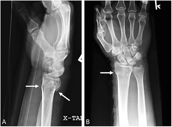
X-rayLateral and anteroposterior wrist X-rays: Colles fracture, arrows on the metaphyseal line of the distal radius with dorsal tilt of the fragment.
Source : Wong et al., International journal of emergency medicine, 2015, figure 5 · CC BY
Four measurements, only one of which appears linked to function in the older person, and a combination that weighs more than either of its parts.
The radiographic assessment of a distal radius fracture rests on simple measurements, made on the AP and lateral views. The physiotherapist does not have to make them, but reading them in a report changes how rehabilitation is conducted, and above all how it is talked about with the patient.
| Parameter | What it measures | Reference | What the evidence says about it |
|---|---|---|---|
| Dorsal tilt (sagittal angulation) |
Orientation of the articular surface on the lateral view. Physiologically angled towards the palm | Physiological volar tilt | No effect on the clinical result at 11-13 years6. Counts at 2 years if combined with shortening5 |
| Ulnar variance (radial shortening) |
Relative height of the ulnar head compared with the radius, on the AP view | 0 mm | In those aged 70 and over, each millimetre towards normal was associated with 10.4 points better on the activities of daily living score (95 % CI 3.86 to 16.84)4 |
| Radial inclination | Obliquity of the articular surface on the AP view | 22° | In those aged 70 and over, each degree of deviation was associated with a loss of strength of 1.1 kg (95 % CI 0.38 to 1.76)4, with no effect on the overall function score |
| Joint involvement (step-off, gap) |
Congruence of the radiocarpal articular surface | A continuous surface | Intra-articular extension influenced neither the clinical result nor the risk of osteoarthritis at 11-13 years6 |
The secondary analysis of the WRIST trial put this question to the test systematically. In 166 participants aged 60 and over (86.7 % women, mean age 70.9), the authors crossed the radiographic measurements with seven functional outcomes at 12 months. Of 84 correlation coefficients calculated, only 2 were statistically significant, both in the subgroup aged 70 and over, and neither concerned the overall hand score nor the function score4. The authors' conclusion is explicit: precise restoration of wrist anatomy is not associated with better outcomes in the older person at 12 months.
The longest prospective follow-up available points the same way and extends it to a wider population. Eleven to thirteen years after the fracture, 292 patients answered the questionnaires and 242 were examined. The median QuickDASH was 5, the EuroQol score 1.0, and both range and grip strength reached 96 % of the contralateral side. Neither radiocarpal osteoarthritis (6 %) nor ulnar styloid non-union (30 %) affected the results. Above all, neither dorsal tilt, nor radial inclination, nor ulnar variance, nor intra-articular extension influenced the long-term clinical result or the risk of osteoarthritis6.
One must nevertheless be careful not to conclude that reduction does not matter. Three reservations are needed, and they are clinically decisive.
First reservation: it is the combination that counts. Brogren's prospective cohort followed 123 patients with the DASH repeated at inclusion, 3 months, 6 months, 1 year and 2 years. Defining malunion as an ulnar variance of at least 1 mm combined with a dorsal tilt greater than 10°, the authors formed three groups: no malunion (n = 35), with one or the other defect (n = 65), with both (n = 23). Only the group combining both defects kept significantly greater disability: 13 DASH points more than the group without malunion (95 % CI 1.4 to 25), and 13 points more than the group with an isolated defect (95 % CI 2.2 to 24)5. An isolated defect behaved no differently from a correctly reduced wrist.
Second reservation: age modulates everything. The most reassuring results come from older populations with moderate functional demand. The 35-year-old who works with their hands is not in that frame, and no data of this level allows the same tolerance thresholds to be applied to them.
Third reservation: this work measures overall function, not rotation. Hand scores such as the DASH or the MHQ include activities that pronation and supination influence, but which they do not determine on their own. That is precisely the subject of the next chapter.
Key points
Supination is the movement these patients recover least well. That is no accident, and it is not the fault of the radiocarpal fracture line.
The distal radioulnar joint joins the sigmoid notch of the radius to the head of the ulna. Together with the proximal radioulnar joint at the elbow it forms the pair that carries pronation and supination : it is the radius that turns around the ulna, not the reverse. Its stability comes from bony congruence, which is weak in itself, and above all from the triangular fibrocartilage complex, whose insertions are on the ulnar fovea and styloid.
Now, the sigmoid notch of the radius is a few millimetres from the usual fracture line. The review by Nypaver and Bozentka devoted to this joint in the context of distal radius fracture sets out its anatomy, biomechanics, patterns of injury, at the time of trauma as well as after treatment, appropriate assessment and treatment options7. The clinical point to keep is simple: a distal radius fracture is almost always also a matter of the distal radioulnar joint, whether the report mentions it or not.
The mechanical reasoning is direct. The radius tilts backwards and shortens; its sigmoid notch then changes orientation and height relative to the ulnar head, which has not moved. Three consequences follow:
This reading sheds light on a clinical fact that long follow-up confirms: in Schmidt's cohort, ulnar styloid non-union concerned 30 % of patients at eleven years without affecting the clinical result6. In other words, an ununited styloid is not in itself a problem; it is instability of the joint it stabilises that would be, and the two are not the same thing.
What the distal radioulnar joint explains in a wrist that does not recover
Three complaints, three distinct mechanisms, three different courses of action
Source of the anatomical and injury framework: Nypaver C, Bozentka DJ, Distal Radius Fracture and the Distal Radioulnar Joint, Hand Clin 2021;37(2):293-307 (PMID 33892882). The associated rehabilitation approaches are a proposal of reasoning, explicitly not drawn from a comparative trial.
What this changes in the session
Faced with a wrist that plateaus in supination at 6 or 8 weeks, the reflex response is to insist on stretching. The useful reasoning is first to distinguish soft tissue stiffness, which gives way progressively, from a bony block or an incongruence, which will not give way. A limitation that no longer moves over three to four weeks of well-conducted work, especially if it comes with pain along the ulnar border, deserves to be reported to the surgeon rather than forced.
The question is not the physiotherapist's to decide, but answering it accurately avoids letting a patient believe they were badly treated.
The WRIST trial recruited 304 adults aged 60 and over with an unstable, isolated distal radius fracture, across 24 health systems in the United States, Canada and Singapore. One hundred and eighty-seven were randomised between volar locking plate (n = 65), external fixation with or without supplementary wires (n = 64) and percutaneous pinning (n = 58); 117 chose non-operative treatment in plaster8.
At 24 months, in 182 patients assessed, the hand scores were as follows: 88 for the volar plate (95 % CI 83 to 92), 83 for external fixation (78 to 88), 85 for pinning (79 to 90) and 85 for plaster (79 to 90), with no clinically relevant difference after adjustment. Pain differed no more. Between 12 and 24 months, the scores had barely changed3.
It is in that same work that one of the most counter-intuitive results in the literature is found: malunion was far more frequent after plaster (59.1 %) than after volar plating (8.0 %), external fixation (17.0 %) or pinning (9.8 %), with a highly significant difference. And yet, that malunion was not associated with the difference in outcome at 24 months3. Surgery straightens better, without that better alignment translating into better function in this population.
In adults operated on for a dorsally displaced fracture, the British DRAFFT trial randomised 461 patients between percutaneous pinning and volar locking plate. More than 90 % were followed to the end. Both groups recovered their function at 12 months, and no clinically relevant difference was found on the PRWE at 3, 6 or 12 months (difference at 12 months of 1.3 points, 95 % CI 4.5 to 1.8, p = 0.398). The number of complications did not differ, and pinning proved £727 cheaper2.
Karantana's Cochrane review, which included 26 trials and 1,946 adults, is more cautious still, and for a methodological reason: all the trials included were at high risk of bias, and allocation concealment was certain in only one. Across the eleven trials comparing pinning with plaster after reduction (917 participants), the quality of the evidence was very low for every outcome reported, to the point that the authors declare themselves uncertain of any effect on patient-reported function9.
Two descriptive figures nevertheless come out of it, useful in everyday practice: redisplacement leading to secondary treatment affected on average 12 % of patients in plaster (3.3 to 75 % across series), and pin-track infection warranting antibiotics on average 7.7 % of pinned patients (0 to 15 %). Two studies also reported digital stiffness after cast removal to be less frequent after pinning (20 % against 36 %)9.
| Study | Population | Comparison | Main result |
|---|---|---|---|
| WRIST 20213 | 304 adults aged 60 and over, unstable fracture, 24 centres | Volar plate, external fixation, pinning, plaster | Comparable hand scores at 24 months (88 / 83 / 85 / 85), p = 0.70 |
| UK DRAFFT 20152 | 461 adults operated on, dorsally displaced fracture | Pinning against volar plate | Identical PRWE at 12 months (difference 1.3; 95 % CI 4.5 to 1.8), pinning cheaper |
| Cochrane 20209 | 26 trials, 1,946 adults, mostly older | Pinning against plaster, and technical variants | Very low quality evidence, uncertainty about function; 12 % redisplacement in plaster, 7.7 % pin-track infection |
| WRIST complications 20198 | 296 adults aged 60 and over | Complication profile by technique | Median compression 18.5 % after plating; pin-track infection 23 to 26 %; plaster: more complications at all severities (RR 1.88) |
It is in this context of equivalence that the American Academy of Orthopaedic Surgeons and the American Society for Surgery of the Hand published updated clinical practice guidelines in 2020 on the evaluation and treatment of acute distal radius fractures, following a formalised methodology: seven recommendations based on the best available data, intended to guide the practitioners who look after these patients19.
No technique has shown itself superior on what the patient feels. The choice therefore turns on the risk profile, the cost and preferences, not on a promise of better function.
Six weeks of waiting are not six weeks of nothing. It is the period in which digital mobility, the shoulder and tissue condition are either prepared or lost.
A below-elbow cast leaves the metacarpophalangeal joints, the thumb, the elbow and the shoulder free. Yet those are precisely the segments where the most costly complications to make up develop: stiffness of the metacarpophalangeal joints in extension, stiffening of the shoulder in the older person, and oedema that settles in for want of a muscle pump.
The Cochrane review devoted to rehabilitation of this fracture examined 26 trials and 1,269 patients, mostly older women, across 23 comparisons of which only 4 were covered by more than one trial. Seven of those trials concerned interventions started during immobilisation. The quality of the evidence was judged low or very low for all the comparisons, which rules out any strong statement11.
What that review reports for the immobilisation period is therefore a signal, not a demonstration: very low quality evidence in favour of hand therapy compared with simple instructions, four days after cast removal and with an effect persisting at one month; very low quality evidence of short-term improvement with early occupational therapy; and no difference between supervised and unsupervised exercises in a trial of 96 participants11.
Where the evidence becomes clearer is after volar plate fixation, precisely because the construct is stable. A randomised trial of 30 patients compared immediate mobilisation after surgery with five weeks of immobilisation, with assessments at 6 weeks, 9 weeks, 3 months, 6 months and 1 year.
The patients mobilised immediately had better range in the sagittal plane and better grip strength up to 6 months, an advantage in the frontal plane up to 9 weeks and in forearm rotation up to 6 weeks. The QuickDASH and PRWE scores were better up to 6 weeks, and the Green O'Brien score remained significantly different up to one year, with 93 % excellent or good results in that group. Above all, no difference was observed in loss of reduction, nor in pain, duration of physiotherapy or time off work10.
This is a pilot study of 30 patients, of level Ib: the signal is consistent and reassuring about safety, but the sample size rules out making a universal rule of it. The transferable message is this: when the construct is stable and the surgeon allows it, waiting brings nothing and costs mobility.
What you work on during immobilisation, and why
Four areas open before the cast comes off, on segments the plaster does not block
Sources: Handoll HH, Elliott J, Cochrane Database Syst Rev 2015, 26 trials, 1,269 patients (PMID 26403335); Karantana A et al., Cochrane Database Syst Rev 2020, digital stiffness 20 % against 36 % in two trials (PMID 32032439); Gutiérrez-Espinoza H et al., J Man Manip Ther 2022, manual lymphatic drainage, mean difference of 13.97 to 17.96 ml depending on the time of measurement (PMID 34668847); Chung KC et al., Plast Reconstr Surg 2019 (PMID 31348349).
This is where the literature is most uncomfortable for our profession, and most useful to read honestly.
One has to start with the least flattering item, because ignoring it would amount to selling the patient a promise the facts do not support.
The WRIST trial analysis of hand therapy followed patients aged 60 and over, with referral and protocol left to the discretion of the surgeon and the therapist. Eighty per cent of participants received care, 70 % combining supervised sessions and home exercises, with on average 9.2 supervised sessions over 14.2 weeks. The results are blunt: no difference in patient-reported scores between those who received care and the others ; the patients not followed up even recovered more grip strength ; and those whose care was shorter reported better function, better work capacity and greater satisfaction15.
This result calls for a methodological reading, not a rejection. It is a secondary observational analysis within a randomised trial: referral to rehabilitation was not randomised. The patients sent to physiotherapy are, by construction, the ones who were doing less well, which is enough to produce an unfavourable association without the treatment being the cause. It is indication bias in its most classic form. The authors put it cautiously: encouraging the resumption of daily activities could be as effective as formal therapy in these patients.
A later randomised trial put the same question with the right method. Seventy-four patients over 60 with an extra-articular fracture were allocated between six weeks of supervised physiotherapy (n = 37) and six weeks of a home exercise programme (n = 37), with two-year follow-up and no losses to follow-up.
The results draw a characteristic curve: at 6 weeks as at one year, the PRWE difference in favour of supervision was 18.6 points (95 % CI 12.8 to 24.3) then 18.5 points (95 % CI 12.7 to 24.2), clinically important gaps. At two years, that difference fell to 3.3 points (95 % CI 2.4 to 9.0), not significant. For the secondary outcomes, effect sizes ranged from medium to large at 6 weeks and one year; at two years, only grip strength kept a large effect in favour of supervision13.
The two pieces of work therefore do not really contradict each other: they describe the same thing from two angles. Supervised rehabilitation substantially speeds up recovery during the first year, and that advantage then fades as the control group catches up. It is honest and usable information: what you sell the patient is time gained and months of trouble avoided, not a different wrist at ten years.
What supervised physiotherapy brings, and for how long
PRWE difference between supervised physiotherapy and a home programme, randomised trial of 74 patients over 60
A single source: Gutiérrez-Espinoza H, Gutiérrez-Monclus R, Román-Veas J et al., Effectiveness of supervised physiotherapy versus a home exercise program in patients with distal radius fracture: a randomized controlled trial with a 2-year follow-up, Physiotherapy 2024;124:93-100 (PMID 38875842). The vertical bars show the 95 % confidence interval. A lower PRWE score means better function: the difference here is in favour of supervised physiotherapy.
The best-supported technique to date in this condition is mobilisation with movement after Mulligan, applied in supination and in wrist extension. A multicentre randomised trial, with concealed allocation, blinding and intention-to-treat analysis, included 67 adults treated in plaster (76 % women, mean age 60). The experimental group received the same exercises and advice as the control group, plus the mobilisations.
The gains are clear in the short term: supination 12° greater (95 % CI 5 to 20) at 4 weeks and 8° greater (95 % CI 1 to 15) at 12 weeks ; extension 14° greater (95 % CI 7 to 20), flexion 9° greater (95 % CI 4 to 15), QuickDASH 11 points lower (95 % CI 3 to 18) and PRWE 13 points lower (95 % CI 4 to 23) at 4 weeks. The benefits persisted at 12 weeks for supination, extension, flexion and the QuickDASH, and patients in the experimental group more often declared themselves improved (risk difference 22 %, 95 % CI 5 to 39). No clear benefit remained on patient-reported measures at 26 and 52 weeks, and no adverse effect was reported12.
The manual therapy meta-analysis that followed confirms that profile. Across eight eligible trials, six of them retained for quantitative synthesis, adding mobilisation with movement to an exercise programme gave at 12 weeks a PRWE 10.2 points lower (p = 0.02) and a DASH 9.86 points lower (p = 0.0001), with no significant effect on grip strength (3.9 %, p = 0.25). By contrast, the comparison between supervised physiotherapy with joint mobilisation and a home programme showed no significant difference in flexion (7.1°, p = 0.20) or extension (11.99°, p = 0.16) at 6 weeks. Manual lymphatic drainage added to conventional treatment reduced oedema consistently and significantly at every measurement point, by 13.97 to 17.96 ml depending on the moment (p between 0.03 and 0.002). The GRADE level of evidence ranged from very low to high depending on the comparison14.
The Cochrane review is the useful reminder of the real state of knowledge. Across 23 comparisons, none reaches a moderate or high level of evidence. It finds no clinically important difference between routine physiotherapy or occupational therapy added to home exercise instructions and those instructions alone, across four heterogeneous trials of 30, 33, 66 and 75 participants (low quality evidence). It does note, in two trials of 46 and 76 participants after volar plating, an advantage for a structured home exercise programme preceded by instruction or accompaniment, also in low quality evidence11.
For the individual modalities, the verdict is starker still: passive mobilisation, ice, pulsed electromagnetic fields, whirlpool baths, dynamic extension splinting for stiffness, none showed a clinically significant difference compared with no intervention, always in very low quality evidence11.
Low quality evidence
Low quality evidence
Supervised physiotherapy against a home programme, person over 60. A clinically important advantage at 6 weeks and 1 year, gone at 2 years except on strength. A randomised trial of 74 patients, with no losses to follow-up13.
Low quality evidence
Structured home exercise programme after volar plating. An advantage over unsupervised exercise in two trials (46 and 76 participants)11. Consistent with the fact that supervision counts for more than the setting.
Low quality evidence
Manual lymphatic drainage for oedema. A significant and consistent reduction, of 13.97 to 17.96 ml depending on the time of measurement14. An effect on an intermediate outcome, not on function.
Very low
Routine physiotherapy added to exercise instructions. No clinically important difference against the instructions alone, across four heterogeneous trials11.
No effect
Individual passive modalities : passive mobilisation, ice, pulsed electromagnetic fields, whirlpool baths, dynamic extension splinting. No clinically significant difference against no intervention, in very low quality evidence11.
Key points
The physiotherapist sees these patients more often than the surgeon does. It is often the physiotherapist who spots the complication as it sets in.
The analysis of complications in the WRIST trial, covering 296 participants aged 60 and over, gives a precise map of what to watch for depending on the technique used8 :
That last point deserves emphasis, because it contradicts a widespread intuition: non-operative treatment is not the risk-free treatment. It shifts the risk, it does not remove it.
This is the complication that worries people most, and the one where the physiotherapist has the clearest lookout role. Meena's meta-analysis is a reminder of the order of magnitude: a reported incidence of 10.5 to 37 % after distal radius fracture depending on the series and the diagnostic criteria used17, a spread that mainly reflects the heterogeneity of the definitions.
On prevention with vitamin C, the literature offers a textbook case of two meta-analyses published in the same year and reaching opposite conclusions.
| Study | Trials included | Result | Heterogeneity | Authors' conclusion |
|---|---|---|---|---|
| Evaniew et al., J Orthop Trauma 201516 | 3 trials, 890 patients | Relative risk 0.45 (95 % CI 0.18 to 1.13) | I² = 70 % | No significant benefit; low quality evidence |
| Meena et al., Eur J Orthop Surg Traumatol 201517 | 4 papers | Risk difference 0.41 (95 % CI 0.19 to 0.92); p = 0.03 | I² = 63 % | Significant reduction; better quality trials needed |
What to make of that contradiction? Read it for what it is. Both teams agree on two points: heterogeneity is high (63 and 70 %), and the overall quality of the evidence is low. Evaniew specifies that the heterogeneity was explained by the diagnostic criteria used, not by the dose or by the treatment of the fracture, and that the result held up under sensitivity analyses on missing data16. Prescribing vitamin C is the doctor's business; the physiotherapist's role is to present this measure neither as established nor as useless, but as debated.
What should raise the suspicion of complex regional pain syndrome
These situations call for a return to the doctor. The management documented in the case reported by Świta combined medication, physiotherapy and active therapy, with notable improvement18, but the authors stress the absence of an established treatment standard.
A published case, with its identifier, rather than an invented vignette: what the literature actually documents, including its grey areas.
A 75-year-old woman, untreated osteoporosis, complex regional pain syndrome at three months
Świta M, Szymonek P, Talarek K et al., J Clin Med 2024;13(4) (PMID 38398434). A clinical case accompanied by a literature review.
History. A 75-year-old patient with untreated osteoporosis sustains a Colles' fracture after a fall. Initial management combines reduction and plaster immobilisation of the forearm, followed by percutaneous Kirschner wire fixation. She then develops signs of complex regional pain syndrome and is admitted to a rehabilitation unit three months after the fracture. The affected forearm is swollen, hot, painful, with severely limited range.
Management and course. Treatment combines medication, physiotherapy and active therapy. The patient shows notable improvement following these interventions18.
What this case teaches, and what it does not establish. It illustrates three useful things: the late onset of this complication, which can appear when the fracture has already healed; the background, untreated osteoporosis in an older woman, which is a reminder that the sentinel fracture had not triggered any bone care; and the fact that combined management can markedly improve the state of a patient admitted at three months. On the other hand, an isolated case demonstrates no therapeutic efficacy, and the authors themselves stress the absence of an established treatment standard and the difficulty of predicting when this complication will occur.
Why there is no second clinical case
The searches conducted for this article did not identify, on PubMed, a published case of rehabilitation of a distal radius fracture teaching something distinct from this one and verifiable on its record. Rather than filling the gap with a vignette composed for the occasion, this chapter sticks to the case that is genuinely documented. A fabricated observation, however plausible, would be exactly the fault this site forbids itself.
What the data change in practice in the assessment, in what is said to the patient and in how sessions are run.
A framework for progression, and what underpins it
The time markers come from the protocols of the trials cited; the framework itself is a matter of clinical reasoning
Milestones from: Quadlbauer S et al., J Wrist Surg 2017, immediate mobilisation after volar plating (PMID 28428911); Reid SA et al., J Physiother 2020, assessments at 4 and 12 weeks (PMID 32291223); Gutiérrez-Espinoza H et al., Physiotherapy 2024, grip strength at 2 years (PMID 38875842); Schmidt V et al., J Hand Surg Eur Vol 2024, follow-up at 11-13 years (PMID 37684021). Assembling them into a single progression is a proposal of clinical reasoning, not a recommendation drawn from a trial.
Also worth reading on the site
Far longer than the bone takes to heal, and that has to be said from the outset. In the WRIST trial, hand scores were still changing between 12 and 24 months, even if the change did not reach the significance threshold3. In the eleven-year prospective follow-up, the authors conclude explicitly that recovery is a process lasting years, and that a decade later, range, strength and QuickDASH had returned to population norms6. In practice, most of it happens in the first six months, but improvement continues well beyond.
On its own, probably less than it looks. At eleven years of follow-up, neither dorsal tilt, nor radial inclination, nor ulnar variance, nor joint involvement was associated with the clinical result or with the risk of osteoarthritis6. In people aged 60 and over, precise restoration of anatomy was not associated with better outcomes at 12 months4. The important nuance is that the combination of shortening and tilt came with persistent disability at 2 years in a prospective cohort5, and that this work mostly covers older patients.
Because it does not depend on the same joint. Pronation and supination are carried by the radioulnar joints, of which the distal one is directly adjacent to the fracture line. Tilt and shortening of the radius change the relationship of its sigmoid notch to the ulnar head, which limits rotation at the end of range7. That is also why mobilisation with movement was studied first on supination, with a gain of 12° at 4 weeks12.
The question is the surgeon's, but the data make it possible to reassure without lying. In patients aged 60 and over, hand scores at 24 months were comparable between volar plate, external fixation, pinning and plaster3. In adults operated on for a dorsally displaced fracture, wires and volar plate gave the same result at 12 months2. Each option has its own complication profile8, and it is on that profile, the cost and the patient's preferences that the choice is made, not on a promise of better function.
Yes, with a nuance that must be carried honestly. A randomised trial in patients over 60 showed a clinically important advantage of supervised physiotherapy over a home programme at 6 weeks and at one year, with a gap of about 18 PRWE points, a difference that disappears at two years except for grip strength13. Conversely, an observational analysis of the WRIST trial found no difference between patients followed up and not followed up15, but that analysis is exposed to indication bias since referral was not randomised. The reasonable message: rehabilitation speeds up recovery, it does not change where you end up in the long term.
The question is not settled. Two meta-analyses published in the same year reach opposite conclusions: Evaniew finds no significant reduction in risk across three trials and 890 patients (relative risk 0.45; 95 % CI 0.18 to 1.13; I² = 70 %)16, while Meena concludes to a significant reduction across four papers (risk difference 0.41; 95 % CI 0.19 to 0.92; p = 0.03; I² = 63 %)17. Both stress high heterogeneity and limited quality of evidence. The decision belongs to the prescriber.
None of the sources selected for this article provides a validated return-to-sport criterion specific to this fracture, and that has to be said rather than a threshold invented. The reasoning therefore rests on union confirmed by the surgeon, freedom from pain during progressive loading, and recovery of grip strength compared with the healthy side, knowing that it remains the parameter that takes longest to come back: it is the only criterion that kept a significant advantage at two years in favour of supervised physiotherapy13.
It deserves to be reported, never played down. In the WRIST trial, median nerve compression concerned 12 of the 65 patients operated on with a volar plate, that is 18.5 %, which made it the commonest complication of that technique8. Paraesthesiae in the median territory that appear, persist or worsen after the operation warrant a medical opinion, all the more so because they may be dealt with by a simple procedure if they are caught early.
Nineteen references, all verified individually on PubMed through the E-utilities interface: identifier resolved, title, journal, year, pagination and author list checked on the record itself, abstract read before citation. The links point to the PubMed record.
A note on method
The bibliographic base was built before writing, subject by subject, by searching PubMed through the E-utilities interface: every identifier was resolved on its XML record, every reference checked for its authors, journal, year and pagination, and every abstract read before being cited. Several display years differ from those commonly cited elsewhere, the record taking precedence: Evaniew and Meena are dated 2015 and not 2016, Quadlbauer 2017, Soerensen 2024. The figures put forward carry their source at the exact place where they are written. The appraisals of the level of evidence follow the GRADE principles and take over, where they exist, the assessments published by the Cochrane reviews cited. Two points are explicitly flagged as unresolved by the sources consulted rather than filled in with a convenient assertion: the absence of a validated return-to-sport criterion specific to this fracture, and the contradiction between the two meta-analyses on vitamin C. The classification codes in the structured data block were checked against the official tables: ICD-11 NC32.5 on the WHO MMS linearisation, ICD-10 S52.5 on the official ICD-10 browser. Article written on 16 August 2026.
Behind this article
An author who explains, a reviewer who checks.
How we write and check our content

Physiotherapist · co-founder of Physio Learning
Marked for life by his first four-hour lecture without a single image, he took a master’s in instructional design so that it would never happen to anyone again. He hunts down publication bias and unreadable slides with the same intransigence.

Head of scientific content
Physiotherapist specialising in neuro-musculoskeletal practice and holder of a master’s in public health. He checks the methodological rigour of every article: primary sources, levels of evidence, no exceptions.
