














On-siteDPCFIFPL

Neurological rehabilitation
Neurological disorders in musculoskeletal practice: screen, assess and treat
Bryan Littré
Go further
Guyon's canal syndrome: a spared back of the hand places the compression at the wrist, not the elbow. The three zones, the causes, what the EMG is worth.
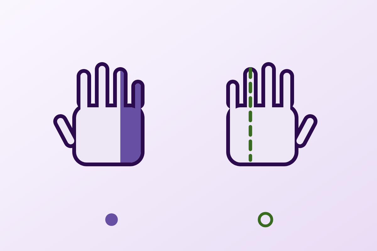
The back of the hand is innervated before the wrist: if it is affected, the compression is not in Guyon's canal but at the elbow or higher.
71.4%of patients operated on for Guyon's canal syndrome are men
What follows covers each of these points in detail, with sources. It is there if you need it.
This topic is taught in a course: Neurological disorders in musculoskeletal practice: screen, assess and treat, on-site with Bryan Littré.
1 / 11
10 article chapters · 55 min in total
Understanding◔ 7 min
Within Guyon's canal, the ulnar nerve divides into a sensory branch and a motor branch.
Locating◔ 7 min
The branch to the back of the hand arises 6.8 cm before the wrist: it does not pass through Guyon's canal.
Causes◔ 6 min
GCS is not mostly idiopathic: the leading cause is a mass, 23 cases out of 56.
The cyclist◔ 6 min
After 600 km, 23 of 25 cyclists have ulnar symptoms; purely motor forms dominate.
Examining◔ 5 min
No quantified diagnostic value exists for Tinel's sign at this level: be wary of it.
Imaging and EMG◔ 6 min
The 80 %, 74 % and 70 % often quoted are yields from an operated series, not sensitivities.
Treating◔ 7 min
There is no randomised trial for this site, whereas the elbow has 15 Cochrane trials.
Clinical cases◔ 4 min
A cyclist's deficit can become permanent: seven years of sequelae without diagnosis, if the exposure continues.
In practice◔ 4 min
A useful request describes the observed picture: purely motor, purely sensory or mixed, and the state of the back of the hand.
Questions◔ 3 min
It is a strong anatomical pointer, never measured for sensitivity, and the two levels can coexist.
Train on this
Neurological rehabilitation
Bryan Littré
Next comes practice: the course that teaches this topic, with Bryan Littré.
Our courses on this topic may be covered by DPC or FIFPL, the French continuing-education funding schemes, depending on your profession and situation.
Entrapment neuropathy · Upper limb
A hand that tingles in its last two fingers makes you think of the elbow. It does so with such reflex speed that the wrist, where the ulnar nerve nevertheless crosses a second narrow passage, is often not even examined. One simple sign separates the two levels, and it costs nothing: sensation on the back of the hand.
Guyon's canal is a passage of 4 to 4.5 cm on the anteromedial aspect of the wrist, closed anteriorly by the palmar carpal ligament, which runs between the pisiform and the hook of the hamate. The ulnar nerve enters it as a trunk and divides within it into a superficial branch, sensory, and a deep branch, motor. That division governs the whole of the clinical reasoning: depending on exactly where the nerve is compressed, the picture is purely motor, purely sensory, or mixed1.
The syndrome is rare, and that has to be said at the outset because it conditions how everything that follows should be read. There is neither a randomised trial nor a meta-analysis of any intervention at this site. The largest published surgical series brings together 56 patients recruited over twenty years across six centres7 ; the broadest literature review on the commonest cause, the ganglion cyst, totals 73 cases reported in 35 articles8. The reference treatment guide is an expert consensus which itself writes that efficacy data are "lacking"15.
The discriminating point. The dorsal cutaneous branch of the ulnar nerve, which supplies the back of the hand on its medial aspect, leaves the nerve trunk at a median distance of 6.8 cm upstream of the tip of the ulnar styloid4. It therefore does not pass through the canal. Compression at the wrist spares the back of the hand ; compression at the elbow does not. This is anatomical reasoning, not a validated test, its sensitivity and specificity have never been measured, but it directs the examination better than any provocation manoeuvre available here.
The causes. Unlike the carpal tunnel, the idiopathic form is not the rule. In Lee's series, the leading cause is a tumour in the broad sense, ganglion cyst foremost, with 23 cases out of 56, ahead of idiopathic (17), trauma (12), anatomical variants (3) and inflammation (3)7. Guyon's canal syndrome therefore requires you to look for a space-occupying cause, which is not true of every entrapment neuropathy.
The cyclist. This is the best documented occupational and sporting cause. After a single 600 km ride, 23 of 25 cyclists have motor symptoms, sensory symptoms or both18. Pressure measurements explain why: without gloves, the hypothenar region takes peaks of 134 to 165 kPa, and the low position on the drops is the worst20.
What the physiotherapist can do. The European HANDGUIDE consensus, built by 35 experts over three Delphi rounds, holds that the patient should always receive instructions, and that those instructions must be combined with something else: a splint, or surgery. The choice is made on severity, duration and treatments already tried15. The level of evidence is that of expert opinion, and it has to be presented as such.
This chapter sets out the anatomy on which all the rest depends. Guyon's canal is not a uniform tube: the nerve divides within it, and the level of that division separates three radically different clinical pictures. A clinician who knows the three zones reads a neurological examination like a map.
Guyon's canal (English speakers also say ulnar tunnel) is a fibro-osseous passage on the anterior and medial aspect of the wrist. It was described in 1861 by Jean Casimir Félix Guyon, a French urological surgeon, and its modern anatomical description, the one practice still rests on, dates from 1985: the work of Gross and Gelberman published in Clinical Orthopaedics and Related Research1.
Those authors measure the tunnel: 4 to 4.5 cm in length. They describe its walls. The floor is formed by the transverse carpal ligament and the pisohamate and pisometacarpal ligaments. The roof is the palmar carpal ligament, a fibrous sheet running from the pisiform. The two lateral pillars are bony: the pisiform medially, and the hook of the hamate laterally. Between these four walls run the ulnar nerve and the ulnar artery, the latter lying lateral to the nerve.
Within the canal, the ulnar nerve divides into two terminal branches. The superficial branch is essentially sensory: it supplies the palmar aspect of the fifth finger and the medial half of the fourth, and gives a motor twig to palmaris brevis. The deep branch is purely motor: it curves around the hook of the hamate, passes through the pisohamate hiatus and reaches the intrinsic muscles of the hand: hypothenar muscles, interossei, the last two lumbricals, adductor pollicis and the deep head of flexor pollicis brevis.
It is that division which grounds Gross and Gelberman's partition into three zones. Zone 1 is the part of the tunnel lying upstream of the bifurcation: there the nerve is still a mixed trunk. Zone 2 surrounds the deep motor branch after the division. Zone 3 surrounds the superficial sensory branch.
Guyon's canal and the sensory territories of the hand
Cross-section of the canal, the division into three zones, and what each territory tells you about the level of the lesion. After Gross & Gelberman 1985 (PMID 3995823) for the zone partition, and Uerpairojkit 2019 (PMID 30518284) for the origin of the dorsal cutaneous branch.
Sources: Gross MS, Gelberman RH. Clin Orthop Relat Res 1985;(196):238-47, PMID 3995823 · Uerpairojkit C et al. J Hand Surg Eur Vol 2019;44(3):263-8, PMID 30518284.
The strength of Gross and Gelberman's paper is not only descriptive. The authors set their anatomical partition against the literature on ulnar compression published at the time, and the result is unusually clear-cut. The 39 cases of combined motor and sensory deficit all sat in zone 1. The 36 zone 2 lesions all produced paralysis of the intrinsic muscles, involvement of the hypothenar muscles depending on the exact level reached within the zone. Zone 3 lesions gave sensory deficits only.
One detail from that review is directly useful at the bedside, and it is almost always left out of second-hand summaries. The mixed deficit, the authors write, most often resulted from compression exerted deep to the nerve; purely sensory deficits, from a lesion sitting superficially. In other words, the clinical picture tells you not only about the longitudinal level of the compression, but also about the position of the cause relative to the nerve: information that counts when you are looking for a ganglion on ultrasound.
Four anatomical measurements that govern the clinical picture
Figures from anatomical studies and one ultrasound meta-analysis. Level of evidence: descriptive anatomy and reference values, not transposable to a given patient.
Sources: PMID 3995823 · PMID 30518284 · PMID 33527596 · PMID 15832354.
A Turkish cadaveric study published in Clinical Anatomy in 2005 dissected 37 hands and forearms from 19 cadavers to look for whatever, within this canal, might serve as a mechanism of compression2. Two findings are worth knowing.
First, a fibrous arch running between the pisiform and the hook of the hamate was found in 21 of 37 hands. In most cases, flexor digiti minimi brevis arose from that arch alone. So it is not an anomaly: it is a common structure, and the passage it delimits, the pisohamate hiatus, is a natural point of constriction.
Second, an accessory muscle was found in 6 hands, including 4 where it crossed that hiatus alongside the deep branch. In two of those four, the superficial branch also accompanied the deep one beneath that anomalous muscle. The authors conclude that the distal portion of Guyon's canal carries a relatively higher risk of ulnar nerve entrapment. That maps exactly onto clinical observation: purely motor zone 2 pictures are not rare even though zone 2 is the shortest.
For the past ten years or so, high-frequency ultrasound has made it possible to measure the cross-sectional area of the nerve, and an increase in that area at the point of compression is the ultrasound argument for entrapment. That still requires knowing the norm. A systematic review with meta-analysis published in the European Journal of Neurology in 2021 gathered 74 studies, 4,186 healthy volunteers and 18,226 nerve sites to establish those reference values3.
At Guyon's canal, the pooled mean cross-sectional area of the ulnar nerve is 4.1 mm², with a 95 % confidence interval of 3.6 to 4.6 mm², over 1,688 measurements. It is a slender nerve: by way of comparison, the median nerve at the wrist measures 8.3 mm² and the ulnar nerve at the elbow 5.9 mm². The authors report a positive effect of height and weight at the various ulnar sites, which is a reason not to apply a single threshold to every build.
This is the most useful piece of reasoning in this article, and the easiest to remember. It rests on a measured anatomical fact, not on a provocation manoeuvre: the branch that supplies the back of the hand leaves the ulnar nerve well before the wrist. This chapter explains why the sign works and, just as importantly, what it does not allow you to assert.
A patient presents with tingling in the fifth finger and the medial border of the fourth, perhaps some clumsiness with fine tasks, perhaps early wasting of the first interosseous space. The dominant reflex is to think of the elbow, and it is not absurd: ulnar neuropathy at the elbow is, according to the 2025 Cochrane review, the second most common entrapment neuropathy after carpal tunnel syndrome16. Statistically, it is the right bet. Clinically, it is a bet that needs checking, and the check comes down to one question.
In the distal forearm, the ulnar nerve gives off a branch called the dorsal cutaneous branch. It curves around the ulna, passes to the back of the wrist and supplies the skin of the dorsal aspect of the hand on its medial side: the fifth finger, the medial half of the fourth, and the region of the fifth metacarpal.
That branch arises upstream of Guyon's canal. A Thai anatomical study published in the Journal of Hand Surgery (European Volume) in 2019 measured it in 44 forearm dissections : the dorsal cutaneous branch takes its origin at a median distance of 6.8 cm upstream of the tip of the ulnar styloid4. Guyon's canal, for its part, occupies the most distal four to four and a half centimetres1. The branch has therefore already left the trunk, by some margin, by the time the trunk enters the passage.
The consequence is direct. A lesion inside Guyon's canal cannot reach a branch that does not pass through it. Sensation on the back of the hand is therefore preserved. A lesion at the elbow, by contrast, sits well upstream of the origin of that branch: it does reach it, and dorsal sensation is impaired.
Where does the lesion sit? The tree that starts from the back of the hand
Anatomical reasoning based on the origin of the dorsal cutaneous branch (Uerpairojkit 2019, 44 dissections) and on the zone partition (Gross & Gelberman 1985). Note: the diagnostic performance of this approach has never been measured.
Sources: PMID 30518284 · PMID 3995823. A tree of anatomical reasoning: no study has measured its sensitivity or its specificity.
Sound anatomical reasoning is not a validated test, and presenting this one as a certainty would be exactly the kind of shortcut this article seeks to avoid. Three caveats go with it.
First caveat: its performance has never been measured. None of the studies gathered for this article provides a sensitivity or a specificity for the examination of dorsal sensation in distinguishing wrist from elbow. The reasoning is anatomically binding; its clinical translation, in a real patient, with a sensory examination of known inter-examiner variability, is not. It is used as a strong pointer, never as proof.
Second caveat: the anatomy of this branch varies. A Japanese study of 30 cadaveric specimens identified two patterns of division: a proximal type, where the branch curves around the ulna upstream of the styloid process, found in 21 of 30 cases, and a distal type, where it curves around it downstream, in 9 of 306. Another study, on 28 fresh specimens, showed that the branch itself subdivides: two twigs on average at the level of the distal ulnar articular surface, up to four5. The authors of the Thai study indeed conclude, on the question of surgical landmarks, that they were unable to define a safe zone4. Anatomical variation does not make the reasoning wrong, but it explains why a case may depart from it.
Third caveat, and the most important in practice: the two levels can coexist. A Korean series published in BMC Musculoskeletal Disorders in 2024 reports seven cases of simultaneous ulnar compression at the elbow and the wrist10. The authors are explicit: "diagnosis by clinical examination alone is difficult", and the usual electromyogram "may not confirm" the diagnosis, because standard protocols do not include segmental testing at the wrist. Their practical recommendation deserves to be kept as it stands: faced with less postoperative improvement than expected after cubital tunnel surgery, double compression should be considered.
The distinction developed here is only of interest if both levels are treated seriously. Compression of the ulnar nerve at the elbow is the subject of a full article on this site: cubital tunnel syndrome, which sets out McGowan's classification, the provocation tests at the elbow, the night splint and the surgical indications. This article does not duplicate it: it deals with the passage sitting fifteen to twenty centimetres lower down, whose causes, imaging and treatment differ.
A reader still hesitating between the two levels after examination has a serious reason to explore them together, and the double compression series recalled above gives them the protocol: segmental electrophysiological testing, including the wrist, and not only the standard study of the elbow.
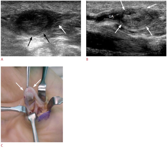
UltrasoundTransverse (A) and longitudinal (B) wrist ultrasound: soft tissue mass in contact with the superficial branch of the ulnar nerve, ulnar artery thrombosis compressing the nerve in Guyon's canal, intraoperative view of the mass (C).
Source : Choi et al., Ultrasonography (Seoul, Korea), 2015, figure 12 · CC BY-NC
The carpal tunnel is, in the great majority of cases, idiopathic. Guyon's canal is not. That is the difference with the heaviest practical consequences between the two, because it makes imaging a useful investigation here, where it is not routinely so there.
The Korean multicentre series published in the Journal of Plastic, Reconstructive & Aesthetic Surgery in 2022 is the best available source on the distribution of causes7. Seven hand surgeons, six centres, a recruitment period running from January 2001 to December 2020: seventy patients screened, fifty-six included. The authors themselves justify their approach by the rarity of the condition: "because of the rarity of the disease, the only published studies on Guyon's canal syndrome are small case series".
The profile of the operated patients: mean age 48.4 years (from 20 to 89), 71.4 % men (40 of 56), mean duration of symptoms before the operation of 18.5 months. Most were office workers. Involvement was unilateral in most. Thirty-eight patients (67.9 %) had an isolated decompression of the canal, eighteen (32.1 %) a nerve decompression combined with another site.
The distribution of causes is the central result:
Causes of Guyon's canal syndrome in the largest published operated series
56 patients, 6 centres, 2001-2020. Note: these are operated patients. Forms that settle without surgery are not in this denominator, so the share of idiopathic cases is probably underestimated in it.
Source: Lee JH et al. J Plast Reconstr Aesthet Surg 2022;75(9):3269-78, PMID 35654688. Level of evidence IV (retrospective case series).
An honest reading of this chart calls for a precaution that second-hand summaries almost always leave out. These 56 patients were all operated on. Mild forms, which settle when the offending activity stops or with a splint, are not in that denominator. Since space-occupying causes lead to theatre more often than functional forms do, the proportion of tumours is mechanically overestimated relative to what a physiotherapy practice sees. What the series establishes solidly is not "ganglion cysts account for 41 % of Guyon's canal syndromes"; it is " among the patients who get operated on, the commonest cause is a mass ", which is amply enough to justify looking for one.
The most complete literature review on this cause was published in the European Journal of Orthopaedic Surgery & Traumatology in 2019: it identifies 73 cases reported in 35 articles of ulnar neuropathy at the wrist secondary to a ganglion cyst8. Seventy-three cases in thirty-five publications: that is the order of magnitude to keep in mind when speaking of the "commonest" cause of an already rare syndrome. The authors distinguish extraneural cysts, by far the more common, from intraneural cysts, much rarer and with a different prognosis.
A Chinese series published in Injury in 2014 provides the most instructive observation for the clinician9. Nine patients, recruited over eleven years, with pure compression of the deep branch by a cyst. The picture is described unambiguously: "varying degrees of muscle atrophy and weakness of the interossei and adductor pollicis were present, with no sensory loss in the hand ". That is Gross and Gelberman's zone 2, in the clinic. Another valuable detail: the cyst arose from the pisohamate joint in eight of nine cases, which tells you where to look on ultrasound.
Mean duration of symptoms before the operation was 16 months there, with a range of 5 to 32 months. That delay recurs in every series (18.5 months in Lee) and it says something important: this diagnosis is made late, because it is not thought of.
Twelve of the 56 patients in the Korean series had a traumatic cause. Two mechanisms dominate the literature. The first is fracture of the hook of the hamate, classic in golfers and in tennis or baseball players, who strike the heel of the hand against the handle. The hook of the hamate forms the lateral pillar of the canal: its fracture, and still more its non-union, keep up direct irritation of the deep branch. Tottas's review lists it among the main causes8. The trap is radiological: this fracture escapes standard wrist views.
The second mechanism is vascular. The ulnar artery runs with the nerve in the canal, and its disease becomes the nerve's. Hypothenar hammer syndrome, from repeated trauma to the heel of the hand, leads to thrombosis or aneurysm of the ulnar artery, which then occupies the space. A case published in the International Journal of Surgery Case Reports in 2024 describes precisely that sequence in a young man, treated by decompression and excision of the aneurysm, with an uneventful recovery23. The authors recall that vascular lesions are "not the usual cause", but that they call for treatment of their own.
Three patients in the Korean series had an anatomical variant, three an inflammatory cause. These are small numbers, but they match what the anatomy predicts. Bozkurt's cadaveric study, already cited, shows that an accessory muscle crosses the pisohamate hiatus with the deep branch in 4 of 37 hands2 : the variant therefore exists in the general population far more often than it becomes symptomatic, which should temper any enthusiasm on finding it incidentally on imaging.
On the inflammatory side, the review by Scarborough and colleagues, published in the British Journal of Hospital Medicine in 2020, lists the secondary causes: fractures, inflammatory conditions, neoplasms, vascular anomalies, aberrant musculature, or a combination of these, noting that the commonest cause remains the ganglion cyst12.
The Physio Learning corpus covers ganglion cysts of the wrist and hand, and that article already cites compression of the ulnar nerve in Guyon's canal as one of the atypical presentations not to be missed. The relationship between the two pages is direct and runs both ways: the cyst is the leading identified cause here, and Guyon's canal is, there, the complication that turns a benign swelling into a surgical indication. A reader arriving from a palpable cyst will find in the dedicated article the data on spontaneous resolution and recurrence; one arriving from an ulnar deficit will find the zone logic here.
This is the only population in which Guyon's canal syndrome has been studied prospectively, with measurements before and after exposure. It deserves its own chapter for that reason, and because it is the only situation where the physiotherapist has levers whose effect has been quantified.
The French term paralysie du cycliste (cyclist's palsy, or handlebar palsy) refers to involvement of the distal ulnar nerve related to prolonged weight-bearing of the hands on the handlebars. It is a long-standing entity, described as early as 198117, but whose true extent was not measured until the early 2000s.
The reference study is prospective and was published in the American Journal of Sports Medicine in 200318. Twenty-five cyclists, road or mountain bike, completed a questionnaire and were then examined and interviewed before and after a ride of 600 kilometres. The authors begin by stating a limitation worth repeating: the true incidence of this condition is unknown.
The result is striking. Twenty-three of twenty-five cyclists experienced motor symptoms, sensory symptoms, or both. The breakdown by hand matters more than that overall figure: motor symptoms alone in 36 % of the hands tested, sensory symptoms alone in 10 %, and both together in 24 %. The predominance of purely motor forms fits the anatomy: it is the deep branch, pressed against the hook of the hamate, that takes the load.
Two comparisons are instructive. Mountain bikers had significantly more sensory deficits than road cyclists. On the other hand, experience did not protect : no significant difference by cyclist experience, either for motor or for sensory symptoms. The idea that a seasoned cyclist "copes" better does not survive measurement.
A second study published in the same journal in 2005 looked for the electrical signature of this phenomenon19. Fourteen subjects, twenty-eight hands, median and ulnar nerve conduction studies carried out before and after a trip of six days and 420 miles, that is about 675 kilometres. This is a level 2 cohort study.
The result is remarkably precise anatomically. The distal motor latencies of the deep branch of the ulnar nerve, recorded at the first dorsal interosseous, were significantly prolonged after the event. By contrast, median nerve motor and sensory conduction, along with ulnar sensory conduction and motor conduction recorded at abductor digiti minimi, did not change significantly.
In other words: it is neither the ulnar trunk, nor the superficial branch, nor even the proximal part of the deep branch that suffers. It is the distal portion of the deep branch, the part that curves around the hook of the hamate and reaches the interossei. Gross and Gelberman's zone 2, measured by electrophysiology forty years after its anatomical description.
The authors also report a side effect: electrophysiological and symptomatic worsening of a carpal tunnel syndrome in three hands, and a new carpal tunnel syndrome in a fourth. Long-distance cycling does not load the ulnar nerve alone.
What to do about it remains. A biomechanical study published in Clinical Biomechanics in 2011 measured what actually happens under the hand20. Thirty-six experienced cyclists rode at constant cadence and power on a turbo trainer, with their hands in three handlebar positions (the tops, the hoods, the drops) and in four conditions: no gloves, unpadded gloves, foam gloves, gel gloves. A high-resolution pressure mat recorded the load, a motion capture system tracked wrist posture, and a laser scan of the hand made it possible to register the pressure maps onto the anatomy.
Hypothenar pressure: what position and glove change
36 experienced cyclists, turbo trainer, constant cadence and power. The no-glove values are measured mean peaks; the reductions are the reported effect of padded gloves, all positions combined.
Source: Slane J, Timmerman M, Ploeg HL, Thelen DG. Clin Biomech (Bristol) 2011;26(6):642-8, PMID 21458120.
Without gloves, mean hypothenar pressure peaks fall between 134 and 165 kPa. The drops position, hands "in the hooks", produces both the highest hypothenar pressure and the most marked wrist extension. Padded gloves reduce the pressure amplitude by 10–28 %, thin foam doing slightly better than gel. The authors' conclusion is directly quotable: these magnitudes are "sufficient to induce ulnar nerve injury if sustained for long periods".
That last sentence contains the most useful piece of advice. The determinant is not pressure alone, it is the product of pressure and duration. A padded glove acts on intensity; changing position acts on duration. The two levers are complementary, and that is exactly how the authors of the 2003 prospective study framed their recommendations: wear gloves, have the bike fitted, and change hand position frequently18.
A Canadian case report published in the Journal of the Canadian Chiropractic Association in 2014 deserves to be read by every practitioner who sees cyclists21. A woman aged 23 years cycles across Canada. After fourteen days, she loses sensation in her left hand, then develops wasting and a claw-hand appearance.
She seeks help. She receives cervical manipulation, soft tissue work, and the advice to wear cycling gloves. The authors are severe, and the abstract quotes them word for word: "no diagnosis was made, and no appropriate steps were taken to make her understand the risks she was taking by continuing to ride".
Seven years later, the anaesthesia in the ulnar territory of the left hand persists. The authors conclude that "treatment of Guyon's canal syndrome may be as simple as stopping cycling until the symptoms resolve". That is the heart of the matter: a permanent deficit arose from a diagnosis that was never made, in a situation where the decisive therapeutic act was free.
For this site there is no provocation test whose sensitivity and specificity are known. That gap is not a reason to examine less; it is a reason to examine differently. The approach that follows rests not on manoeuvres but on mapping: which muscle, which territory, what is spared.
Let us say it at the outset to avoid any misunderstanding: none of the sources gathered for this article provides a quantified diagnostic value for Tinel's sign at the wrist, for the Guyon's canal compression test, or for any other provocation manoeuvre at this level. That is not an oversight in the search, it is the state of the literature on a rare condition. Any source announcing "Tinel at Guyon's canal: sensitivity 70 %, specificity 90 %" would be quoting a figure that does not exist.
What the literature does support is more solid than an isolated test. Scarborough's review puts it this way: "the level of compression can be estimated clinically on examination, by assessing the motor and sensory changes in the hand"12. That is Gross and Gelberman's partition used as a clinical tool.
First question: is the back of the hand involved? That was the subject of the previous chapter. Dorsal sensation preserved, you stay at the wrist; impaired, you move up to the elbow. It is the question that has to come first because it is the only one that completely changes the territory to explore.
Second question: is there a motor deficit, and which one? The ulnar intrinsic muscles have to be tested one by one, rather than settling for an impression of overall strength. The first dorsal interosseous is the most informative: it is the one the cyclist electrophysiology finds affected first19, and its wasting is visible to the naked eye in the first space. Adductor pollicis is tested by Froment's sign: the patient holds a sheet of paper between thumb and index finger, and compensates for the weakness of the adductor by flexing the thumb interphalangeal joint, through flexor pollicis longus supplied by the median nerve. The interossei are tested by spreading and closing the fingers against resistance.
Third question: are the hypothenar muscles involved? That question is not redundant with the previous one, and it is a point the original 1985 description makes valuable. Gross and Gelberman note that in zone 2 lesions, whether or not the hypothenar muscles were affected depended on the location of the lesion within zone 2 »1. A deficit of the interossei with hypothenar muscles spared signals a more distal lesion than a deficit that takes them all.
Fourth question: is ulnar palmar sensation affected? Fifth finger and medial half of the fourth, palmar aspect. Its involvement, together with a motor deficit, points to zone 1; on its own, to zone 3; absent despite a motor deficit, to zone 2.
| Picture observed | Back of the hand | Intrinsic motor function | Ulnar palmar sensation | Most likely location |
|---|---|---|---|---|
| Mixed | normal | impaired | impaired | Zone 1 : trunk, upstream of the bifurcation. All 39 cases of mixed deficit in the 1985 review sat there. |
| Purely motor | normal | impaired | normal | Zone 2 : deep branch. All 36 zone 2 lesions gave paralysis of the intrinsics. |
| Purely sensory | normal | normal | impaired | Zone 3 : superficial branch. Compression tends to be superficial relative to the nerve. |
| Mixed + back of hand affected | impaired | impaired | impaired | Elbow or more proximal : the dorsal branch has arisen upstream. See the cubital tunnel. |
| Extending beyond the ulnar territory | variable | variable | extends beyond | Plexus or C8-T1 root : step outside the entrapment frame. Cited as confounding by Chen & Tsai 2014. |
In the carpal tunnel, tests exist whose diagnostic values have been measured in large numbers, and it is legitimate to use them. Here, those values do not exist. Three reasons nonetheless make the mapping approach more reliable than it looks.
First, it is based on a near-perfect anatomical correspondence in the only series that examined it: 39 out of 39, and 36 out of 36. No provocation test in hand pathology shows such consistency, even in small numbers.
Second, it produces usable information. A positive Tinel says "the nerve is irritable somewhere". A purely motor deficit with intact sensation says "the deep branch, in zone 2, look for a cyst arising from the pisohamate joint", which is very precisely what Wang's series found in eight cases out of nine9.
Third, it has been confirmed by an independent modality. The Korean segmental conduction study, published in the Journal of Clinical Neurology in 2022, compared its electrophysiological localisation with that of ultrasound in fourteen patients: "the five proximal and six distal lesions seen on ultrasound all corresponded to the locations found by the segmental studies"13. Fourteen patients is not many; but two independent methods agreeing eleven times out of eleven support the principle of localisation by level.
Palpation keeps a role, not as a diagnostic test but as a search for a cause. Three landmarks deserve to be palpated systematically.
The prominence of the pisiform and the space just lateral to it, where the canal projects: a swelling, however small, however immobile, should prompt a request for ultrasound. Lee's series recalls that about a third of operated patients were operated on for a mass, and Tottas's review notes that cysts may be impalpable while still compressing the nerve8 : the absence of a palpable mass therefore rules out nothing.
The hook of the hamate, over the hypothenar eminence, about a centimetre medial and distal to the scaphoid tubercle. Exquisite pain on pressure there, especially in a racquet, golf or bat athlete, should prompt imaging suited to looking for a fracture or non-union of the hook.
The course of the ulnar artery : a pulsatile mass, a bruit, or a change in the colour of the fingers on Allen's test points to arterial disease, and completely changes the downstream pathway.
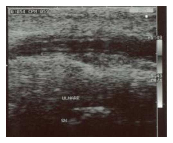
UltrasoundLongitudinal ultrasound of the ulnar artery at the wrist in a patient with hypothenar hammer syndrome: thickened, thrombosed artery, a vascular cause of ulnar nerve compression in Guyon's canal.
Source : Cigna et al., Plastic Surgery International, 2010, figure 1 · CC BY
This is the chapter where the figures circulating second-hand are most misleading. One commonly reads that "the EMG confirms the diagnosis". What the series show is more nuanced, and the nuance changes what to do when the study comes back normal.
Lee's multicentre series reports the yield of each investigation in its 56 operated patients7. The figures are as follows: ultrasound positive in 7 of 10 patients assessed, CT in 2 of 5, MRI in 17 of 23, and electrodiagnostic studies in 35 of 44.
Yield of the investigations, and why this is not a sensitivity
Proportion of positive studies among the patients in the series who had that investigation. The population consists entirely of operated patients, so the diagnosis is established: there is no unaffected subject in the denominator, so no specificity can be calculated.
Source: Lee JH et al. J Plast Reconstr Aesthet Surg 2022;75(9):3269-78, PMID 35654688. Small numbers tested (5 to 44 depending on the investigation).
Two precautions make these figures usable, and their absence makes them misleading.
These are not sensitivities. A sensitivity is calculated in a population where the diagnosis is established by an independent reference standard, with affected subjects and unaffected ones for the specificity. Here, every patient was operated on: the population is selected by the surgical indication itself. The 80 % figure for electrodiagnosis is a yield within an operated series, which is real information but a different kind. There is, to our knowledge, no published sensitivity or specificity for electrodiagnosis in Guyon's canal syndrome.
The numbers are tiny. Seven out of ten for ultrasound: ten patients. Two out of five for CT: five patients. One patient more or less shifts these proportions by ten to twenty points. They should be read as orders of magnitude, not as performance figures.
That is the clinical message of this chapter. Nine of forty-four patients had a normal electrodiagnostic study, and they were operated on, with a cause found. A patient whose EMG comes back normal when the clinical picture is convincing is not a patient without Guyon's canal syndrome: it is a patient in whom the study did not show what surgery found.
The Korean series on combined elbow-wrist compression gives a technical explanation for part of these false negatives10. Standard electrophysiological protocols do not include segmental testing at the wrist : the "inching test", which stimulates the nerve in small increments along its course to localise the point of slowing. A study that does not look in the right place does not find.
The study published in the Journal of Clinical Neurology in 2022 assessed precisely that technique in fourteen patients with typical, unilateral symptoms13. The ulnar nerve was stimulated at three points (3 cm distal to, level with, and 2 cm proximal to the pisiform), recording at the first dorsal interosseous and at the fifth finger.
The results establish two things. First, a correspondence between electrophysiological type and level: proximal lesions came with involvement of all the deep motor fibres and the superficial sensory fibres (type I), whereas distal lesions affected more selectively the motor fibres destined for the first dorsal interosseous (type III). Second, an agreement: the five proximal and six distal lesions visualised on ultrasound all corresponded to the location found by the segmental studies.
Fourteen patients, eleven lesions localised: that is little, and the authors claim no more than to propose a "useful assistive technique". But for a clinician writing a request for investigation, the operational conclusion is clear: specify the question asked. Requesting "search for an ulnar neuropathy at the wrist, with segmental study around the pisiform" is not the same as requesting "upper limb EMG".
Since the commonest cause in the operated patient is a mass, imaging here does not have the status of a confirmatory test that it has elsewhere: it is the search for the cause. Scarborough's review gives a division of roles consistent with the yield data12 : radiographs and CT serve to exclude a fracture ; ultrasound serves to diagnose ganglion cysts and vascular anomalies, and it can localise the level of compression ; nerve conduction studies serve to support the diagnosis and to look for a more proximal compression.
MRI is presented as the reference for soft tissue lesions by the authors of the 2023 case report, who write that "if soft tissue lesions compress the ulnar nerve in Guyon's canal, MRI is considered the gold standard for diagnosis"22. That is an assertion by case report authors, not comparative data: the 17/23 yield in Lee's series remains the best available measurement, and it is not 100 %.
As for ultrasound, the reference cross-sectional area quoted in the first chapter, 4.1 mm² (95 % CI 3.6-4.6) at Guyon's canal3, makes it possible to interpret a report. A nerve measured clearly above that range at the point of compression is an argument; a nerve within the norm does not rule out a dynamic compression or a very distal lesion.
| Investigation | What it looks for | Measured yield | The limitation to know about |
|---|---|---|---|
| Electrodiagnosis | Confirm the involvement, localise it, look for a second site | 35/44 (80 %) in an operated series | Neither sensitivity nor specificity published. 9 normal studies out of 44 in operated patients. The standard protocol does not include segmental testing at the wrist. |
| Ultrasound | Cyst, vascular anomaly, level of compression, cross-sectional area | 7/10 (70 %), very small numbers | Operator-dependent. Norm at the canal: 4.1 mm², modulated by height and weight. A cyst may be impalpable but remains visible. |
| MRI | Soft tissue masses, intraneural cysts | 17/23 (74 %) | Presented as the reference for masses by case report authors; the measured yield is not 100 %. |
| Radiograph / CT | Fracture of the hook of the hamate, non-union | 2/5 for CT | Fracture of the hook of the hamate escapes standard wrist views : it has to be requested explicitly. |
We have to start with an unpleasant, true sentence: at this site, therapeutic evidence is almost non-existent. The chapter that follows does not hide that void; it shows what can be done with it, which is more useful than a GRADE table built to look complete.
The comparison is instructive. For ulnar neuropathy at the elbow, there is a Cochrane review, updated for the fourth time in 2025, bringing together 15 randomised trials and 970 participants16. In it you find quantified comparisons between simple decompression and transposition, between endoscopic and open approaches, with confidence intervals and a GRADE rating of certainty.
For the wrist, nothing of the kind exists. No Cochrane review, no randomised trial, no meta-analysis of any intervention. This is not an oversight by methodologists: it is the arithmetical consequence of rarity. When the largest operated series recruits 56 patients over twenty years across six centres, an adequately powered two-arm randomised trial is out of reach.
For want of trials, the best available source is a formal consensus. The European HANDGUIDE study, published in the British Journal of Sports Medicine in 2013, brought together 35 experts (hand surgeons and hand therapists nominated by the national associations belonging to their European federations, along with physical and rehabilitation medicine physicians), in a three-round Delphi procedure15.
The authors set out the frame themselves, in a sentence worth quoting: " effectiveness data for interventions in this disorder are lacking ". The consensus is therefore explicitly a substitute for evidence, not a synthesis of it.
What the experts agreed on after three rounds:
Modalities and the level of evidence actually available
Cards stacked in decreasing order of certainty. No modality reaches the level of randomised trials for this site: the top card is an expert consensus.
Sources: PMID 23902776 (HANDGUIDE) · PMID 24774037 (series of 9 cases) · PMID 21458120 and PMID 12860549 (cyclist).
It is the only element the consensus makes invariable, and it is also the one that falls most directly to the physiotherapist. The content is not detailed in the study abstract, but the data on causes make it possible to reconstruct it without extrapolating: it means identifying and removing the mechanical load bearing on the heel of the hand.
In the cyclist, the content is documented and quantified: padded gloves, 10 to 28 % less pressure, bike fitting, and frequent changes of hand position, avoiding prolonged holding of the drops, the position identified as the most demanding20,18.
In the worker, the same logic applies to the tool, to resting the wrist on a work surface, to using a hammer or a power screwdriver. Brown's case is a reminder that an instruction not given has a cost: the patient carried on riding for want of understanding the risk, and the deficit became permanent21.
The consensus retains "instructions + splint" as an appropriate option, without the abstract specifying its type, its duration or its wearing schedule. No study of splint effectiveness in Guyon's canal syndrome was identified. The mechanical reasoning is coherent (limiting wrist extension, the position associated with the highest hypothenar pressure in Slane's study), but it remains reasoning.
You have to resist the temptation to fill that void with cubital tunnel data, where the night splint has a literature of its own. The passage, the mechanism and the causes differ: an elbow extension splint has no functional equivalent at the wrist.
The consensus retains "instructions + surgery" as the second appropriate option, the choice being made on severity, duration and previous treatments15. Scarborough's review is more direct: mild symptoms can be managed without operating, but "surgical exploration and decompression are the treatment of choice for nerve-compressive causes, with largely good results"12. The logic is clear: when a mass occupies the canal, no instruction will make it disappear.
The best documented functional results come from the series of nine patients operated on for a cyst compressing the deep branch9. At a mean follow-up of 23 months : grip strength went from 63 %–88 % of the unaffected side, thumb-index pinch from 61 %–87 %. On the modified Bishop scale, six patients (67 %) excellent, two (22 %) good, one (11 %) fair. The authors conclude that early surgical treatment allows satisfactory functional recovery.
On timing, the 2023 case report proposes a threshold worth knowing even though it does not come from a comparative study: if symptoms persist or worsen beyond three months, early intervention is presented as the treatment of choice22. Set that against the delays actually observed (16 to 18.5 months before the operation in the series), to gauge the gap between what is recommended and what happens.
The 2023 case report mentions that physiotherapy was prescribed after the excision to speed up nerve recovery, with two years of follow-up and a very good recovery on the modified Bishop score22. It is a single case, and it does not demonstrate the effectiveness of rehabilitation: it documents a practice.
In the absence of a validated protocol for this site, the sensible approach is to treat what is measurable: the strength of the intrinsics, for which the series give quantified targets (88 % of grip and 87 % of pinch of the unaffected side, at 23 months), fine dexterity, and the protection of the operated area. That programme has to be presented to the patient for what it is: management grounded in reasoning and in the functional targets measured in the series, not in trials.
The four cases that follow are genuine indexed case reports, cited with their PubMed identifier and, where one exists, their PubMed Central reference. None is reconstructed. They were chosen because each contradicts a different received idea about this syndrome.
Source: Brown CK, Stainsby B, Sovak G. J Can Chiropr Assoc 2014;58(4):413-20 : PMID 25550666, PMC4262815.
A woman of 23 cycles across Canada. After fourteen days, she loses sensation in her left hand. Wasting and a claw-hand appearance follow. She consults several therapists. She receives cervical manipulation, soft tissue work, and the advice to wear cycling gloves.
The authors write that " no diagnosis was made, and no appropriate steps were taken to make her understand the risks she was taking by continuing to ride". Seven years after the initial episode, the loss of sensation in the ulnar territory of the left hand persists.
What the case contradicts. The idea that cyclist's palsy is always transient. The authors stress that treatment "may be as simple as stopping cycling until the symptoms settle": that is precisely what makes this case painful to read. The decisive step was free and was never offered, for want of a diagnosis. For a physiotherapist, the lesson is not therapeutic, it is diagnostic: cervical manipulation was treating a hypothesis that had never been tested.
Source: Jakirlic M, Salihagic S, Katica N, Dujso V. Acta Inform Med 2023;31(4):326-8 : PMID 38379688, PMC10875934.
A 45-year-old patient is managed in plastic and reconstructive surgery for release of the ulnar nerve in Guyon's canal. The diagnosis is made on history and physical examination, then ultrasound and MRI are used to determine the origin of the neuropathy. A ganglion cyst is confirmed on histopathology one month after excision. Physiotherapy is prescribed to speed up nerve recovery, and the patient is followed for two years. At the end of follow-up, functional recovery of the hand is judged very good on the modified Bishop score.
What the case illustrates. The complete sequence as it ought to unfold: clinical assessment first, imaging for the cause, surgery on the cause, rehabilitation afterwards, long follow-up. It is also the case already cited by the site's article on ganglion cysts of the wrist and hand, one and the same reference linking the two pages, which is no accident: the cyst is the leading identified cause here.
Source: Zied M, Farouk C, Mohsen C, Wajdi C, Ali H, Wael G. Int J Surg Case Rep 2024;121:109978 : PMID 38954970, PMC11263623.
A young man is admitted with Guyon's canal syndrome due to an aneurysm of the ulnar artery. He undergoes surgical decompression; recovery is uneventful and the result satisfactory. The authors recall that "vascular lesions are not the usual cause" of ulnar compression at the wrist, and that "Guyon's canal syndrome is less common than cubital tunnel syndrome or carpal tunnel syndrome".
What the case contradicts. The reflex of looking only for a cyst. The ulnar artery shares the passage with the nerve: its disease becomes the nerve's. In practice, that justifies palpating the course of the artery and being concerned by a pulsatile mass or a colour change in the fingers, which points to a vascular pathway and not to rehabilitation.
Source: Khajeh R, Farzan M, Moazen Jamshidi SMM, Moharrami A. Arch Bone Jt Surg 2021;9(5):598-600 : PMID 34692944, PMC8503765.
A 33-year-old man presents with a wrist mass and moderate pain, sitting in zone 3 of the ulnar nerve, and with no neurological signs at all. It is a schwannoma, successfully excised; the patient becomes asymptomatic.
What the case illustrates. A mass in the canal does not mechanically produce a deficit. Zone 3 surrounds only the superficial sensory branch: a lesion developing there without compressing gives a mass, not a neuropathy. The clinical corollary is useful in both directions: a mass without a deficit is no reassurance against the need to investigate, and a deficit without a palpable mass does not rule out a space-occupying cause, Tottas's review recalling that a cyst may be impalpable8.
| Case | Profile | Cause | Outcome | The received idea it corrects |
|---|---|---|---|---|
| 1 · Brown 2014 PMID 25550666 | Woman, 23, long-distance cyclist | Compression by the handlebars, 14 days | Permanent sensory deficit at 7 years | "Cyclist's palsy always settles." Not when it goes undiagnosed. |
| 2 · Jakirlic 2023 PMID 38379688 | Woman, 45 | Ganglion cyst (histology) | Very good recovery at 2 years (modified Bishop) | "Imaging is superfluous." Here it gives the cause, and therefore the treatment. |
| 3 · Zied 2024 PMID 38954970 | Young man | Aneurysm of the ulnar artery | Decompression, uneventful recovery | "It is a cyst or nothing." The artery shares the passage. |
| 4 · Khajeh 2021 PMID 34692944 | Man, 33 | Schwannoma, zone 3 | Excision, patient asymptomatic | "A mass in the canal produces a deficit." Not if it sits where there is only uncompressed sensory tissue. |
This chapter brings everything above back to a physiotherapy session. It does not offer a protocol, none is validated for this site, but a reasoned approach, with the points at which you have to hand over.
Go back over the history of the loading. Since when, on which hand, and above all: what is pressing on the heel of that hand? Bike and mileage, vibrating tool, crutches, wheelchair, wrist resting on a work surface, racquet or bat sport. The delays in the series (16 to 18.5 months between the first symptoms and the operation) indicate that this diagnosis is made late: the question does not ask itself, it has to be asked.
Carry out the four-question examination, in the order of the chapter devoted to examination: back of the hand, detailed intrinsic motor function, hypothenar muscles, ulnar palmar sensation. Record the results separately rather than as an overall impression: it is the combination that localises.
Palpate the three landmarks : the pisiform region looking for a mass, the hook of the hamate looking for exquisite tenderness, the course of the ulnar artery looking for abnormal pulsatility.
Decide on the direction. At the end of that first session, only three situations:
The chapter on investigations showed that a standard electrophysiological protocol does not explore the wrist by segmental testing10. A vague request therefore produces a study that may miss it. Framing the question, by contrast, is within everyone's reach:
The useful content of a request
When conservative management is chosen, the content is simple and its justification honest. Removing the load is the only measure the consensus makes invariable, and the only one whose mechanical effect is quantified in the cyclist. The rest (dexterity work, maintaining range, progressive strengthening of the intrinsics as reinnervation allows) is justified by reasoning and by the functional targets of the operated series, not by trials.
The milestones count for more than the content. An improvement should be perceptible within a few weeks of removing the load. The three-month threshold proposed in the literature for surgical referral22 gives a clear limit: beyond it, without improvement, you do not extend, you redirect.
After surgery, the quantified targets from Wang's series9 give usable markers of progress: grip strength and thumb-index pinch expressed relative to the unaffected side, with values reached of 88 % and 87 % at a mean follow-up of 23 months. Measuring rather than estimating tells you whether the trajectory is that of the published series.
Treating the neck by default. That is the error in Brown's case, and it is instructive because it was not absurd: tingling along the ulnar border can come from C8-T1. But the cervical hypothesis has to be tested, not assumed, and examining the back of the hand takes thirty seconds.
Concluding from a normal EMG. Nine of forty-four patients had a normal electrodiagnostic study in Lee's series, and they were operated on with a cause found7. A negative study does not close the file when the clinical picture is convincing.
Extending conservative treatment in the face of a motor deficit. Recovery after decompression is good when it is early; sequelae are possible when it is not. A motor deficit that is setting in is a reason to refer, not to intensify rehabilitation.
Without hedging: a mass identified, a progressive motor deficit, wasting, vascular suspicion, symptoms beyond three months without improvement, or disappointing recovery after cubital tunnel surgery. In each of these situations, the physiotherapist's role is to write a precise letter, not to carry on.
By examining sensation on the back of the hand, on its medial border. The branch that supplies it arises on average 6.8 cm upstream of the ulnar styloid and does not cross Guyon's canal: if that sensation is normal, the lesion is at the wrist; if it is impaired, it is at the elbow or higher. It is a strong anatomical pointer, whose sensitivity and specificity have nonetheless never been measured, and the two levels can coexist.
No. In the largest operated series available, 9 patients out of 44 had a normal electrodiagnostic study and were nonetheless operated on with a cause found. Part of these false negatives is explained by the protocol: standard studies do not include segmental testing at the wrist.
The reasoning leans strongly that way, because the commonest cause in operated patients is a mass : 23 cases out of 56. Unlike the carpal tunnel, the idiopathic form is not the rule here. Ultrasound is the first-line investigation for looking for a cyst or a vascular anomaly; MRI adds more on soft tissue masses.
Yes, and it is a classic trap recalled by the literature review on this cause. The absence of a palpable swelling does not rule out a space-occupying cause, which is one more argument for imaging when the clinical picture is convincing.
No. It often settles once the exposure stops: the authors of the Canadian case write that treatment may be as simple as stopping riding until the symptoms subside. But the same article reports a young woman whose sensory deficit still persisted seven years later, for want of a diagnosis and of the advice to stop.
They help, without being enough. Padded gloves reduce hypothenar pressure by 10–28 %, thin foam doing a little better than gel. But the authors insist on the pressure × duration product: the glove acts on intensity, frequent changes of hand position act on duration. You need both, and you need to avoid holding the drops for long stretches.
Yes, it is rare but documented. A 2024 series reports seven cases and stresses that diagnosis by clinical examination alone is difficult. The most useful warning sign is less improvement than expected after cubital tunnel surgery : the wrist should then be explored with segmental testing.
Three things. Diagnostic recognition, which is probably its most important contribution given the delays observed. Instructions, the only measure the European consensus makes invariable, that is identifying and removing the mechanical load. And postoperative rehabilitation, with measurable functional targets. By contrast, no passive technique, no neurodynamic mobilisation and no injection has been assessed at this site: that has to be said to the patient rather than transposing the elbow data.
The threshold proposed in the literature is three months of persisting or worsening symptoms. Immediately, by contrast, in the face of an identified mass, a progressive motor deficit, wasting, or vascular suspicion.
Twenty-four references, resolved one by one through the PubMed E-utilities API. For each, the abstract was read in full and the figures quoted in the article checked at source. The year retained is that of the issue, re-read on the XML record and cross-checked with esummary : the first "year" field of a PubMed record is sometimes a revision date, and shifts the citation by a year.
What this evidence base does not allow you to assert
The level above is the subject of a separate article: cubital tunnel syndrome, ulnar compression at the elbow. The commonest cause identified here is covered in its own right in ganglion cysts of the wrist and hand. The commonest entrapment neuropathy of the wrist, with which coexistence is possible, is described in carpal tunnel syndrome.
Behind this article
An author who explains, a reviewer who checks.
How we write and check our content

Physiotherapist · co-founder of Physio Learning
Marked for life by his first four-hour lecture without a single image, he took a master’s in instructional design so that it would never happen to anyone again. He hunts down publication bias and unreadable slides with the same intransigence.

Head of scientific content
Physiotherapist specialising in neuro-musculoskeletal practice and holder of a master’s in public health. He checks the methodological rigour of every article: primary sources, levels of evidence, no exceptions.
