














On-siteDPCFIFPL

Neurological rehabilitation
Neurological disorders in musculoskeletal practice: screen, assess and treat
Bryan Littré
Go further
Functional neurological disorder: the positive signs (Hoover, abductor) are 92 to 100 % specific, and rediagnosis has run at 4 % since 1970.
Functional neurological disorder is a positive diagnosis: it rests on an internal inconsistency at examination, not on a negative work-up.
4%of functional disorder diagnoses are revised, a rate stable since 1970
What follows covers each of these points in detail, with sources. It is there if you need it.
This topic is taught in a course: Neurological disorders in musculoskeletal practice: screen, assess and treat, on-site with Bryan Littré.
1 / 11
10 article chapters · 30 min in total
Understand◔ 5 min
FND is a real and common disorder: 16 % of new neurology patients, the 2nd most common reason for referral.
Diagnose◔ 4 min
Up to DSM-IV a psychological factor was required; that is no longer true, replaced by positive clinical signs.
Examine◔ 5 min
Fourteen clinical signs are validated, with a specificity of 92 to 100 % and a sensitivity of 8 to 100 %.
Explain◔ 2 min
Making a firm diagnosis brings the cost of the condition down by 9 to 90.7 % depending on the study.
Rehabilitate◔ 4 min
The central principle is to divert attention away from the limb, never to bring it back to it.
Physio4FMD◔ 2 min
The primary outcome is not significant (3.5 points), compared with local physiotherapy, not with nothing.
Manage a seizure◔ 2 min
Functional seizures are the subtype the physiotherapist meets least and dreads most.
Prognosis◔ 2 min
At an average of 7.4 years, 39 % stay the same or worsen, with wide variation across studies.
In practice◔ 2 min
The first session serves to check the explanation and to show a sign, before any technique.
FAQ◔ 2 min
FND denotes involuntary symptoms: a positive sign shows a movement that the patient is not commanding.
Train on this
Neurological rehabilitation
Bryan Littré
Next comes practice: the course that teaches this topic, with Bryan Littré.
Our courses on this topic may be covered by DPC or FIFPL, the French continuing-education funding schemes, depending on your profession and situation.
Functional neurology · Rehabilitation
A leg that will no longer lift, a tremor that disappears when the conversation moves on, a gait that collapses under the slightest gaze. The reflex is to look for what the MRI is missing. The right reasoning is the opposite: to go and look, at examination, for signs you do find, whose specificity exceeds 92 %.
Functional neurological disorder (FND) denotes real, involuntary neurological symptoms, characterised by a failure of voluntary control and of perception, while the basic structure of the nervous system is normal14. ICD-11 classifies it under the label dissociative neurological symptom disorder, code 6B60, with a subtype for each dominant symptom.
It is not a diagnosis of exclusion, and that has changed officially. DSM-5 and then ICD-11 removed the requirement for a psychological precipitant and based the diagnosis on demonstrating an internal inconsistency at examination: the symptom behaves differently depending on whether it is observed directly or indirectly. That inconsistency is looked for; it is not inferred from a negative work-up3.
The signs have been measured. A systematic review retained 14 signs validated in controlled studies, 7 motor, 5 sensory and 2 of gait. Their sensitivity is low and highly variable (8 to 100 %), their specificity is high and consistent: 92 to 100 %6. A positive sign therefore points strongly, a negative sign rules nothing out.
The fear of missing a disease is no longer the argument it was. Across 27 studies and 1,466 patients followed for a median of five years, the proportion of patients who ultimately receive another diagnosis has fallen from 29 % in the 1950s to 4 % (95 % CI 2 to 6) since 1970, with no further fall since5. That is the order of magnitude of rediagnosis in many other neurological conditions.
The physiotherapist has a described role, and it is central. An international physiotherapy consensus, published in 2015, sets the frame: education about the mechanism, movement retraining that diverts attention away from the limb, self-management strategies, all within an explicitly non-blaming setting9. A twin consensus exists for occupational therapy13.
And the level of evidence has to be stated as it is. The phase 3 randomised trial Physio4FMD, 355 patients across 11 centres, did not show specialist physiotherapy to be superior to local neurological physiotherapy for physical function at 12 months (adjusted difference 3.5; 95 % CI −2.3 to 9.3; p = 0.23). More patients reported themselves improved and mental health scores were better, and both approaches proved safe10. Read correctly, that result does not say physiotherapy is useless: it says the specialist protocol did no better than ordinary neurological physiotherapy, in patients whose diagnosis had been made and explained.
This chapter sets out the vocabulary and the order of magnitude. Both count: a practitioner who thinks they are seeing a rare curiosity will treat as a rare curiosity something they meet several times a year, and a badly chosen word at the moment of diagnosis costs months of rehabilitation.
Functional neurological disorder produces genuine and involuntary neurological symptoms: weakness, tremor, dystonia, gait disturbance, seizures, sensory disturbance, speech disturbance. The wording used by the systematic review of the cost of the condition, published in Neurology in 2023, is useful because it is workable: it is a problem of voluntary control and perception, while the basic structure of the nervous system is normal14.
Two words in that sentence deserve emphasis. Involuntary : the patient is not feigning, and letting them think otherwise is the most costly error in their care. Basic structure normal : conventional imaging shows no lesion, which does not imply that the brain is working normally. The 2022 Lancet Neurology review describes precisely the mechanisms shared across subtypes, around attention directed to the limb, prior expectations and the sense of agency4.
“Hysteria”, “conversion disorder”, “psychogenic”, “non-organic”, “functional”: all five denote overlapping territories at different times, and they do not carry the same charge. ICD-11 settles on dissociative neurological symptom disorder, code 6B60, broken down by dominant symptom: weakness or paresis (6B60.6), gait disturbance (6B60.7), non-epileptic seizures (6B60.4), tremor (6B60.82), and a dozen others. In clinical practice as in the rehabilitation literature, it is functional neurological disorder that has taken hold, because it describes the mechanism rather than assigning a cause.
ICD-11 places these subtypes in chapter 06 (mental, behavioural and neurodevelopmental disorders), under the dissociative disorders block. The physiotherapist mostly meets the first four in the table below.
| ICD-11 code | Official label | What the rehabilitation clinician sees |
|---|---|---|
| 6B60 | Dissociative neurological symptom disorder | The parent category, all symptoms taken together |
| 6B60.6 | … with paresis or weakness | Motor deficit of one or more limbs, the target of Hoover's sign and the abductor sign |
| 6B60.7 | … with gait disturbance | Unsteady gait, giving way, a “skating” appearance, often without genuine falls |
| 6B60.82 | … with tremor | Tremor variable in frequency, sensitive to entrainment and distraction |
| 6B60.83 | … with dystonia | Fixed postures, often of sudden onset, frequently painful |
| 6B60.4 | … with non-epileptic seizures | Paroxysmal seizures with no electroencephalographic correlate |
| 6B60.3 | … with other sensory disturbance | Reduced or absent sensation in a non-anatomical distribution |
| 6B60.5 | … with speech disturbance | Dysarthria, aphonia, stammering of sudden onset |
The most telling figure for a clinician does not come from a population study but from a census of referral reasons. A Scottish study included 3,781 new patients referred to neurology clinics, in every neurology centre in the country, over fifteen months. Functional and psychological symptoms accounted for 16 % of the initial diagnoses, the second category behind headache (19 %) and ahead of epilepsy (14 %)2.
What actually fills a neurology clinic
Initial diagnoses made by neurologists across 3,781 new patients, every neurology centre in Scotland, 15 months of collection. After Stone 2010 (PMID 20646830).
A physiotherapist who receives patients referred from neurology statistically meets more functional disorders than multiple sclerosis.
Population measurements, harder to establish, converge. A systematic review published in 2025 in the Journal of Neurology, Neurosurgery and Psychiatry gathered 39 articles: incidence is estimated at between 10 and 22 cases per 100,000 person-years, and minimum prevalence at between 80 and 140 per 100,000, with a possible range of 50 to 1,600 depending on method. The authors stress that most studies underestimate, and that FND is probably more common than several neurological diseases better served by research1.
The cost deserves a word, because it documents what is at stake in diagnostic delay. The systematic review in Neurology retained 16 economic studies: the additional annual cost associated with FND ranges from 4,964 to 86,722 in 2021 US dollars, most of it indirect (sick leave, lost activity). Above all, several interventions reduce that cost by 9 to 90.7 %, and among them is simply making a firm diagnosis14.
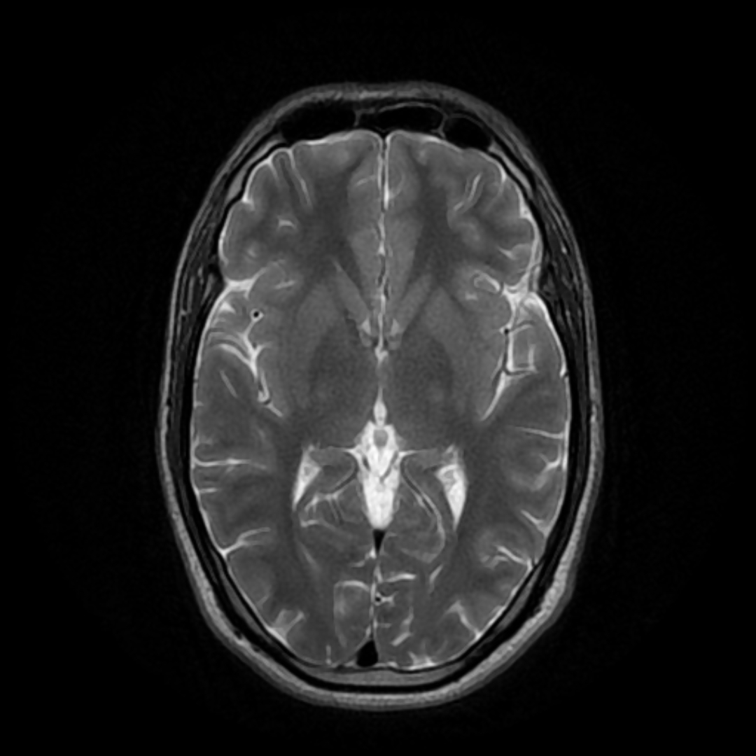
MRIAxial T2-weighted brain MRI, with no visible abnormality: illustrates the normal imaging expected in functional neurological disorders.
Source : 511KeV, Wikimedia Commons · CC BY-SA
This is the pivot of the whole article. As long as we reason by exclusion, the patient waits, investigations pile up, cost rises and the prognosis worsens. Positive reasoning comes down to one sentence: we do not look for what the work-up is missing, we look for an inconsistency at examination.
Up to DSM-IV, a diagnosis of conversion disorder required a psychological factor to be identifiable in connection with the onset of the symptom. That requirement has been removed. The central criterion has become the clinical demonstration of an incompatibility between the symptom and recognised neurological disease. The concepts review published in JAMA Neurology in 2018, signed by fifteen authors in the field, states the principle without hedging: the diagnosis rests on positive clinical features, not on the absence of findings3.
In practice, an internal inconsistency is observed in three ways: the symptom varies with the attention paid to the limb, it disappears during an automatic movement while persisting on command, or it matches no recognised anatomical distribution. All three are looked for at examination, with the hands.
Two lines of reasoning, two patient trajectories
On the left, the reasoning by exclusion, as diagnostic delay produces it. On the right, positive reasoning, the kind DSM-5 and ICD-11 describe. The consequences at the foot of each column are documented by Gelauff 2014 (PMID 24029543) for prognosis and O'Mahony 2023 (PMID 37339887) for cost.
Both columns start from the same patient. What separates them is an intention at examination, not a piece of equipment.
The classic objection is the fear of being wrong: what if a disease revealed itself later? It has been quantified. A systematic review published in the BMJ gathered 27 studies and 1,466 patients with unexplained motor or sensory symptoms, with a median follow-up of five years. The proportion of patients who ultimately receive another diagnosis fell from 29 % in the 1950s to 4 % since the 1970s, and that figure has not moved across three decades. The authors attribute the fall to improved study quality, and not to the arrival of the CT scanner5.
The rediagnosis rate, decade by decade
Proportion of patients initially labelled with “conversion symptoms” who subsequently receive another diagnosis explaining their presentation. Random-effects meta-analysis, 27 studies, 1,466 patients, median follow-up 5 years. After Stone 2005 (PMID 16223792).
The plateau reached in the 1970s has not moved since: the fear of the missed diagnosis belongs to a poor-quality literature, not to current practice.
This is the nuance that guards against the opposite misreading. Finding a positive Hoover's sign establishes that a functional component exists, not that no other disease is present. The two regularly coexist, and FND readily arises in patients with a neurological disease. Clinical reasoning therefore remains: which part of this picture is functional, and which part is not.
The practical chapter. The signs are numerous in the literature, only fourteen have been validated in controlled studies, and their performance is very asymmetrical: they serve to confirm, never to exclude. This chapter describes those a physiotherapist can perform in a session, and what each allows you to conclude.
Daum, Hubschmid and Aybek searched Embase, Medline and PsycINFO from 1965 to June 2012, retained 11 controlled studies from 147 eligible articles, and documented 14 signs : 7 motor, 5 sensory, 2 of gait. The result comes down to two numbers: sensitivity of 8 - 100 %, specificity of 92 - 100 %. The authors set out the limitations without softening them: class III level of evidence, only two blinded studies, and no data at all on inter-rater reproducibility6.
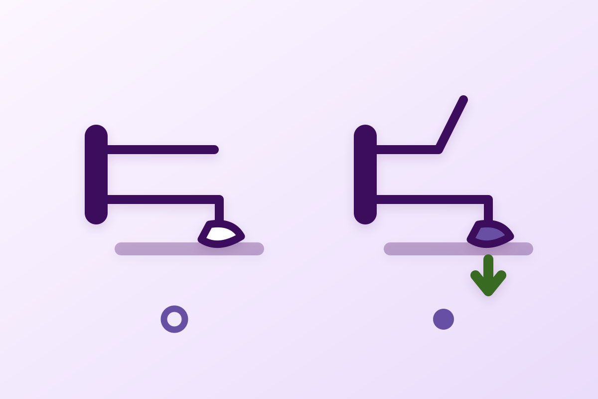
It is the best known and the most useful in practice. The principle is this: hip extension is weak when asked for directly, and becomes normal when it happens automatically, in synergy with flexion of the opposite hip against resistance.
Hoover's sign: two steps, one single difference
Diagram of the principle. The performance figures come from the prospective cohort of McWhirter 2011 (PMID 22118379), 337 patients referred with suspected stroke, of whom 124 had a lower limb deficit.
The specificity, measured across 116 patients without a functional disorder, is the solid figure. The sensitivity, calculated on 8 patients, is not.
The prospective cohort of McWhirter and colleagues remains the best available measurement in real conditions. The sign was tested in 337 consecutive patients referred with suspected stroke; among them, 124 had a lower limb deficit, of whom 8 ultimately had a functional disorder. Sensitivity 63 % (95 % CI 24 to 91), specificity 100 % (95 % CI 97 to 100). The authors themselves conclude that the sign is moderately sensitive and highly specific, and call for larger series as well as a study of inter-observer variability7.
Less well known, it answers a real weakness of Hoover's: in a patient with powerful hip extensors, Hoover's becomes uninterpretable. Sonoo described it in 33 patients with unilateral paresis of one lower limb, 17 of structural origin and 16 functional. The examiner asks for abduction of each hip while resisting it; it is the behaviour of the opposite leg that settles it. The sign correctly classified all 33 cases, whereas Hoover's was uninformative in 16 patients because the extensors were too strong, and in 2 others because the flexors were too strong8.
Two reservations are needed. The series is small and single-centre, and it dates from 2004: that perfect result has not been replicated at this scale. It remains that the sign is simple, quick, and that it covers exactly the case in which Hoover's fails.
For the upper limb and the trunk, validated signs are rarer. Horn and colleagues prospectively compared 30 patients with a functional motor disorder and 40 controls with a unilateral structural deficit. The sternocleidomastoid sign (weakness of head rotation towards the affected side, when that muscle turns the head to the opposite side and should not be involved) had a specificity of 90 % (95 % CI 77 to 96) for FND. Conversely, the platysma sign reached 100 % specificity for a structural origin. The combination of a positive SCM sign and a negative platysma sign carried a specificity of 95 % and a sensitivity of 63 %11.
Faced with a tremor, two manoeuvres are classic: asking the patient to tap a voluntary rhythm with the unaffected limb (entrainment shows itself as a tremor that adopts that frequency or stops), and diverting attention with a cognitive task ( distractibility shows itself as a clear reduction). It is worth knowing what these manoeuvres are worth when instrumented: a systematic review of 38 diagnostic accuracy studies in clinical neurophysiology concludes that baseline features and task-related features overlap widely between tremor syndromes, and that no single feature distinguishes them all. It is the combined analyses that add something12.
In other words: at the bedside, entrainment and distractibility are arguments, not proof. In case of doubt, multimodal electrophysiology exists, and it is the neurologist who requests it.
| Sign | What you observe | Measured performance | Source |
|---|---|---|---|
| Hoover | Weak hip extension on command, normal in synergy with resisted contralateral flexion | Se 63 % (24 to 91), Sp 100 % (97 - 100) | McWhirter 2011, 337 patients7 |
| Abductor | The opposite leg goes into hyperadduction instead of staying fixed | 33 / 33 cases correctly classified, single series | Sonoo 2004, 33 patients8 |
| Sternocleidomastoid | Weakness of head rotation towards the affected side, incompatible with the muscle's action | Sp 90 % (77 - 96) | Horn 2017, 70 patients11 |
| SCM positive + platysma negative | Combining the two manoeuvres of the same examination | Sp 95 % (83 to 99), Se 63 % (44 - 80) | Horn 201711 |
| Entrainment, distractibility | The tremor adopts the imposed frequency, or gives way to a cognitive task | Considerable overlap between syndromes; no single feature settles it | van der Veen 2021, 38 studies12 |
| The 14 validated signs, together | 7 motor, 5 sensory, 2 of gait | Se 8 - 100 %, Sp 92 - 100 %, class III | Daum 2014, systematic review6 |
This chapter deals with the one intervention whose effect is documented both on the patient's experience and on the cost of the condition: saying the diagnosis, and saying it well. A physiotherapist is not the one who makes the diagnosis, but they are almost always the one who has to explain it again.
The 2015 physiotherapy consensus makes it a treatment step in its own right, before any technique. An effective explanation has four elements: naming the disorder, affirming that the symptoms are real, explaining how they arise (a problem of functioning and not of structure), and saying that it is reversible9.
Demonstrating the positive sign to the patient is the most powerful tool in that explanation. Showing someone that their leg contracts normally when they are not looking at it turns an assertion into something they can feel. It is the opposite of “your tests are normal”, which leaves the patient with an absence in place of a diagnosis.
The systematic review of economic cost concludes that interventions, including the simple act of making a firm diagnosis, reduce the cost of the condition by 9 to 90.7 %14. And the prognosis review shows that diagnostic delay is negatively correlated with outcome15. Those two results meet: time spent without a name costs, in money as in recovery.
| To avoid | Why | Say instead |
|---|---|---|
| “Your tests are normal” | States an absence, not a diagnosis. The patient concludes that nothing has been found yet. | “Your examination shows something specific, and I am going to show it to you.” |
| “It's psychological”, “it's stress” | Assigns a cause the diagnosis has not assumed since DSM-5, and which is often wrong. | “It is a disorder of movement control. The circuit is intact, the command is not getting through properly.” |
| “There's nothing wrong” | Contradicts the patient's experience, since something is indeed wrong. | “There is a real disorder, and it has a name.” |
| “You could do it if you wanted to” | Implies feigning. It is the most destructive error in their care. | “This is not voluntary. That is exactly what the sign demonstrates.” |
| “We'll see if it passes” | Diagnostic delay is a documented unfavourable prognostic factor. | “We start now, and here is what with.” |
The content of physiotherapy for FND is described in an international consensus, and it differs markedly from usual neurological rehabilitation. This chapter describes what is recommended, then says exactly where that recommendation comes from.
Fourteen authors, physiotherapists, neurologists and neuropsychiatrists, produced a consensus recommendation in 2015 published in the Journal of Neurology, Neurosurgery and Psychiatry. The frame is explicitly biopsychosocial, and treatment turns on four axes: working on illness beliefs, correcting self-directed attention towards the limb, retraining movement patterns that have become habitual, and passing on self-management strategies, all in a positive and non-blaming context. The authors themselves write that the data were then insufficient to produce evidence-based recommendations9.
Three principles set this rehabilitation apart from classic neurological rehabilitation.
Divert attention away from the limb, instead of bringing it back to it. In neurological rehabilitation after a stroke, the patient is asked to watch their movement, to feel it, to control it. Here it is the opposite: self-directed attention is part of the mechanism. We work through automatic tasks, rhythmic movements, indirect instructions.
Look for the moments when the movement works. The patient almost always has situations in which the symptom disappears: a step backwards, walking sideways, a movement made without thinking about it. Those moments are the starting point for retraining, and demonstrating them to the patient is in itself therapeutic.
Set a frame and an end point. The consensus insists on constructing the pathway: referral criteria, content, and how it ends. Rehabilitation with no planned end sustains the idea of an irreversible chronic disease.
Before the phase 3 randomised trial, several cohorts had documented clear results. The intensive 5-day programme described by the same team included 47 patients whose mean symptom duration was 5.5 years and of whom 64 % were out of work for health reasons: 65 % reported themselves “much” or “very much” improved at the end of the programme, 55 % at three months, with significant gains on the physical domains of the SF-36, the Berg Balance Scale and the 10-metre walk test. Mental health measures, on the other hand, did not move16.
In the United States, the hospital multidisciplinary programme Motor Retraining (one week, with daily physiotherapy, occupational therapy, speech therapy and psychotherapy) reported across 32 consecutive patients 86.7 % reporting improvement at discharge and 69.2 % at six months, with a 59.1 % fall in the PMDRS score rated by a physician on standardised video. A notable result: neither symptom duration, nor a history of abuse, nor psychiatric comorbidity predicted failure17.
Finally, the 2017 randomised feasibility study, which preceded Physio4FMD, produced clear figures: across 57 patients analysed, 72 % of the intervention group reported themselves improved at six months against 18 % of controls, with an adjusted difference of 19.8 points (95 % CI 10.2 to 29.5) on SF-36 physical function, and a cost per QALY of £12,08718.
Rehabilitation modalities for FND and the associated level of evidence
Each card carries the source that supports it, and the study type. The level does not judge clinical usefulness: it says what the literature allows us to state.
No rehabilitation modality for FND today reaches the level of a recommendation based on high-quality evidence. Saying so to the patient is part of the care.
It is the most important trial in the field, and its primary result is negative. Passing over it would be dishonest; reading it as a condemnation of physiotherapy would be wrong. This chapter reads it line by line.
A pragmatic, multicentre, phase 3 trial conducted in 11 hospitals in England and Scotland. Adults with a clinically established diagnosis of functional motor disorder, made by a neurologist. Randomisation 1:1 between protocolised specialist physiotherapy (nine sessions plus a follow-up) and usual care (referral to local neurological physiotherapy). Primary outcome: the “physical function” domain of the SF-36 at 12 months. Recruitment from October 2018 to March 2020, interrupted by the pandemic, resumed from August 2021 to January 202210.
355 patients included, 179 in specialist physiotherapy and 176 in usual care. 89 patients had to be excluded from the primary analysis because the suspension of care during the pandemic prevented them from receiving the allocated treatment (27 in the specialist arm, 62 in the control arm). The primary analysis finally covered 241 patients.
Adjusted difference on SF-36 physical function at 12 months: 3.5 points (95 % CI −2.3 to 9.3; p = 0.23). Not significant. No serious adverse events related to the interventions; 35 serious events in 24 patients in the specialist arm (17.0 %) and 24 in 18 patients in the control arm (17.0 %), all judged unrelated to physiotherapy10.
What it allows. The specialist nine-session protocol did no better, on self-reported physical function at one year, than referral to the neurological physiotherapy available locally. The comparator was not doing nothing: both arms received physiotherapy.
What it does not allow. Concluding that physiotherapy is of no use in FND. There was no arm without physiotherapy. The authors themselves write that more patients in the specialist arm rated their motor symptoms as improved and scored better on subjective mental health measures, and that both forms of physiotherapy appear safe and are valued in selected patients. They call for the interventions to be refined and for evidence-based referral methods to be built10.
An important methodological reservation. The 89 pandemic-related exclusions are heavily unbalanced: 62 out of 176 in the control arm against 27 out of 179 in the specialist arm. A loss of that size and that asymmetry affects the initial comparability, whatever the quality of the analysis plan.
Psychogenic non-epileptic seizures (ICD-11 6B60.4) are the subtype the physiotherapist meets least and dreads most. Two things to know: what the best available trial says, and what to do when a seizure happens in a session.
CODES is a pragmatic multicentre randomised trial conducted in 27 neurology or epilepsy services in the United Kingdom, then 17 liaison psychiatry services. 368 adults who had had dissociative seizures in the preceding eight weeks and no epileptic seizure for twelve months were randomised between specific cognitive behavioural therapy plus standardised medical care and standardised medical care alone.
Primary outcome, monthly seizure frequency at 12 months: not significant (median 4 seizures against 7; estimated IRR 0.78; 95 % CI 0.56 to 1.09; p = 0.144). Several secondary outcomes were, by contrast, favourable: seizures rated less bothersome, a longer seizure-free period over the last six months (IRR 1.64; 1.22 to 2.20; p = 0.001), better quality of life, better psychosocial functioning, less psychological distress and fewer somatic symptoms19.
The transferable lesson is the same as for Physio4FMD: in this field, the “hard” primary outcomes hold out, and the outcomes describing daily life move.
The physiotherapist does not have to make the diagnosis, but it is in their interest to know what to do. The principles taken from management guidance are simple: make the environment safe, do not restrain, do not apply painful stimulation, speak calmly and briefly, let the seizure end, resume the session when the patient can. A known and documented seizure does not call for routine emergency services; a new picture, or a seizure unlike the previous ones, does.
The prognosis of FND is often presented in two opposite and equally false ways: “it goes away on its own” or “it is permanent”. The data say something else, and above all they point to levers.
The reference systematic review retained 24 studies and 10,491 patients with functional motor symptoms, with a weighted mean follow-up of 7.4 years. The result is bleak and has to be given as it is: on average, 39 % of patients are the same or worse at follow-up, with considerable spread across studies (10 to 90 %). Levels of physical disability and of psychological comorbidity at follow-up remain high15.
The prognostic factors identified are few but clinically usable. Associated with a better outcome: a short duration of symptoms, an early diagnosis, and high satisfaction with the care received. Associated with a poorer outcome: diagnostic delay and a personality disorder. Sex has no effect. Other factors (age, comorbid anxiety and depression, IQ, education, marital status, ongoing litigation) varied from one study to another15.
What weighs on prognosis, and what makes no difference
Prognostic factors for functional motor symptoms. Systematic review of 24 studies, 10,491 patients, weighted mean follow-up 7.4 years. After Gelauff 2014 (PMID 24029543).
Three of the five factors identified depend on how care is conducted, not on the patient. That is what makes this table usable.
This chapter summarises the course of care, from first contact to discharge, distinguishing what falls to the physiotherapist from what must be referred on.
No. Feigning exists, it is rare, and it belongs to another diagnosis (factitious disorder or malingering in the medico-legal sense). FND denotes involuntary symptoms. That is exactly what a positive sign demonstrates: the movement happens when the patient is not commanding it, which no one feigning would set out to produce.
No, and that is a fundamental change in DSM-5 and then ICD-11. A psychological factor may exist and matter in management, but its absence in no way rules out the diagnosis. Looking for it in order to validate the diagnosis leads to interviews that are pointless and sometimes hurtful.
No. It establishes that a functional component is present. The two can coexist, and McWhirter's cohort was precisely made up of patients referred with suspected stroke. Faced with a sudden deficit, the emergency pathway takes priority, whatever functional signs are observed.
No number is validated. The Physio4FMD protocol had nine sessions plus a follow-up; the published intensive programmes last five days. What is documented is the value of stating a frame and an end point, not an optimal duration.
No. The physiotherapy consensus describes care that starts as soon as the diagnosis is made, within a multidisciplinary frame where one exists. The Motor Retraining series showed that psychiatric comorbidity did not predict failure of the motor programme.
That the symptom is real, that it comes from a problem in the way motor control works, and that the structure of the nervous system is intact. The clearest comparison is software and hardware: nothing is broken, something is running badly, and that can be retrained.
No, outside the seizure itself. They do mean knowing what to do if a seizure happens in a session, and referring on to specific care, which the CODES trial documents on its secondary outcomes.
With caution. The long-term prognostic data are unfavourable (39 % the same or worse at more than seven years), and the available studies cover populations seen in specialist centres, so severe ones. It is, on the other hand, accurate and documented to say that the disorder is reversible and that an early diagnosis improves the chances.
Twenty-two references, resolved one by one through the PubMed E-utilities API. For each of them the abstract was read in full and the figures cited in the article checked at source. The year kept is that of the issue, re-read on the XML record: the first “year” field of a PubMed record is sometimes a revision date, and shifts the citation by a year.
Three neighbouring articles in the corpus shed light on the pictures FND is most often discussed against. Complex regional pain syndrome is the other diagnosis on this site made on positive clinical criteria, with the same requirement of demonstration. Fibromyalgia shares the vocabulary of explanation and the same difficulty in delivering the diagnosis. Finally, faced with an isolated gait disturbance in an older person, normal pressure hydrocephalus is the diagnosis not to miss, and cerebellar ataxia the one that examination separates best from it.
Behind this article
An author who explains, a reviewer who checks.
How we write and check our content

Physiotherapist · co-founder of Physio Learning
Marked for life by his first four-hour lecture without a single image, he took a master’s in instructional design so that it would never happen to anyone again. He hunts down publication bias and unreadable slides with the same intransigence.

Head of scientific content
Physiotherapist specialising in neuro-musculoskeletal practice and holder of a master’s in public health. He checks the methodological rigour of every article: primary sources, levels of evidence, no exceptions.
