

















In brief
The physiotherapist is not the first-line treatment for trigeminal neuralgia, and this article will not pretend otherwise. Its value rests on three actions: recognising the presentation (a flash of pain, one-sided, within a trigeminal territory, set off by innocuous actions such as speaking, chewing or shaving); settling the differential that actually walks into the clinic (temporomandibular disorder, dental pain, occipital neuralgia, cluster headache); and recognising the red flags of a secondary form. The reference treatment is carbamazepine, prescribed by a doctor. What physiotherapy contributes is real but modest, and the level of evidence is weak: on 15 August 2026, the PubMed query crossing the descriptors Trigeminal Neuralgia and Exercise Therapy returns no study at all, and the word “physiotherapy” does not appear once in the reference European guideline.
A clinical synthesis based on the European Academy of Neurology guideline (Bendtsen, Eur J Neurol 2019), the ICHD-3 international classification of headache disorders currently in force, the reference review in Lancet Neurology (Bendtsen 2020), the worldwide incidence and prevalence meta-regression 1945-2024 (Jeong, J Clin Neurol 2026) and on real published cases indexed in PubMed: 35 references checked one by one against PubMed and CrossRef metadata.
Clinical synthesis
A woman of 58 comes in for “jaw pain”. She describes shocks lasting two seconds, on the right, from the corner of her lip up to her cheekbone, so violent that she stops speaking in mid-sentence. She no longer puts make-up on that side. Between attacks she feels nothing. Her dentist root-treated a premolar eight months ago; the pain has not shifted. This presentation is not muscle tension, and neither is it a disorder of the temporomandibular joint. It is, until proven otherwise, trigeminal neuralgia.
The diagnosis is clinical, and it rests on four features. The international classification of headache disorders, in its 3rd edition (still the version in force in August 2026) requires recurrent paroxysms of unilateral facial pain, confined to the territory of one or more trigeminal branches, meeting all of the following characteristics: a duration “from a fraction of a second to two minutes”, severe intensity, an electric shock-like, shooting or stabbing quality; and precipitation by innocuous stimuli within the affected territory (ICHD-3 13.1.1,PMID 29368949). This last criterion is the most discriminating and the most often forgotten: the classification states that some attacks may appear spontaneous, but that there must be a history or a finding of pain provoked by an innocuous stimulus for the criterion to be met.
The physiotherapist does see these patients, almost never for the right reason. Of 104 patients at a neurosurgical centre in Baghdad, 88 (84.6 %) had seen a dentist before the diagnosis, the commonest intervention being tooth extraction, and about 67 % had drawn no benefit at all from that care (Badran, BMC Neurol 2026,PMID 41663983). In a specialist orofacial pain clinic, 22.2 % of patients had undergone an extraction and 2.8 % a root canal treatment before the diagnosis, without relief (Limrachtamorn, J Oral Facial Pain Headache 2026,PMID 42220298). The same mechanism operates in rehabilitation: facial pain labelled “masseter tension” can fill months of sessions.
Three signs should send you looking for a secondary cause, and this article devotes a whole chapter to them. The European guideline has measured their value: an objective sensory deficit in the trigeminal territory carries a positive likelihood ratio of 20.6 for a secondary form, bilateral involvement one of 9.5, and age at onset is significantly lower in secondary forms (Bendtsen, Eur J Neurol 2019,PMID 30860637). The trap lies elsewhere: these signs are specific but poorly sensitive. Their absence is no reassurance. The guideline says so bluntly: no clinical feature is sensitive enough to rule out a secondary form, and MRI is strongly recommended in the work-up of every patient.
The reference treatment is drug treatment: carbamazepine (200 to 1200 mg/day) or oxcarbazepine (300 to 1800 mg/day) first line. These are medical prescriptions, and this article goes no further into them than what the physiotherapist needs in order to follow their patient. When drug control fails or is not tolerated, surgery is indicated; microvascular decompression is the first surgical line in classical forms, with 68 to 88 % of patients pain-free and off treatment at one to two years.
What physiotherapy can contribute is real, and it has to be stated at its true size. As of 15 August 2026 there is no randomised trial of therapeutic exercise in this condition. Of the 22 randomised trials that PubMed indexes under the descriptor Physical Therapy Modalities, a title-by-title reading shows that fifteen are percutaneous radiofrequency procedures or related neuromodulation, five acupuncture, one motor cortex stimulation, and a single one concerns TENS, with no control group without TENS. The physiotherapist's value lies upstream: recognising, not treating in error, referring quickly, and supporting a patient whose psychological burden is major. The European guideline, which never uses the word “physiotherapy”, does on the other hand explicitly recommend offering these patients psychological and nursing support.
- A facial pain that comes as a flash, one-sided, lasting under two minutes, set off by an innocuous action, is not muscle tension.
- The physiotherapist is not the first line. Their decisive role is to recognise and refer, not to treat the paroxysmal pain.
- A young patient, bilateral involvement or an objective sensory deficit: a secondary cause until proven otherwise. But their absence rules nothing out.
- The level of evidence for physiotherapy in this condition is weak, and measurable: saying so is more useful to the patient than hiding it.
How is trigeminal neuralgia recognised from the patient's story alone?
The four features, and nothing else
The 3rd edition of the international classification of headache disorders (ICHD-3) remains the version in force in August 2026. It has to be said, because an ICHD-4 is circulating in conversation: an “alpha” version was indeed announced in 2020 by Goadsby and Evers, but the work is explicitly under way, with two progress reports published in Cephalalgia in 2024 and then 2025 (PMID 38388354). Citing “the ICHD-4” as an authoritative reference would be a mistake today: it is the 2018 ICHD-3 that stands (PMID 29368949).
The diagnosis, under code 13.1.1, rests on recurrent paroxysms of unilateral facial pain, in the territory of one or more branches of the trigeminal nerve, with no radiation beyond it, and meeting all of the following characteristics:
- a duration running from a fraction of a second to two minutes;
- a pain of severe intensity;
- a quality that is electric shock-like, shooting, stabbing or sharp;
- precipitation by innocuous stimuli within the affected trigeminal territory.
The fourth criterion is the pivot. The classification takes care to state that some attacks may be, or may seem, spontaneous: there must then be a history or a finding of pain provoked by an innocuous stimulus for the criterion to hold. It adds that the clinician should ideally try to reproduce the triggering phenomenon, while acknowledging that this is not always possible, whether the patient refuses or the trigger zone is awkward to locate. In other words: the trigger is something you look for, not something you infer.
The duration of the paroxysm, and what it really separates
Logarithmic scale. Durations as defined by the ICHD-3 criteria for each entity, not observed durations
Sources: ICHD-3 diagnostic criteria 13.1.1, 13.4, 3.1 and 2.1 (Headache Classification Committee, Cephalalgia 2018,PMID 29368949). Temporomandibular disorder and dental pain do not appear on this chart: their pain is not defined by an attack duration, and that is precisely what sets them apart.
The trigger: the most useful sign in the presentation
What strikes you in the story is how ordinary the triggers are. In a retrospective series of 36 patients from a tertiary orofacial pain clinic, pain was set off by light touch to the face, washing, tooth brushing, speech and chewing (Limrachtamorn 2026,PMID 42220298). None of these actions hurts a healthy person: that is the very definition of an innocuous stimulus. A patient who describes pain brought on by a sustained chewing effort, or by firm pressure on the muscle, is describing something else.
Two details of the story are worth an examination manoeuvre. First, avoidance: the patient no longer shaves one side, no longer wears make-up, eats on the other side, speaks less. Then, the refractory period: ICHD-3 notes that after a paroxysm there is usually a period during which the pain cannot be triggered. That detail, reported spontaneously, is hard to invent and has no equivalent in myofascial pain.
Purely paroxysmal, or with concomitant continuous pain
ICHD-3 distinguishes, for the classical form as for the idiopathic form, a purely paroxysmal variant and a variant with concomitant continuous pain (a background pain of moderate intensity, burning in quality, between the shocks, in the same territory). The old terminology spoke of “typical” and “atypical” neuralgia; it has been abandoned.
The distinction is not cosmetic. In a surgical series of 330 patients, 31 (9.4 %) had a form with concomitant continuous pain (Segura-Lozano, Cureus 2024,PMID 38855490). These patients respond less well: after Gamma Knife radiosurgery, the rate of complete long-term pain relief was 15.2 % in patients with continuous background pain against 35.7 % without it (Dong, Medicine 2024, 46 vs 112 patients,PMID 39705465). For the physiotherapist the point is twofold: continuous background pain does not disqualify the diagnosis, a frequent error, and it signals a harder road ahead.
| ICHD-3 code | Entity | What defines it | What it changes |
|---|---|---|---|
| 13.1.1.1 | Classical trigeminal neuralgia | Neurovascular compression of the root with morphological change to the nerve (displacement, indentation, atrophy) | Microvascular decompression is the first surgical line |
| 13.1.1.2 | Secondary trigeminal neuralgia | An identifiable neurological disease: multiple sclerosis (13.1.1.2.1), a space-occupying lesion (13.1.1.2.2), another cause (13.1.1.2.3) | Calls for treatment of the cause; that is the subject of chapter 5 |
| 13.1.1.3 | Idiopathic trigeminal neuralgia | No cause found, including after MRI | Neuro-ablative treatments are to be preferred if MRI shows no neurovascular contact |
| Each classical and idiopathic form then comes in two variants | |||
| ….1 | Purely paroxysmal | No pain between attacks | Better response to interventional treatments |
| ….2 | With concomitant continuous pain | Background pain of moderate intensity between the shocks, same territory | Lesser response: 15.2 % vs 35.7 % complete relief after Gamma Knife |
Sources: ICHD-3 (PMID 29368949); classification by Cruccu et al., Neurology 2016 (PMID 27306631); EAN 2019 guideline (PMID 30860637); Gamma Knife figures from Dong 2024 (PMID 39705465).
- Four features, all of them mandatory: under two minutes, severe, flash-like, set off by an innocuous action.
- The trigger is looked for at examination; it is not inferred from a story of pain “on chewing”.
- An ongoing background pain does not rule out the diagnosis: it is a recognised variant, with a poorer prognosis.
- The version in force is the ICHD-3 of 2018. The ICHD-4 is an announced work in progress, not an authoritative reference.
What do you need to know about the trigeminal nerve to understand where the pain sits?
Three branches, and one precise limit
The trigeminal is the fifth cranial nerve and the main sensory nerve of the face. It divides into three branches: V1 ophthalmic (forehead, upper eyelid, bridge of the nose, cornea), V2 maxillary (lower eyelid, cheekbone, ala of the nose, upper lip, upper teeth and gum, palate) and V3 mandibular (lower lip, chin, lower teeth and gum, anterior two thirds of the tongue, pre-auricular region), V3 being the only branch that also carries motor innervation, for the muscles of mastication.
The limit that matters in practice lies elsewhere: the angle of the mandible and the region behind the ear are not trigeminal. That territory belongs to the upper cervical roots, C2 and C3, of which the greater occipital nerve is the posterior branch. A flash of pain sitting there is not trigeminal neuralgia, and the question to ask becomes the one in the chapter of our article on occipital neuralgia.
The trigeminal territories, and where the nerve stops
A clinical orientation diagram. Real boundaries vary from one person to another and overlap at the edges
Territories after the classical anatomical description of the trigeminal nerve; distribution of affected branches after Badran et al., BMC Neurology 2026, a retrospective series of 104 patients (PMID 41663983). An orientation diagram: it does not replace a comparative sensory examination.
Which branch, and what that does not tell you
The two lower branches dominate. In the Badran series, joint involvement of V2 and V3 concerned 40.5 % of patients, a single branch 31.7 % and all three branches 12.5 %; the right side of the face was affected in 67 % of cases (PMID 41663983). It is this V2-V3 predominance that explains the dental detour: the pain projects exactly where the patient and their practitioner are looking for a tooth.
One belief deserves correcting here, because it circulates and because it has been measured. You often read that involvement of the first branch should raise the fear of a secondary cause. The meta-analysis in the European guideline calculated the value of that sign: sensitivity 27 % (17-39), specificity 72 % (64-79), and a positive likelihood ratio of 1.0 (PMID 30860637). A likelihood ratio of 1.0 literally means that the sign does not change the probability. V1 involvement is not a red flag; sensory deficit and bilaterality are, and that is the subject of chapter 5.
The mechanism, in two steps
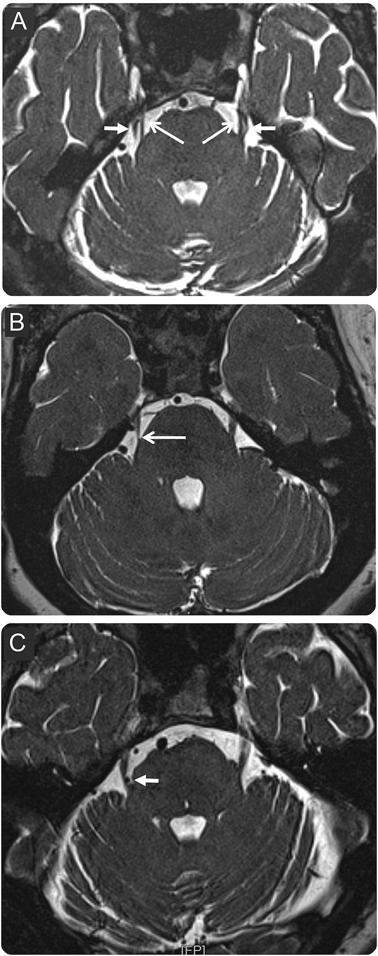
MRIAxial CISS MRI at the entry of the trigeminal nerve root into the pons: bilateral neurovascular contact without root deformation (A), unilateral contact with root deformation on the symptomatic side (B), contact on the symptomatic side without deformation and contralateral deformation without contact (C), arrows on the points of conflict.
Source : Cruccu et al., Neurology, 2016, figure 3 · CC BY-NC-ND
The reference review in Lancet Neurology describes a two-stage mechanism (Bendtsen et al. 2020,PMID 32822636). Contact between an artery and the trigeminal root at its entry into the brainstem produces, when it is marked enough to deform the nerve, a focal demyelination. Demyelinated fibres become the seat of ectopic discharges and of cross-talk from one fibre to another: an innocuous tactile input then recruits nociceptive fibres. This is the most widely accepted explanation of the clinical paradox, that a light touch produces an unbearable shock.
Which artery? A recent series of 412 patients whose conflict was confirmed in theatre during a microvascular decompression gives a solid answer: an offending vessel was found intraoperatively in 407 of them (98.8 %), and the superior cerebellar artery was the commonest cause (Huang et al., J Neurol 2026,PMID 42342895).
Two practical consequences follow. The first: in the form secondary to multiple sclerosis, the lesion is not vascular but a demyelinating plaque in the root entry zone or in the pontine trigeminal complex, the same mechanism with a different cause (Di Stefano, J Headache Pain 2019,PMID 30782116). The second, more important still for a physiotherapist: the lesion sits at the root of the nerve, in the posterior fossa, not in the tissues of the face. No technique applied to the masseters, the pterygoids or the peri-orbital tissues reaches the site of the problem. That is a mechanistic reason, not an opinion, for not expecting facial manual therapy to treat the paroxysms.
One nuance deserves stating: the mere existence of a neurovascular contact on MRI is not enough. The European guideline is explicit: demonstrating a neurovascular contact must not be used to confirm the diagnosis; it serves to decide whether and when to refer a patient for microvascular decompression. Contacts exist in people who have no symptoms; only those that deform the nerve count.
- V2 and V3 dominate (hence the dental detour: the pain projects exactly where a tooth is being looked for).
- The angle of the mandible and the back of the head are not trigeminal: they are C2-C3. That is the anatomical boundary that separates this presentation from occipital neuralgia.
- V1 involvement is not a red flag: likelihood ratio measured at 1.0.
- The lesion sits at the root of the nerve, in the posterior fossa. Nothing applied to the face reaches it.
Who is affected, and how often?
What the widest available measurement says
The most complete synthesis to date is a systematic review with meta-regression covering 1945 to 2024: 17 eligible studies, a cumulative population of 174,485,958 people including 109,070 affected patients, across twelve countries and four continents (Jeong et al., J Clin Neurol 2026,PMID 41517817). It gives a pooled worldwide incidence of 25.33 per 100,000 person-years, an annual prevalence of 45.38 per 100,000 and a lifetime prevalence of 108.43 per 100,000.
These three numbers must never be quoted on their own. Their 95 % confidence intervals are respectively 11.87-54.02, 15.41-133.61 and 30.54-384.18: the upper bound is worth, depending on the parameter, four to twelve times the lower one. That spread is not a flaw in the review, it is its result: the primary studies differ too much, in method as in populations, for a single value to mean anything. The seminal Rochester study, still the historical reference, found a crude incidence of 4.3 per 100,000 over forty years of follow-up (Katusic, Ann Neurol 1990,PMID 2301931), six times less than the modern pooled estimate. A recent Swedish population study confirms that the order of magnitude is still debated (Svedung Wettervik, Eur J Pain 2023,PMID 36680398).
Worldwide incidence and prevalence, and the uncertainty that goes with them
Logarithmic scale. The dot is the pooled estimate, the bar its 95% confidence interval
Single source: Jeong YD, Jo Y, Son Y et al. Global Incidence and Prevalence of Trigeminal Neuralgia, 1945-2024. J Clin Neurol 2026;22(1):102-112 (PMID 41517817 ·DOI 10.3988/jcn.2025.0433). The very wide intervals are the result of the review, not an artefact of the figure.
Age, and why it is a signal
Trigeminal neuralgia is a disease of the second half of life. The mean age was 52.5 years in Badran's neurosurgical series (range 17-85 years,PMID 41663983) and 66.8 ± 10.7 years in a tertiary orofacial pain clinic (PMID 42220298). That distribution is what gives weight to the first red flag: the meta-analysis in the European guideline established that patients with a secondary form are significantly younger (p < 0.0001) than those with a primary form (PMID 30860637). A typical presentation in a thirty-year-old is not good news; it is an indication for imaging.
Multiple sclerosis is the commonest identifiable secondary cause. Patients with multiple sclerosis carry a risk of trigeminal neuralgia twenty times higher than the general population, and 1.9-4.9 % of them have it; conversely, multiple sclerosis is found in 2-14 % of patients with trigeminal neuralgia. In these forms, age at onset lies between 40 and 50 years, and bilateral involvement is reported in about 18 % of patients (Di Stefano, J Headache Pain 2019,PMID 30782116).
A rare disease inside a common complaint
The two scales have to be put side by side. An annual prevalence of 45 per 100,000 means, in simple arithmetic from the pooled figure, about nine patients a year in a town of 20,000 inhabitants (an order of magnitude, not a local measurement). Meanwhile orofacial pain is an everyday reason for consulting: a recent review in Practical Neurology recalls that it can arise from more than 160 different conditions, and that trigeminal neuralgia is the rarest form of all, far behind temporomandibular disorders and post-traumatic neuropathic pain linked to dental care (Renton, Pract Neurol 2026,PMID 42320903).
This asymmetry dictates the clinical stance. Faced with facial pain, the prior probability leans massively towards something other than the trigeminal nerve; it is the presentation, not the frequency, that should make you raise a hand. And the reverse is true: it is because the disease is rare that those who have it pass through so many clinics before being recognised.
- Pooled incidence 25.3 per 100,000 person-years, but with an interval of 11.9 to 54.0: never quote the bare figure.
- Twice as many women as men, on incidence as on prevalence.
- A disease of the second half of life: a young patient should send you looking for a secondary cause.
- It is the rarest form of orofacial pain: temporomandibular disorders and post-dental neuropathic pain are far commoner.
How do you avoid confusing it with a TMJ disorder, dental pain, occipital neuralgia or cluster headache?
The differential table for facial pain
Each row carries the code of the corresponding international classification, so that the reader can go back to the criteria themselves rather than rely on a summary.
| Presentation | Site | Rhythm and duration | Trigger | The sign that settles it |
|---|---|---|---|---|
| Trigeminal neuralgia ICHD-3 13.1.1 | Face, one-sided, V1, V2 or V3 territory, with no radiation beyond it | Paroxysms from a fraction of a second to 2 minutes; refractory period after the attack | An innocuous stimulus: light touch, speech, chewing, tooth brushing, a draught | The trigger zone on skin or mucosa, reproducible, and the absence of pain between attacks |
| Temporomandibular disorder ICHD-3 11.7 · DC/TMD | Pre-auricular region, masseter, temporalis; often bilateral | A dull pain, daily or near-continuous, with no electric paroxysm | A chewing effort that is sustained, yawning, parafunctions; not a light touch | The pain is reproduced by palpation of the masticatory muscles and by mandibular movements |
| Dental pain ICHD-3 11.6 | One tooth, one sector; the patient often points to the spot with a finger | Provoked and then prolonged (pulpitis), or continuous and growing (abscess) | Heat, cold, sweet things, percussion of the tooth | The pulp vitality test and the radiograph; this is a dentist's examination, not a physiotherapist's |
| Occipital neuralgia ICHD-3 13.4 | Back of the head, upper neck, territory of the occipital nerves; uni- or bilateral | Paroxysms of a few seconds to a few minutes (a duration that overlaps that of the trigeminal nerve) | Neck movements, pressure over the course of the nerve | Tenderness on palpation of the nerve, scalp dysaesthesia, and relief from an anaesthetic block: three requirements that trigeminal neuralgia does not have |
| Cluster headache ICHD-3 3.1 | Orbital, supraorbital or temporal, strictly one-sided | Attacks of 15 to 180 minutes untreated, from one every other day to eight a day | Often nocturnal; alcohol during an active period. No cutaneous trigger | The ipsilateral cranial autonomic signs (lacrimation, rhinorrhoea, ptosis) and the restlessness (the patient cannot keep still) |
| Cervicogenic headache ICHD-3 11.2.1 | Posterior, then referred towards the forehead or the orbit, one-sided without changing side | Continuous or fluctuating, over hours or days | Cervical spine movements, sustained postures | The pain is reproduced by cervical movement; spinal mobility is limited |
| Post-traumatic trigeminal neuropathy ICHD-3 13.1.2.3 | Territory of the injured nerve, often V3: after extraction, implant, surgery | Pain that is continuous, burning in quality, with allodynia | Touch, but the pain is there at rest too | An objective sensory deficit and a dated history of a dental procedure; this is not a purely paroxysmal presentation |
| Persistent idiopathic facial pain ICHD-3 13.12 | Poorly delimited, does not follow a nerve territory | Daily, for most of the day | No reproducible trigger | Absence of a trigger and of anatomical systematisation; a diagnosis of exclusion |
Codes and criteria: ICHD-3, Cephalalgia 2018 (PMID 29368949). Diagnostic criteria for temporomandibular disorder: DC/TMD, Schiffman & Ohrbach, JADA 2016 (PMID 26922248). International Classification of Orofacial Pain (ICOP), Cephalalgia 2020 (PMID 32103673). On the boundary between genuine neuralgia and post-dental trigeminal pain, a diagnostic pathway study of 672 patients from a tertiary centre distinguishes five phenotypes, among them confirmed alveolar neuropathy and simple odontogenic misclassification (Shemesh et al., Diagnostics 2026,PMID 42279542).
Trigeminal and occipital: two nerves, two presentations
The two conditions share a word and an attack duration, and nothing else. Occipital neuralgia affects the greater occipital nerve and hurts at the back of the head; trigeminal neuralgia affects the fifth cranial nerve and hurts in the face. That is the sentence to remember.
Three requirements in the ICHD-3 13.4 criteria complete the separation of the two presentations, and they are absent in trigeminal neuralgia: occipital pain comes with dysaesthesia or allodynia of the scalp, it is associated with tenderness on palpation of the nerve branches concerned or with trigger points at the emergence of the greater occipital nerve, and it is temporarily relieved by an anaesthetic block of the nerve. A patient whose pain yields to thumb pressure below the nuchal line does not belong in this article; they belong in our synthesis on occipital neuralgia.
The confusion nevertheless runs both ways, and it is documented. Anatomical variations in the course of the greater occipital nerve make some occipital pain radiate towards the maxilla or the external ear, which can wrongly point to the trigeminal nerve. Symmetrically, a pure V3 trigeminal presentation projects close to the angle of the mandible, at the border of the cervical territory. The practical rule: look for the trigger. A trigger on the skin or mucosa of the face, set off by a light touch, points to the trigeminal nerve; a pain on palpation of the nerve trunk at the neck points to occipital neuralgia.
The TMJ: the commonest neighbour, and the costliest in sessions
This is the confusion that costs the patient most, because temporomandibular disorder is itself a legitimate indication for physiotherapy: the patient enters a coherent pathway, but for the wrong disease. The reference review by Maarbjerg, Di Stefano, Bendtsen and Cruccu stresses that confusion between the two persists in practice despite distinct criteria (Cephalalgia 2017,PMID 28076964).
Three discriminators are usually enough. The rhythm: temporomandibular disorder gives a dull pain, present most days; the neuralgia gives shocks of a few seconds separated by strictly pain-free periods. The trigger: the first responds to a sustained mandibular effort and to firm palpation of the muscles, the second to a light touch that ought to produce nothing. Reproducibility at examination: TMJ pain is reproduced by palpating the masseter and moving the mandible, which is part of the standardised assessment; a neuralgia is reproduced by brushing a trigger zone a few millimetres across, often perioral or nasolabial.
One point deserves saying bluntly: the two can coexist, and a genuine temporomandibular disorder does not rule out an associated trigeminal neuralgia. If a patient treated for the TMJ keeps having paroxysmal shocks set off by a light touch, the question must be reopened. Our synthesis on TMJ syndrome covers the temporomandibular side, and the feature on bruxism and temporomandibular disorders the parafunctional side.
Decision tree for facial pain
An orientation tool for the first contact. None of these branches removes the need for a medical opinion
Built from the ICHD-3 criteria of the entities cited (PMID 29368949) and from the review in Lancet Neurology by Bendtsen et al. 2020 (PMID 32822636). Proportion of forms with concomitant continuous pain: Segura-Lozano 2024 (PMID 38855490). An orientation tool, not a validated diagnostic algorithm.
- Occipital neuralgia is the back of the head; trigeminal neuralgia is the face. Attack duration does not separate them; the territory does.
- Temporomandibular disorder is reproduced by palpation of the muscle; the neuralgia is set off by a light touch.
- Dental pain calls for a pulp vitality test, not for a physiotherapy assessment.
- Cluster headache is recognised by its cranial autonomic signs and its restlessness, not by the pain alone.
When should you suspect a secondary cause?
About one form in seven is not primary
Secondary trigeminal neuralgia, that is to say attributable to an identifiable neurological disease other than a simple vascular conflict, accounts for up to 15 % of cases. Multiple sclerosis plaques are the abnormality most often identified; then come space-occupying lesions of the cerebellopontine angle, arteriovenous malformations and, more rarely, inflammatory or ischaemic lesions of the brainstem (Di Stefano, J Headache Pain 2019,PMID 30782116).
These forms do not merely have another cause: they respond less well. A retrospective observational study matching 36 patients with multiple sclerosis and trigeminal neuralgia to 36 patients without multiple sclerosis, matched for age and sex, found in the multiple sclerosis group higher pain scores, significantly more frequent recourse to blocks and to radiofrequency, and a markedly higher prevalence of refractory forms, correlated with the EDSS score (Yön & Ulusoy, Noro Psikiyatr Ars 2026,PMID 41613036).
Red flags in paroxysmal facial pain
- A young patient. Patients with a secondary form are significantly younger than those with a primary form (p < 0.0001 on the pooled data of the European guideline). A typical presentation before the age of 40 calls for imaging.
- Bilateral involvement. Specificity of 100 % (99-100), positive likelihood ratio of 9.5. A bilateral neuralgia should send you looking for multiple sclerosis, in which bilateral involvement is reported in about 18 % of cases.
- An objective sensory deficit in the trigeminal territory: hypoaesthesia to light touch or to pinprick, found on comparative examination. Specificity of 98 % (96-99), positive likelihood ratio of 20.6: this is the most informative clinical sign of all. ICHD-3 itself states that a sensory deficit should prompt a search for a cause.
- Other associated neurological signs: weakness of the masticatory muscles, involvement of another cranial nerve, hearing loss, balance disturbance, cerebellar signs. They point to a lesion of the cerebellopontine angle.
- Resistance to well-conducted medical treatment. Specificity of 93 % (81-99) but a likelihood ratio of only 2.1: a weak sign, not to be over-read on its own.
- Pain that changes character: a purely paroxysmal presentation that becomes continuous, or an extension of the territory, deserves reporting again to the treating doctor.
The limit to know, and to state. No clinical feature is sensitive enough to rule out a secondary form. The European guideline writes it explicitly and draws the consequence: MRI, combining three high-resolution sequences, must be part of the work-up of every patient with trigeminal neuralgia, not only of those who carry a red flag (Bendtsen et al., Eur J Neurol 2019,PMID 30860637).
Highly specific, barely sensitive: what that means at the bedside
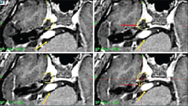
MRIMRI showing a tortuous vascular loop compressing and distorting the right trigeminal nerve, yellow arrows, extending down to the trigeminal ganglion.
Source : Somaza et al., Surgical neurology international, 2014, figure 2 · CC BY-NC-SA
The chart below sets side by side, for each sign, its sensitivity and its specificity as the meta-analysis in the European guideline calculated them. The shape of the chart is the message: every specificity bar is high, every sensitivity bar is low.
Diagnostic value of the clinical signs for a secondary form
Pooled data from seven studies, meta-analysis in the EAN 2019 guideline
Single source: tables 1 and 2 of Bendtsen L, Zakrzewska JM, Abbott J et al. European Academy of Neurology guideline on trigeminal neuralgia. Eur J Neurol 2019;26(6):831-849 (PMID 30860637 ·DOI 10.1111/ene.13950). Pooled data from seven studies for the clinical signs, eight for the trigeminal reflexes.
The consequence for the physiotherapist
It is simple to state and sometimes hard to hold to: it is not for the physiotherapist to decide between a primary and a secondary form. That requires an MRI, and imaging is not within their scope. What is within their scope is to do three things.
First, look for a sensory deficit, because it is the most informative sign of the lot and because it is tested with a wisp of cotton wool and a blunt point, comparing the two sides in the three territories. Then, record what you find and what you do not find: a sensory examination recorded as normal has value for the doctor who will see the patient. Finally, write. A letter that says “paroxysms of less than two minutes, right V2 territory, reproducible nasolabial trigger, normal comparative sensory examination, no improvement after six sessions” allows an immediate referral; a letter that says “persistent facial pain” allows nothing.
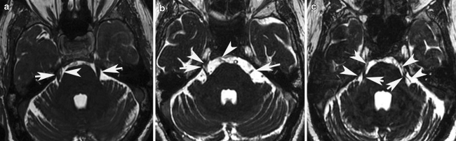
MRIAxial FIESTA MRI sequence showing a superior cerebellar artery loop in contact with the trigeminal nerve root, arrows, on the right side then on both sides.
Source : Paliwal et al., The Journal of Headache and Pain, 2012, figure 1 · CC BY
- Up to 15 % of trigeminal neuralgias are secondary; multiple sclerosis is the most frequently identified cause.
- The objective sensory deficit is the most informative sign: positive likelihood ratio of 20.6.
- These signs are specific but poorly sensitive: their absence rules nothing out, and MRI is recommended in everyone.
- The physiotherapist's role is to examine sensation, to record, and to write precisely (not to decide).
What can medical treatment and surgery do?
Carbamazepine, a solid reference on fragile evidence
The European guideline is clear: for maintenance treatment, carbamazepine (200 to 1200 mg a day) or oxcarbazepine (300 to 1800 mg a day) remain the most effective drugs, particularly at early stages; higher doses are sometimes needed (PMID 30860637). In an acute exacerbation, admission to hospital may be needed in order to rehydrate and to give intravenous fosphenytoin or lidocaine; a recent case documents exactly this course during a crisis in a neuralgia secondary to a petroclival meningioma, with almost complete relief after the loading dose (Mochizuki et al., Front Pain Res 2026,PMID 42100134). These choices belong to the doctor; they appear here so that the physiotherapist understands their patient's pathway and recognises the adverse effects that will explain the cancelled sessions.
The evidence does have to be faced, however. The Cochrane review devoted to carbamazepine in chronic neuropathic pain included ten studies and 480 participants, among them patients with trigeminal neuralgia. Its authors write that no study provided first- or second-tier evidence for any efficacy outcome, and conclude that carbamazepine is “probably effective in some people”, with this explicit caveat: no trial lasted longer than four weeks, none had satisfactory reporting quality, and none used an outcome equivalent to substantial clinical benefit (Wiffen et al., Cochrane Database Syst Rev 2014,PMID 24719027).
This does not mean the drug does not work: clinical experience and the strong stance of the European guideline say the opposite. It means that the reference treatment for a disease described since the 18th century rests on old, short and small trials. A physiotherapist wondering why the literature of their own discipline is so thin on this subject will find part of the answer here: it is thin everywhere.
The adverse effects, for their part, are well documented and frequent. In four studies of that review, 65 % of patients on carbamazepine (113 of 173) had at least one adverse effect, against 27 % on placebo: for every five patients treated, two suffer an adverse effect they would not have had on placebo. Drowsiness, dizziness, ataxia and impaired concentration explain many a missed session, and sometimes a complaint of “imbalance” wrongly brought to the physiotherapist.
The second lines, and botulinum toxin
When first-line treatments fail for lack of efficacy or of tolerance, the European guideline gives weak recommendations, based on low to very low quality evidence, to lamotrigine, gabapentin, botulinum toxin type A, pregabalin, baclofen and phenytoin, alone or in combination (PMID 30860637).
Botulinum toxin type A has been the most studied since. An updated meta-analysis of 23 studies, four of them randomised trials, found a significant reduction in the visual analogue scale score from baseline in the randomised trials (effect size −4.05; 95 % CI −6.13 to −1.97; p = 0.002), concluding that it is an effective and safe option in refractory patients while calling for better quality studies (Hu et al., Clin J Pain 2024,PMID 38385501). Four randomised trials out of twenty-three studies: the caution of the conclusion is justified.
Surgery: when, and which one
The guideline is explicit about timing: it is recommended to offer surgery if the pain is not sufficiently controlled by drugs or if they are poorly tolerated, and there is no need to have tried every drug before asking for a neurosurgical opinion. This is information many patients do not have, and which a physiotherapist may legitimately remind them of while sending them back to their doctor. A prospective study cited by the guideline shows, moreover, that 65 % of patients referred to a specialist centre were still adequately controlled on drugs two years later, while 35 % were sent on to surgery.
In classical forms (those in which MRI shows a neurovascular conflict deforming the nerve), microvascular decompression is recommended as the first surgical line, with a strong recommendation despite low quality evidence. The historical series by Barker and Jannetta, covering 1,185 patients operated on over twenty years, reported 70 % excellent results at ten years, that is to say patients pain-free and off medication, plus 4 % with occasional pain not requiring long-term treatment (N Engl J Med 1996,PMID 8598865). The same series puts a number on the price to be paid, and it must be given alongside the benefit: two deaths shortly after the operation (0.2 %), one brainstem infarct (0.1 %) and sixteen ipsilateral deafnesses (1 %). Thirty per cent of patients relapsed during the study period, most of them within the first two years, and 11 % were reoperated on; beyond ten years the annual recurrence rate falls below 1 %.
Pain-free patients at long-term follow-up, by technique
Medians and ranges from 45 non-randomised cohort studies analysed by the EAN 2019 guideline
Single source: table 9 and text of the EAN 2019 guideline (Bendtsen et al., Eur J Neurol 2019;26(6):831-849,PMID 30860637), a synthesis of 45 non-randomised cohorts. The 1-2 year comparison comes from the same document.
| Technique | Principle | Pain-free in the long term | What you need to know |
|---|---|---|---|
| Microvascular decompression | Retrosigmoid craniotomy: a material is interposed between the artery and the nerve. The nerve is not damaged. | 77 % (62-89) | A recommendation graded strong for first line in classical forms. The only technique with a reported mortality. |
| Internal neurolysis | Longitudinal dissection of the fascicles of the root. | 72 % | Only one study retained: the figure carries no range and must be read with reservation. |
| Balloon compression | A balloon inflated in Meckel's cave, percutaneously. | 68 % (55-80) | An ablative technique: postoperative facial hypoaesthesia is expected, not accidental. |
| Gamma Knife radiosurgery | Focused irradiation of the root, with no opening. | 58 % (30-66) | The guideline stresses the low number of complications of this technique. The effect is delayed by several weeks. |
| Radiofrequency thermocoagulation | Thermal lesioning of the Gasserian ganglion, percutaneously. | 58 % (26-82) | The widest range of the six: results depend heavily on the operator and on the series. |
| Glycerol rhizotomy | Injection of glycerol into the trigeminal cistern. | 28 % (18-59) | The weakest long-term result of the six techniques analysed. |
Single source: EAN 2019 guideline, a synthesis of 45 non-randomised cohorts (PMID 30860637). No randomised trial has compared these techniques with each other: these figures are not direct comparisons.
In idiopathic forms, on the other hand, no recommendation can be made for choosing between the neuro-ablative techniques, nor between those and decompression: neuro-ablative treatments are to be preferred when MRI shows no neurovascular contact. Finally, in secondary forms, treatment generally follows the same principles as in primary forms, and the guideline adds that, on top of medical and surgical management, one should offer these patients psychological and nursing support. That is the only non-drug, non-surgical opening in the document, and the next chapter draws the consequences.
- Carbamazepine or oxcarbazepine first line: this is the reference treatment, and it does not belong to the physiotherapist.
- Cochrane is severe: not one trial longer than four weeks, no first- or second-tier evidence. The drug remains recommended; its evidence base is thin.
- Two patients in five suffer an adverse effect attributable to the treatment: that explains many a cancelled session.
- Surgery does not have to be earned by exhausting every drug first: microvascular decompression gives 77 % of patients pain-free in the long term.
What can physiotherapy actually do, and at what level of evidence?
Measurement, rather than impression
Rather than assert that “the evidence is limited”, here is the measurement, made on 15 August 2026 on PubMed by crossing MeSH descriptors. The descriptor Trigeminal Neuralgia holds 8,451 references. Crossed with Physical Therapy Modalities, 267 remain, of which 22 randomised trials. Crossed with Exercise Therapy: zero. With Musculoskeletal Manipulations: nine references, no randomised trial.
The 22 trials deserve reading one by one, because the MeSH descriptor Physical Therapy Modalities does not cover what a physiotherapist calls their practice. Going through the 22 title by title gives: fifteen trials of percutaneous radiofrequency or related interventional neuromodulation (thermocoagulation or pulsed radiofrequency of the Gasserian ganglion, that is to say pain-specialist procedures), five trials of acupuncture or electroacupuncture, one trial of motor cortex stimulation, and a single trial of TENS. Fifteen plus five plus one plus one: all 22 are accounted for, none is left aside. No trial of exercise, none of manual therapy, none of patient education. A scoping review published in 2026, which mapped the interventions used in this condition between 2020 and 2025 from five databases, arrives at the same landscape: pharmacological, surgical and non-invasive interventions, with rehabilitation forming no documented category at all (Mateos-Romero et al., Pain Manag Nurs 2026,PMID 42315398).
The cross-check confirms this void on the guideline side. In the full text of the European Academy of Neurology guideline, the terms “physiotherapy”, “physical therapy”, “exercise”, “rehabilitation”, “manual therapy” and “massage” do not appear a single time. By contrast, “psychological” appears three times, “nursing” three times and “self-management” twice, and the document explicitly recommends offering patients psychological and nursing support (PMID 30860637). So an indication does exist: it simply is not the one we would have wanted.
What the only TENS trial contains, and why it is not enough
This single trial deserves describing precisely, because it is regularly cited as proof of the efficacy of TENS in this indication. It covers 31 patients who were refractory or partial responders to drug treatment, randomly allocated between two modes of TENS, continuous and burst, with pain assessed before treatment and fifteen days later. Twenty-six patients out of 31 (83.7 %) improved, the continuous mode doing slightly better than the burst mode (Yameen et al., J Pak Med Assoc 2011,PMID 22204173).
The figure of 83.7 % is impressive, and it says nothing about the efficacy of TENS. There was no group without TENS. Both arms received the treatment; the comparison was about the mode, not about the principle. An improvement rate without a comparator, in a disease that has spontaneous remissions (the European guideline does note that periods of partial or complete remission occur), measures the natural course as much as the treatment. Thirty-one patients, fifteen days of follow-up: this is a pilot study, and it should be cited as such.
Electroacupuncture: the only methodologically solid trial
A recent trial changes the landscape, and honesty requires reporting it as carefully as the previous one, in the other direction. It is a multicentre randomised trial with a 2×2 factorial design, in 120 participants, comparing active electroacupuncture against sham electroacupuncture, and low-dose carbamazepine (300 mg/day) against placebo, over four weeks, with follow-up at 28 weeks (Li et al., J Neurol 2024,PMID 38816482).
The main effects of electroacupuncture and of carbamazepine were significant (p < 0.001), with a significant interaction between the two (p = 0.041). At four weeks, the reduction in the visual analogue scale score was −1.6 for electroacupuncture alone, −0.9 for carbamazepine alone and −3.7 for the combination, each compared with its own control group. Adverse effects related to electroacupuncture were less frequent than those of carbamazepine (10.2 % against 25.4 %). One point deserves noting: at 28 weeks, the effect of carbamazepine alone was no longer distinct from its placebo, whereas that of electroacupuncture persisted.
Three reservations, though. It is a single trial, run in a single country, on a practice, electroacupuncture, that is neither taught nor authorised within the usual scope of physiotherapy practice in France. The result is interesting for understanding the disease; it does not amount to a transferable indication for rehabilitation.
What is left, and what it is worth
Non-drug, non-surgical interventions, by level of evidence
A reading of the evidence base as of 15 August 2026. The levels are those the sources allow, not published GRADE ratings
The author's synthesis from the sources cited on each card: EAN 2019 guideline (PMID 30860637), Li 2024 (PMID 38816482), Yameen 2011 (PMID 22204173), Badran 2026 (PMID 41663983), and PubMed MeSH counts of 15 August 2026. The level labels are an editorial reading of this evidence base, not published GRADE ratings.
The physiotherapist's real place, in five points
Recognise, and say so. This is the main contribution, and it is considerable given the documented delay. A physiotherapist who identifies the presentation and writes a precise letter saves their patient months, and sometimes a tooth.
Do not treat what is not yours to treat. Refusing six weeks of sessions on masseters that have nothing to do with the pain is a professional act, not a surrender.
Explain. Many patients arrive convinced they have “something wrong with their teeth” or a psychiatric illness. Naming the disease, describing its mechanism (an irritated nerve root, fibres that short-circuit, a light touch that recruits pain fibres) and warning that spontaneous remissions occur is fully part of patient education.
Treat what coexists, provided you name it separately. A patient can have trigeminal neuralgia and neck pain, and a genuine temporomandibular disorder. These last two belong to rehabilitation. The mistake is not to treat them: it is to let the patient believe that this treatment will act on the shocks.
Support the impact. That is the subject of the next chapter, and it is the part of the European guideline where the physiotherapist has most to do, even though the document does not name them.
- Zero randomised trials of exercise and zero mention of physiotherapy in the European guideline: that is measured, not estimated.
- TENS rests on a study of 31 patients with no control group: you may offer it, you can promise nothing.
- The only solid trial concerns electroacupuncture, outside the French scope of practice.
- The physiotherapist's value lies upstream of treatment: recognising, not treating in error, explaining, referring, supporting.
Why is this pain still called the “suicide disease”?
What a national registry says over seventeen years
The phrase had long circulated without solid data. A Swedish national retrospective cohort study has just documented it: 3,531 adults diagnosed between 2001 and 2005, followed for a median of more than seventeen years from the national health and death registers (Ehinger, Kronvall & Uvelius, J Headache Pain 2026,PMID 42001017).
The standardised incidence ratios are significantly raised for cerebrovascular disease (1.4 to 1.8 depending on the analysis), myocardial infarction (1.3), external causes of injury (1.5) and intentional self-harm (2.1). Men had an increased risk of infarction and of ischaemic stroke, women an increased risk of self-harm and of unintentional poisoning. The most striking result comes in two statements that must be quoted together: overall mortality was not increased, but the risk of suicide was more than doubled in women (standardised mortality ratio of 2.5). The authors conclude that these data support calling this disease a genuine “suicide disease”.
Excess risks measured in a national cohort of 3,531 patients
Standardised incidence and mortality ratios. The vertical line at 1.0 is the absence of excess risk
Standardised ratios: Ehinger E, Kronvall E, Uvelius E. J Headache Pain 2026;27(1):117 (PMID 42001017). Clinic data: Limrachtamorn, J Oral Facial Pain Headache 2026 (PMID 42220298). The values 1.4-1.8 are a range across analyses, not a confidence interval.
What that looks like in a clinic
These figures translate into behaviours that are observed without being named: a patient who speaks little and quietly, who refuses to let a hand come near their face, who arrives masked or with a scarf pulled up for fear of a draught, who cancels when the weather turns cold. These are not personality traits, they are strategies for avoiding a pain that the slightest stimulation sets off.
The specialist clinic series already cited gives the measure of the intensity: 8.17 ± 1.08 out of 10 at the first assessment, with moderate to moderately severe depressive symptoms, moderate to severe anxiety and moderate perceived stress frequently found on the PHQ-9, GAD-7 and PSS-10 scales (PMID 42220298). An American study of patients referred for pre-surgical assessment of facial pain has also examined the link between the dimensions of facial pain and suicidal ideation (Campbell et al., Pain Rep 2026,PMID 42147755).
Two practical consequences. The first: adapt the session by announcing every contact before touching, avoiding the painful territory, not persisting with a palpation that has already set off an attack, accepting that a patient speaks little. The second, more serious: know that the distress is real and do not leave it unanswered. A patient who says they “cannot go on like this” is not exaggerating; the excess risk of self-harm is measured at 2.1. The physiotherapist is not the mental health professional, but they are often the one who sees the patient most often, and the European guideline does explicitly call for psychological support.
- A measured excess risk of intentional self-harm (2.1) and of suicide in women (2.5) in a national cohort of 3,531 patients.
- Overall mortality is not increased: this is a disease that wears you down, not one that kills.
- Mean intensity at the first specialist consultation: 8.17 out of 10, with frequent anxiety and depressive symptoms.
- Adapt the session: announce every contact, avoid the territory, do not repeat a palpation that has set off an attack.
What do published case reports teach us?
Published case no. 1: the sensory deficit that changes everything
A man of 59, with a history of hyperlipidaemia, hepatic steatosis and hyperuricaemia, presents with progressive numbness and a sensation of swelling of the left gum and the left side of the face, present for twenty days. He is first seen at a dental practice, where a diagnosis of trigeminal neuritis is made; he is given prednisone, with no effect whatsoever.
Neurological examination finds reduced sensitivity to pinprick and to temperature in the left trigeminal territory, with normal masseter power. MRI shows a diffusion-restricted lesion along the trigeminal sensory pathway, in the left pontine tegmentum: this is a subacute pontine infarct selectively involving the trigeminal sensory fibres, sparing the motor fibres, a presentation the authors call extremely rare and prone to diagnostic error (Wang, Niu & Wang, Front Pain Res 2026,PMID 42317569 ·PMC13272495).
What this case teaches. The objective sensory deficit was there from the start, and it was the most informative sign of the presentation, a likelihood ratio of 20.6 for a secondary cause. A comparative sensory examination, with a wisp of cotton wool and a blunt point, in the three territories and on both sides, takes two minutes and needs no equipment. The patient was treated with corticosteroids at a dental practice while he was having a brainstem stroke.
Published case no. 2: a V3 numbness that was not a neuralgia
A man of 45 has had, for several months, discomfort in the right side of the face and numbness in the mandibular territory (V3), a presentation initially put down to trigeminal neuralgia. Brain MRI reveals a strongly enhancing lesion in the right Meckel's cave, along the nerve. Surgical exploration of the skull base and histological analysis conclude that this is a neurosarcoidosis of the trigeminal nerve, with non-caseating granulomas, no neoplastic or infectious lesion, an isolated cranial nerve lesion that closely mimics a trigeminal schwannoma (Msherghi et al., J Med Case Rep 2026,PMID 41814347 ·PMC13088403).
What this case teaches. Two red flags coexisted: a sensory deficit and an unusually young age. It also teaches a point of vocabulary that has consequences: facial pain with permanent numbness is not trigeminal neuralgia in the ICHD-3 sense, but a painful trigeminal neuropathy (code 13.1.2), a neighbouring and distinct entity whose discovery calls for imaging.
Published case no. 3: the perfectly typical presentation that hid a malformation
A man of 54 presents with severe paroxysmal facial pain, compatible with trigeminal neuralgia. Magnetic resonance imaging reveals an arteriovenous malformation of the trigeminal nerve, an exceptional cause of which this was the first manifestation (Kadiri et al., Radiol Case Rep 2025,PMID 41035745 ·PMC12481911).
What this case teaches. This is the most unsettling of the four, because it carried no obvious red flag. It illustrates exactly the limit measured in chapter 5: the clinical signs are highly specific and barely sensitive, and it is for that reason that the European guideline asks for an MRI in all patients, not only in suspicious cases. A typical presentation is no guarantee: it is an indication for imaging like any other.
Published case no. 4: two years of wandering for a pain in the tongue
A woman of 63 has had, for two years, isolated pain in the left half of the tongue. Glossopharyngeal neuralgia is suspected first; oxcarbazepine brings insufficient relief. Referred to a specialist centre, she has a high-resolution MRI which shows vascular compression of the trigeminal nerve; a glossopharyngeal nerve block rules out the other hypothesis, and microvascular decompression of the trigeminal nerve confirms the diagnosis (Xu et al., Front Pain Res 2026,PMID 42181615 ·PMC13190604).
What this case teaches. The anterior two thirds of the tongue belong to the lingual nerve, a branch of V3: tongue pain can therefore be trigeminal, even if it first suggests the glossopharyngeal nerve. Above all it is a reminder of how long these delays run, two years, and of the fact that they happen even within sound medical pathways.
The fifth case, the one the physiotherapist will meet most often
This is not an isolated case but a profile, documented in a case review from a dental practice: a man of 72 with chronic stabbing pain, intermittent, electric shock-like in quality, sitting in a maxillary tooth and radiating along the trigeminal distribution; carbamazepine alone had not been enough, and the pain did not recur after management combining a psychological intervention with antidepressant and anxiolytic treatment (Tizzoni, Tizzoni & Clerici, F1000Research 2021,PMID 35966965 ·PMC9345266).
What this case teaches. Dental pain that yields to no dental care should make you reconsider the organ. And the psychological dimension is not a fallback diagnosis: it is part of the picture, as the previous chapter put into figures.
What the five cases have in common
- In four cases out of five, the first professional consulted was not a neurologist: a dentist, a pain specialist, a GP. That is the ordinary pathway of this disease, not a series of exceptions.
- In two cases, an objective sensory deficit was present at the very first examination and was not looked for until several weeks later.
- In one case, no red flag was present and yet the cause was secondary: that is the argument for routine MRI.
- Level of evidence of a case report: 5, the weakest there is. These five stories serve to recognise, not to decide. Decisions are made on the European guideline and on the systematic reviews cited above.
How do you apply this in the clinic?
The five questions that do the sorting
They take three minutes, in this order, and most of the time they are enough to point the way.
- “How long does an attack last? A few seconds, a few minutes, a few hours?” An answer in seconds points to a neuralgia, trigeminal or occipital. An answer in hours rules both out.
- “Between two attacks, do you have pain?” “Nothing at all” is highly suggestive. Background pain does not rule out the diagnosis, but it points to the form with concomitant continuous pain.
- “What sets it off? Is a simple light touch enough?” This is the central question. Light touch, speech, tooth brushing, a draught: innocuous stimuli. Prolonged chewing and firm pressure: you are somewhere else.
- “Show me exactly where”, asking the patient to point with a finger. Forehead, cheekbone, lip, jaw: trigeminal. Back of the head, neck: occipital. A single tooth: a dental cause until proven otherwise.
- “Have you changed anything in your daily life because of this pain?” No longer shaving, no longer wearing make-up, eating on one side only, avoiding speech: these avoidances are almost pathognomonic and are not met in myofascial pain.
The four examination manoeuvres
1. The comparative sensory examination. A wisp of cotton wool and a blunt point, the three territories, both sides, every time. It is the highest-yield manoeuvre in the whole examination: the objective sensory deficit carries a likelihood ratio of 20.6 for a secondary cause. A normal examination is recorded too: it has value for the doctor who comes next.
2. The search for the trigger zone, once only, with the patient's explicit agreement and after warning them. You brush very lightly, with a fingertip or a wisp of cotton wool, the perioral, nasolabial and chin areas. If the light touch sets off a shock, the ICHD-3 criterion is met. You do not do it again: the refractory period is no reason to test the limit.
3. Palpation of the masticatory muscles and mandibular movement, for the temporomandibular hypothesis. What you are looking for here is to reproduce the patient's usual pain, not simply to find a tender muscle: a masseter that hurts on palpation is commonplace.
4. Upper cervical examination and palpation of the greater occipital nerve, for the occipital hypothesis. Pain reproduced by pressure over the course of the nerve, scalp dysaesthesia: the picture tips over.
The pathway these manoeuvres can shorten
Two separate series, two different denominators. The figures do not add up
Sources: Badran SA, Qasim AM, Alhamandi F et al. BMC Neurology 2026;26(1):253 (PMID 41663983); Limrachtamorn T. J Oral Facial Pain Headache 2026;40(3):167-175 (PMID 42220298). The recruitment bias of tertiary centres is explicit: these proportions are not generalisable to the general population.
The letter, and why its precision changes everything
A generic letter produces a generic referral. Here is what, in a letter, lets a doctor decide immediately on an MRI or a neurological opinion:
The six items to include
- The duration of the paroxysms, in seconds or in minutes, as the patient describes it.
- The precise territory, naming the branch: “right V2 territory”, not “cheek pain”.
- The trigger, and whether it was reproduced at examination: “light touch to the right nasolabial region, shock reproduced”.
- The result of the comparative sensory examination, including when it is normal.
- The presence or absence of red flags: bilaterality, sensory deficit, other neurological signs, age.
- What has already been tried, and without effect: dental care, rehabilitation sessions, analgesics.
Knowing when to stop
It is the hardest decision, because it goes against professional reflex. Three situations require suspending rehabilitation and writing:
The presentation is typical and has never been investigated. No session should come before the medical opinion. This is not a condition where you try six sessions “to see”.
A red flag appears during treatment. A sensory deficit that was not there at the first session, an extension of the territory, the appearance of signs from another cranial nerve: you stop and you refer the same day.
Rehabilitation of a coexisting problem changes nothing about the shocks. If you are treating a genuine temporomandibular disorder and the paroxysms persist unchanged, that confirms they belong to something else, not that the treatment should be intensified.
Running the session, when there is one
When rehabilitation is justified for a coexisting reason, a few adaptations change the patient's experience: announce every contact before touching the face; work on the pain-free side when possible; avoid draughts, blowing air conditioning and manoeuvres that require long stretches of speech; offer to postpone a session during a flare rather than impose it; and never persist with an area that has set off a shock, even “to desensitise it”. There is no data supporting progressive desensitisation of the trigger zone in this condition, and the mechanism, demyelinated fibres that short-circuit, gives no grounds to hope for it.
- Five questions: duration, pain between attacks, trigger, site pointed to with a finger, avoidances.
- The highest-yield manoeuvre is the comparative sensory examination: two minutes, no equipment, a likelihood ratio of 20.6.
- Look for the trigger once only, warning the patient first. Do not work it again in session.
- A letter that names the branch, the duration and the trigger triggers an MRI; a letter that says “facial pain” triggers nothing.
References
Every reference was resolved on 15 August 2026 against PubMed metadata through the E-utilities API or Europe PMC: journal, year, volume, pagination, author list, DOI and PMC identifier. The abstracts were read, and the figures quoted in the article checked at source, in full text when it was accessible. PMID links point to PubMed, DOI links to doi.org.
Classifications and guidelines
- Headache Classification Committee of the International Headache Society (IHS). The International Classification of Headache Disorders, 3rd edition. Cephalalgia 2018;38(1):1-211.PMID 29368949 ·DOI 10.1177/0333102417738202
- Goadsby PJ, Evers S, Gelfand AA, Lipton RB, May A, Pozo-Rosich P, Schoenen J, Schwedt TJ, Tassorelli C, Terwindt G, Wang SJ. International Classification of Headache Disorders-4: Work in Progress 1. Cephalalgia 2024;44(2):3331024241233937.PMID 38388354 ·DOI 10.1177/03331024241233937
- International Classification of Orofacial Pain, 1st edition (ICOP). Cephalalgia 2020;40(2):129-221.PMID 32103673 ·DOI 10.1177/0333102419893823
- Bendtsen L, Zakrzewska JM, Abbott J, Braschinsky M, Di Stefano G, Donnet A, Eide PK, Leal PRL, Maarbjerg S, May A, Nurmikko T, Obermann M, Jensen TS, Cruccu G. European Academy of Neurology guideline on trigeminal neuralgia. Eur J Neurol 2019;26(6):831-849.PMID 30860637 ·DOI 10.1111/ene.13950
- Cruccu G, Finnerup NB, Jensen TS, Scholz J, Sindou M, Svensson P, Treede RD, Zakrzewska JM, Nurmikko T. Trigeminal neuralgia: New classification and diagnostic grading for practice and research. Neurology 2016;87(2):220-228.PMID 27306631 ·DOI 10.1212/WNL.0000000000002840
- Schiffman E, Ohrbach R. Executive summary of the Diagnostic Criteria for Temporomandibular Disorders for clinical and research applications. J Am Dent Assoc 2016;147(6):438-445.PMID 26922248 ·DOI 10.1016/j.adaj.2016.01.007
Reference reviews
- Bendtsen L, Zakrzewska JM, Heinskou TB, Hodaie M, Leal PRL, Nurmikko T, Obermann M, Cruccu G, Maarbjerg S. Advances in diagnosis, classification, pathophysiology, and management of trigeminal neuralgia. Lancet Neurol 2020;19(9):784-796.PMID 32822636 ·DOI 10.1016/S1474-4422(20)30233-7
- Maarbjerg S, Di Stefano G, Bendtsen L, Cruccu G. Trigeminal neuralgia: diagnosis and treatment. Cephalalgia 2017;37(7):648-657.PMID 28076964 ·DOI 10.1177/0333102416687280
- Renton T. Orofacial pain beyond trigeminal neuralgia. Pract Neurol 2026.PMID 42320903 ·DOI 10.1136/pn-2026-005154
- Elsaraj SM, Kasha-Blois R, Wint-White R, Iacopino AM, Shoppoff L, Cioffi I. Trigeminal neuralgia: a primer for dental practitioners. Quintessence Int 2026.PMID 42484626 ·DOI 10.3290/j.qi.b7060092
- Mateos-Romero M, Compañ-Gabucio L, Company V, Puelles E, Quinones S, Andreu-Cervera A. Current Landscape of Clinical Interventions for Trigeminal Neuralgia: A Scoping Review. Pain Manag Nurs 2026.PMID 42315398 ·DOI 10.1016/j.pmn.2026.05.025
Epidemiology
- Jeong YD, Jo Y, Son Y, Kim DK, Kim TH, Cho J, Kim S, Kang J, Smith L, Woo HG, Yon DK. Global Incidence and Prevalence of Trigeminal Neuralgia, 1945-2024: A Systematic Review and Meta-Regression Analysis. J Clin Neurol 2026;22(1):102-112.PMID 41517817 ·DOI 10.3988/jcn.2025.0433
- Katusic S, Beard CM, Bergstralh E, Kurland LT. Incidence and clinical features of trigeminal neuralgia, Rochester, Minnesota, 1945-1984. Ann Neurol 1990;27(1):89-95.PMID 2301931 ·DOI 10.1002/ana.410270114
- Svedung Wettervik T, Snel D, Kristiansson P, Ericson H, Hariz M. Incidence of trigeminal neuralgia: A population-based study in Central Sweden. Eur J Pain 2023;27(5):580-587.PMID 36680398 ·DOI 10.1002/ejp.2081
Secondary forms and multiple sclerosis
- Di Stefano G, Maarbjerg S, Truini A. Trigeminal neuralgia secondary to multiple sclerosis: from the clinical picture to the treatment options. J Headache Pain 2019;20(1):20.PMID 30782116 ·DOI 10.1186/s10194-019-0969-0
- Yön Mİ, Ulusoy EK. Clinical Characteristics and Treatment Resistance of Trigeminal Neuralgia in Multiple Sclerosis Patients: A Retrospective Observational Study. Noro Psikiyatr Ars 2026;63:108-111.PMID 41613036 ·DOI 10.29399/npa.29088
Clinical forms and concomitant continuous pain
- Segura-Lozano MA, Carranza-Rentería O, Velázquez-Delgado G, Munguía-Rodríguez AG. Histochemical Analysis of Altered Arachnoid Tissue in Patients With Paroxysmal Trigeminal Neuralgia With Concomitant Continuous Pain. Cureus 2024;16(6):e61502.PMID 38855490 ·DOI 10.7759/cureus.61502
- Dong G, Li Q, Sun J, Chen E, Lin X, Tong J, Chen H, Yao X, Wang H, Tian X. Long-term outcomes of Gamma Knife radiosurgery for trigeminal neuralgia patients with or without concomitant continuous pain. Medicine (Baltimore) 2024;103(51):e41026.PMID 39705465 ·DOI 10.1097/MD.0000000000041026
Medical treatment
- Wiffen PJ, Derry S, Moore RA, Kalso EA. Carbamazepine for chronic neuropathic pain and fibromyalgia in adults. Cochrane Database Syst Rev 2014;(4):CD005451.PMID 24719027 ·DOI 10.1002/14651858.CD005451.pub3
- Hu X, Xia Y, Li J, Wang X, Liu H, Hu J, Bi J, Wu J, Wang T, Lin Z, Xiong N. Efficacy and Safety of Botulinum Toxin Type A in the Treatment of Trigeminal Neuralgia: An Update on Systematic Review With Meta-analyses. Clin J Pain 2024;40(6):383-392.PMID 38385501 ·DOI 10.1097/AJP.0000000000001207
- Mochizuki Y, Kobayashi M, Hirata S, Takabatake K, Mine Y, Fujimaki T. Case Report: Intravenous fosphenytoin successfully treated acute exacerbation of secondary trigeminal neuralgia due to petroclival meningioma. Front Pain Res 2026;7:1817373.PMID 42100134 ·DOI 10.3389/fpain.2026.1817373
Surgery
- Huang Y, Huang Y, Xiao C, Huang Q, Chai X. Preoperative imaging evaluation of primary trigeminal neuralgia using 3D TOF-MRA and 3D FIESTA-c: A retrospective study of 412 cases. J Neurol 2026;273(7):418.PMID 42342895 ·DOI 10.1007/s00415-026-13893-z
- Barker FG 2nd, Jannetta PJ, Bissonette DJ, Larkins MV, Jho HD. The long-term outcome of microvascular decompression for trigeminal neuralgia. N Engl J Med 1996;334(17):1077-1083.PMID 8598865 ·DOI 10.1056/NEJM199604253341701
Non-drug treatments
- Yameen F, Shahbaz NN, Hasan Y, Fauz R, Abdullah M. Efficacy of transcutaneous electrical nerve stimulation and its different modes in patients with trigeminal neuralgia. J Pak Med Assoc 2011;61(5):437-439.PMID 22204173
- Li R, Sun J, Luo K, Luo N, Sun R, Gao F, Wang Y, Xia Y, Li X, Chen L, Ma R, Shao X, Liang Y, Fang J. Electroacupuncture and carbamazepine for patients with trigeminal neuralgia: a randomized, controlled, 2 × 2 factorial trial. J Neurol 2024;271(8):5122-5136.PMID 38816482 ·DOI 10.1007/s00415-024-12433-x
Diagnostic delay and impact
- Badran SA, Qasim AM, Alhamandi F, Shahadha AA, Alsamok AS, Al-Taie RH, Al-Juboori AA. Diagnostic challenges of trigeminal neuralgia in dental settings: a retrospective study. BMC Neurol 2026;26(1):253.PMID 41663983 ·DOI 10.1186/s12883-026-04674-2
- Limrachtamorn T. Clinical features and diagnostic pathways of trigeminal neuralgia: a retrospective study in a tertiary orofacial pain clinic. J Oral Facial Pain Headache 2026;40(3):167-175.PMID 42220298 ·DOI 10.22514/jofph.2026.043
- Shemesh SZ, Kelmer P, Asprilla J, Hadari Y, Goor Aryeh I, Ungar L. Post-Dental and Alveolar Nerve-Related Trigeminal Pain in Patients Referred with Trigeminal Neuralgia Terminology: A Retrospective Tertiary-Center Diagnostic Pathway Study. Diagnostics (Basel) 2026;16(11):1674.PMID 42279542 ·DOI 10.3390/diagnostics16111674
- Ehinger E, Kronvall E, Uvelius E. Trigeminal neuralgia is associated with increased risk of cerebrovascular disease, myocardial infarction and suicide: a nationwide Swedish study. J Headache Pain 2026;27(1):117.PMID 42001017 ·DOI 10.1186/s10194-026-02368-1
- Campbell JM, Caston R, Shoraka O, Rolston JD, Rahimpour S. Suicidal ideation among patients with facial pain disorders. Pain Rep 2026;11(3):e1415.PMID 42147755 ·DOI 10.1097/PR9.0000000000001415
Published case reports
- Wang L, Niu Y, Wang L. Pontine infarction isolated to trigeminal sensory fibers: a case report. Front Pain Res 2026;7:1770129.PMID 42317569 ·PMC13272495 ·DOI 10.3389/fpain.2026.1770129
- Msherghi A, Taslicay CA, Al Qudah H, Salim HA, Alizada S, Elhadi M, Wintermark M, Gule-Monroe M. A case of trigeminal nerve neurosarcoidosis initially presenting with isolated neurological findings mimicking trigeminal schwannoma: a case report and review of the literature. J Med Case Rep 2026;20(1):205.PMID 41814347 ·PMC13088403 ·DOI 10.1186/s13256-026-05927-x
- Kadiri SE, Andour H, Qajia H, Izi Z, Jiddane M, Touarsa F. Unveiling a rare cause of trigeminal neuralgia: Arteriovenous malformation diagnosed via MRI. Radiol Case Rep 2025;20(12):6062-6065.PMID 41035745 ·PMC12481911 ·DOI 10.1016/j.radcr.2025.08.065
- Xu Y, Li Y, Zhang Y, Chen Y, He Z, Zhu S, Luo Z. Case Report: Isolated lingual pain: a rare and atypical presentation of trigeminal neuralgia successfully treated with microvascular decompression. Front Pain Res 2026;7:1780537.PMID 42181615 ·PMC13190604 ·DOI 10.3389/fpain.2026.1780537
- Tizzoni R, Tizzoni M, Clerici CA. Atypical odontalgia and trigeminal neuralgia: psychological, behavioral and psychopharmacological approach in a dental clinic: an overview of pathologies related to the challenging differential diagnosis in orofacial pain. F1000Research 2021;10:317.PMID 35966965 ·PMC9345266 ·DOI 10.12688/f1000research.51845.3
On the same axis, in our resources
- Occipital neuralgia (greater occipital neuralgia) is the other neuralgia of the head. Same attack duration, opposite territory: back of the head, not the face.
- Temporomandibular joint (TMJ) syndrome is the commonest differential, and the only one on this list that fully belongs to rehabilitation.
- Bruxism and temporomandibular disorders covers the parafunctional side of jaw pain.
- Cervicogenic tension-type headache explains the convergence of the C1-C3 afferents and of the trigeminal nerve in the trigeminocervical nucleus.
- Rehabilitation after facial palsy is about the other nerve of the face, the seventh: motor, and that one really is an indication for rehabilitation.
- Cluster headache: the neighbour from the differential table above, covered in full. Attacks of 15 to 180 minutes against paroxysms of a few seconds, cranial autonomic features and restlessness against a trigger zone.
- Migraine: the commonest primary headache, and the page that carries the red flags for headache.

