














On-siteDPCFIFPL

Sports physiotherapy
From injury to return to play: structure every stage of reconditioning
Maxence Ponthus
Go further
Jones fracture: zone 2 against styloid avulsion, two neighbouring fractures with opposite prognoses. Non-union, surgery and return to sport, sourced.

A fracture of the base of the 5th metatarsal is not a single entity: the prognosis depends on whether the line reaches the joint between the 4th and 5th metatarsals.
97.3%bone union after screw fixation of a Jones fracture in athletes
What follows covers each of these points in detail, with sources. It is there if you need it.
This topic is taught in a course: From injury to return to play: structure every stage of reconditioning, on-site with Maxence Ponthus.
1 / 12
11 article chapters · 39 min in total
Classify◔ 3 min
The Lawrence and Botte classification distinguishes three zones by the fracture line, not its appearance.
Vascular supply◔ 2 min
The proximal diaphysis depends mainly on a nutrient artery, unlike the tuberosity.
Epidemiology◔ 3 min
Proximal 5th metatarsal fractures account for 61 to 78 % of all foot fractures.
Diagnose◔ 4 min
One question alone decides it: does the line reach the joint between the 4th and 5th metatarsals?
Distinguish◔ 2 min
The classic Jones fracture is acute: weight bearing, immediate pain and inability to walk.
Surgery◔ 7 min
The Jones fracture carries an increased risk of non-union even with well-conducted surgery.
Prognosis◔ 5 min
Only midfoot adductus is confirmed as a risk factor in meta-analysis.
Rehabilitate◔ 4 min
No source describes a validated rehabilitation protocol: caution comes before protocol.
Case studies◔ 2 min
The authors report three Jones fractures in sumo wrestlers.
In practice◔ 4 min
Everything starts with one question: does the line reach the joint between the 4th and 5th metatarsals?
FAQ◔ 3 min
Every Jones fracture is a fracture of the base of the 5th metatarsal, but the reverse is false.
Train on this
Sports physiotherapy
Maxence Ponthus
Next comes practice: the course that teaches this topic, with Maxence Ponthus.
Our courses on this topic may be covered by DPC or FIFPL, the French continuing-education funding schemes, depending on your profession and situation.
Two fractures a few millimetres apart, on the same bone, after the same misstep. One heals on its own in six weeks and deserves little more than a rigid shoe. The other sits in a zone where two vascular networks meet without overlapping, fails to heal in nearly one case in three without surgery, and refractures one time in ten even when operated on. Telling them apart on the radiograph is the central act of this article.
A synthesis written from primary sources verified one by one on PubMed: identifier resolved, journal, year and author list checked, abstract read before citation. Full bibliography at the end of the article.
The Jones fracture in three figures
Three independent pieces of work that explain why this fracture is not an ankle sprain that went wrong
Sources: Attia AK et al., Am J Sports Med 2021, meta-analysis of 22 studies and 646 Jones fractures in athletes (PMID 33740393); Yates J et al., Foot (Edinb) 2015, meta-analysis of 6 studies and 237 patients (PMID 26481787). These figures concern sporting populations: they do not transfer as they stand to the sedentary patient.
What to have in mind
Three red flags in pain along the lateral border of the foot
At the base of the fifth metatarsal, the prognosis is read not in the appearance of the fracture line but in its position. A few millimetres separate a fracture treated in a shoe from a fracture that gets a screw.
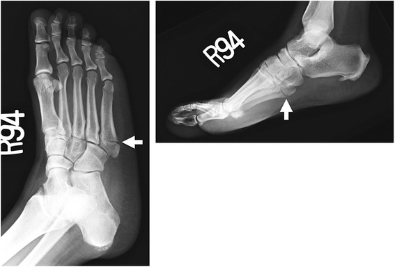
X-rayAnteroposterior and lateral X-rays of the foot: transverse Jones fracture, about 2 cm distal to the base of the fifth metatarsal, arrows on the fracture line.
Source : Wong et al., International journal of emergency medicine, 2015, figure 6 · CC BY
The classification is not an academic exercise here: it is the only thing that distinguishes two opposite kinds of management.
The most widely accepted classification is that of Lawrence and Botte, which divides the proximal portion of the fifth metatarsal according to the location of the fracture line, and not according to its appearance. Bušková et al., in their critical review published by JBJS Reviews, are a reminder that it was indeed the potential for union that guided that division1 :
The three zones of the base of the fifth metatarsal
The decisive landmark is the joint between the fourth and fifth metatarsals: zone 2 extends to it, zone 3 is beyond
Diagram built from the description of the Lawrence and Botte classification as reported by Bušková K, Bartoníček J, Rammelt S, JBJS Rev 2021 (PMID 34673663) and Cheung CN, Lui TH, Arch Trauma Res 2016 (PMID 28144601). The insertions of peroneus brevis and of the lateral band of the plantar fascia are shown after the anatomical study by DeVries JG et al. on 10 cadaveric specimens, J Foot Ankle Surg 2015 (PMID 25441854). The proportions are schematic and are not a measuring template.
The anatomical study by DeVries et al., conducted on ten frozen cadaveric specimens, measured precisely the insertions in the proximal 1.5 cm of the bone. From it they draw a subdivision into three zones A, B and C, whose value is that it ties each site to a mechanism :
This reading by mechanism sheds light on an everyday clinical observation: the fracture most often occurs on a foot in forced inversion, a movement that simultaneously tensions peroneus brevis, which opposes inversion, and the lateral plantar fascia. It is the same movement that produces the lateral ankle sprain, which is why the two injuries are so often confused in practice.
Key points
It is one of the rare occasions, in foot trauma, where an anatomical explanation was demonstrated experimentally before being invoked clinically.
The founding work is that of Smith, Arnoczky and Hersh, published in 1992 in Foot & Ankle. Ten fresh cadaveric or amputation specimens were studied after arterial injection of India ink or barium sulphate. The result comes down to three observations, which must be read in order3 :
The authors conclude that a relative deficiency of blood supply following a proximal diaphyseal fracture may contribute to delayed union and non-union. The term is not in their abstract, but that is the very definition of a watershed zone: a territory served by the far ends of two networks, where each arrives at the limit of its reach.
The vascular watershed of the base of the fifth metatarsal
Two networks, one junction, and a fracture line that falls precisely on it
Built after Smith JW, Arnoczky SP, Hersh A. The intraosseous blood supply of the fifth metatarsal: implications for proximal fracture healing. Foot Ankle 1992;13(3):143-52 (PMID 1601342), a study on 10 cadaveric specimens after arterial injection. The diagram illustrates the authors' description; it does not reproduce a plate from their paper.
It would be convenient to stop the reasoning here. Honesty requires it to be pointed out that the vascular argument has never been linked directly to a measured non-union rate : Smith describes an anatomy, the clinic observes failures, and matching the two remains a hypothesis, a solid, coherent one, but not tested as such.
That matters all the more because the systematic review by Polzer et al. disputes, on prospective data, the clinical description usually drawn from that anatomy. We come to it in the treatment chapter.
Key points
Two very different populations share the same bone: the woman over forty who twists her foot, and the pivoting athlete.
Four figures that place fifth metatarsal fractures
Data from two population series and a critical review
Sources: Bušková K et al., JBJS Rev 2021, for the proportion of foot fractures (PMID 34673663), Petrisor BA, Ekrol I, Court-Brown C, Foot Ankle Int 2006, 411 metatarsal fractures (PMID 16539897), Kane JM et al., Foot Ankle Spec 2015, 1,275 fifth metatarsal fractures (PMID 25666689); Schwagten K et al., Foot Ankle Surg 2021 (PMID 33229215).
One clarification first: out of 411 metatarsal fractures recorded in one year in a Scottish series, of mean age 42, the fifth metatarsal is the most often fractured of all, with an increasing proportion of women in the higher age bands20. It is this bone that is seen, and it is at its base that the prognostic distinction is played out.
The series by Kane et al., which reviewed 1,275 fifth metatarsal fractures in a multicentre orthopaedic practice, delivers a result directly usable in the clinic: the injury mechanism predicts the location. Twisting injuries are a statistically significant predictor of zone 1 injuries. Women sustain 75 % of zone 1 injuries and 84 % of dancer's fractures. In young patients, men predominate; in older patients, it is women19.
That age-related swing is not trivial. Schwagten et al., who studied the dancer's fracture specifically (a spiral fracture of the distal shaft, distinct from the three proximal zones), find that in the group over 40, all patients had a low-energy mechanism, against only 27 % in those under 40. From that they draw a strong clinical conclusion: treat this fracture as a fragility fracture17.
A low-energy 5th metatarsal fracture after the age of 50 is a bone signal
An ordinary twist that breaks a bone in a patient over fifty deserves a question about bone quality, as with any fragility fracture. That reasoning is developed in our article on osteoporosis and the prevention of fragility fractures. The physiotherapist who receives the patient for return to weight bearing is often the first clinician to have the time needed to ask the question.
The true Jones fracture, the zone 2 one, preferentially affects the pivoting athlete: football, basketball, American football. Attia's meta-analysis, which includes only studies covering athletes exclusively, brought together 646 Jones fractures from 22 studies, with return-to-sport rates detailed by discipline: 99.0 % in American football, 96.6 % in football, 91.1 % in basketball5.
That gap between disciplines, modest but consistent, draws out the logic of the injury: basketball, with its pivoting on a foot planted on the floor and its landings from jumps, loads the lateral border of the foot harder than running in a straight line.
Key points
This is the central point of this article. The two fractures look alike on a poorly read radiograph, and what you do differs completely.
One question alone decides it: does the fracture line extend to the joint between the fourth and fifth metatarsals? If yes, it is a zone 2: a Jones fracture. If the line stays proximal to it, confined to the tuberosity, it is a zone 12.
Two errors are made in opposite directions, and they must be named:
| Criterion | Zone 1: avulsion | Zone 2: Jones fracture | Zone 3: diaphyseal |
|---|---|---|---|
| Site of the fracture line | Tuberosity (styloid) | Metaphyseal-diaphyseal junction, extends to the 4th-5th joint | Proximal diaphysis, beyond the joint |
| Dominant mechanism | Twisting, inversion: a statistical predictor19 | Adduction load on a fixed forefoot | Repeated loading: stress fracture |
| Background | 75 % women19, all ages | Pivoting athlete, young man | Endurance or loading athlete |
| Prodromal pain | Absent | Sometimes | Frequent, suggestive |
| Prognosis without surgery | Excellent, even when displaced or intra-articular4 | Union 71.4 % in athletes5 ; 76 % in systematic review6 | The worst: significantly higher failure without surgery4 |
| Usual approach | Functional treatment, weight bearing allowed4 | Functional treatment possible; screw fixation from the outset in athletes15 | Early screw fixation recommended4 |
Three images can be mistaken for a fracture, and a fourth for a zone 1 when it is something else:
This is an accessory ossicle at the lateral border of the foot, immediately adjacent to the base of the fifth metatarsal. The most recent meta-analysis of it, covering 21,312 feet from 22 studies, puts the pooled prevalence at 0.6 % (95 % CI 0.4-0.9), with a comparable distribution between the sexes and a prevalence of 0.6 % on radiographic examinations. The authors conclude explicitly that it must be differentiated from fifth metatarsal fractures and from Iselin's disease15.
What distinguishes it: smooth, corticated margins, a constant position, and above all, when doubt persists, its usual presence on the opposite foot, which a comparative film reveals.
In the adolescent, the secondary ossification centre of the tuberosity appears on the radiograph as a separate fragment. It runs parallel to the axis of the metatarsal, whereas a zone 1 fracture line is usually transverse, that is, perpendicular to that axis. That difference in orientation is the simplest reading criterion. The subject is developed in our article on growth conditions of the lower limb in children and adolescents.
This is not a trap of interpretation but a neighbouring entity, often confused in everyday language. It is a spiral, oblique fracture of the distal shaft, therefore far further out than the three proximal zones. O'Malley, Hamilton and Munyak described the original series of 35 dancers from two national ballet companies: the line starts distal-lateral and runs proximal-medial. Treatment was mainly non-operative, including for displaced fractures, with pain-free walking at a mean of 6.1 weeks, return to the barre at 11.6 weeks and return to performance at 19 weeks. All the dancers returned to their professional activity without limitation18.
The distinction matters because the prognosis is the opposite of that of zone 2: the dancer's fracture is a fracture with a good prognosis, whatever its displacement.
The error that costs the most
Pain along the lateral border of the foot after an inversion is labelled “ankle sprain” in a great many cases. The mechanism is the same, the pain sometimes sits in the same place, and the examination concentrates on the anterior talofibular ligament. Palpation of the base of the fifth metatarsal is part of the Ottawa rules for the foot, and it is precisely by that route that the case report published in JOSPT led a direct-access physiotherapist to request imaging that revealed a fracture27. Our article on the lateral ankle sprain sets out that approach.
Key points
Two different clinical histories can produce the same image, and they do not call for the same strategy.
The classic Jones fracture is acute: a step, immediate pain, inability to bear weight. The stress fracture of the fifth metatarsal is something else entirely: prodromal pain along the lateral border of the foot, increasing over weeks, which ends by completing itself, sometimes abruptly, on an ordinary step that gives the illusion of trauma.
This is the distinction Polzer makes: zone 3 fractures are explicitly described as “diaphyseal stress fractures”, at the distal limit of the intermetatarsal joint or just beyond, and it is they that show a significantly higher failure rate when treated without surgery in a non-weight-bearing boot4.
Our article devoted to stress fracture in the athlete deals with the general mechanism, the imbalance between microdamage and bone repair under cyclical loading, with imaging and with relative energy deficiency. This article does not repeat it : it deals with one particular site, where the acute fracture and the stress fracture coexist and get confused, and where the site of the fracture line governs the prognosis more than the mechanism does.
The question to ask is simple and rarely asked: “Did it hurt there before?” A positive answer changes three things:
Read the odds ratio of 23.3 with caution
This is a case-control study covering 37 athletes in total, of evidence level III. An odds ratio of that size in such a small sample necessarily has a wide confidence interval, and the association measured does not establish causality: an athlete in energy deficiency readily combines vitamin D deficiency, excessive load and low bone density. What the result justifies: measuring vitamin D in a fifth metatarsal stress fracture. What it does not justify: promising that supplementation will prevent recurrence.
Key points
This is the question where the literature frankly contradicts itself, and where the contradiction itself is instructive.
Three pieces of work converge, with different methodologies. The review published by the Journal of the American Academy of Orthopaedic Surgeons sums up the current position: the Jones fracture, sitting at the metaphyseal-diaphyseal junction, carries an increased risk of non-union and persistent pain, and delayed union and refracture can occur despite excellent surgical technique and postoperative management16.
Attia 2021, American Journal of Sports Medicine. A meta-analysis conducted according to PRISMA, 22 eligible studies, 646 Jones fractures exclusively in athletes. The results:
The authors recommend surgical fixation for all Jones fractures in athletes5.
Roche and Calder 2013, KSSTA. A systematic review of 26 studies, of which 22 were level 4 and only one a randomised trial. Union of acute fractures: 76 % non-operatively, 96 % after screw fixation. For delayed unions: 44 % against 97 %. For screw-fixed non-unions: 97 %. Return to sport after screw fixation of an acute fracture: 4 to 18 weeks. The authors explicitly advise against returning before complete radiological union, because of the risk of refracture6.
Yates 2015, The Foot. A meta-analysis of 6 studies and 237 patients: the non-operative group had significantly higher odds of non-union, OR 5.74 (95 % CI 2.65-12.40; p < 0.001), with a longer time to union and to return to sport7.
Polzer et al. published in Injury a systematic review whose conclusion directly contradicts the three-zone reading. Their method is explicit: include only prospective trials comparing either the same treatment for different fractures, or different treatments for the same fracture. Six prospective trials met that criterion. Their conclusions4 :
The authors therefore propose reducing the classification to two entities : metaphyseal fractures not going beyond the distal end of the intermetatarsal joint, to be treated functionally whatever the number of fragments, the displacement and the joint involvement; and metadiaphyseal fractures sitting at that limit or just beyond, which require early screw fixation.
How to hold both together without picking a side
The contradiction is less radical than it looks, and it resolves through the population and through the outcome measure.
Attia speaks only of athletes, and his primary outcome is return to sport and its timing. Polzer speaks of the general population treated in prospective trials, and his outcome is union. A sedentary patient who unites in fourteen weeks instead of nine has been well treated; a professional footballer in the same situation has missed half a season, with a probability of returning to play twenty-seven points lower.
A second factor separates them: the surgical series often cover complete, displaced fractures, whereas the prospective trials of functional treatment readily include incomplete lines. It is not the same fracture.
The honest reading is therefore not “Attia is right and Polzer is wrong”, but: the stronger the requirement on timing, the more surgery is justified; the more distal the fracture line, the more it becomes necessary whatever the patient.
Union and return to sport, surgery against non-operative treatment
Figures from two syntheses covering sporting populations, not transferable to the sedentary patient
Source: Attia AK, Taha T, Kong G, Alhammoud A, Mahmoud K, Myerson M. Am J Sports Med 2021;49(12):3422-3436, meta-analysis of 22 studies and 646 Jones fractures in athletes (PMID 33740393). An exclusively sporting population: these values do not apply to the sedentary patient, in whom Polzer's 2012 review finds good results with functional treatment (PMID 22465516).
A fourth synthesis, not restricted to athletes, points the same way without having the same force: Wang's meta-analysis, on 11 papers and 404 participants of mean age 29.8, finds a medium to large favourable effect of surgery on the non-union rate, the time to union and the time to resuming activity22. Its value is that it covers a less selected population; its limitation is that it mixes the zones.
The comparison by Valkier et al. covers 51 feet with an avulsion fracture, of which 31 treated without surgery and 20 operated on. An unexpected and instructive result: 11 of the 31 non-operated patients (35.5 %) developed a non-union, against none of the operated ones (p = 0.004). But (and this is the second half of the sentence, which people often forget to quote), all the patients were asymptomatic at one year, whichever group they were in. The authors recommend surgery for avulsions displaced by more than 2 mm, and insist on realistic expectations13.
It is a useful reminder: a radiological non-union is not a clinical non-union. On the tuberosity, a fragment that never fuses can remain perfectly painless. A Chinese randomised trial in 46 young or sporting patients with an avulsion displaced by 2 to 3 mm points the same way: the functional score was better at 6 months in the operated group, but no longer differed at 12 months ; only the time to full weight bearing and to returning to work were significantly shortened by surgery25.
Treatment strategies and level of evidence
An appraisal of the level of evidence following the GRADE principles, from the sources cited in this chapter
Intramedullary screw fixation from the outset for a zone 2 fracture in an athlete : union, rate and timing of return to sport all superior, across 646 fractures and 22 studies, with three concordant direct comparisons5, corroborated by two other syntheses67.
Functional treatment of zone 1 avulsions, including displaced and intra-articular ones : six concordant prospective trials; a plaster boot significantly delays return to the previous level4.
Early screw fixation of zone 3 stress fractures : significantly higher non-operative failure rate, shorter time to union and to return, across few prospective trials4.
Lateral off-loading insole after a zone 2 fracture on a varus hindfoot : 100 % union and no refracture in a series of 21 fractures, but with no control group8.
Very early return after screw fixation, before radiological union : a series of 26 fractures in 25 collegiate athletes, mean return at 3.6 weeks with no major complication23, but a systematic review explicitly advises against this practice6.
Treating a zone 2 as a zone 1 : that is, allowing free weight bearing without monitoring union. No source consulted supports this course, which arises from a reading error and not from a therapeutic choice.
An appraisal built following the GRADE principles from the sources cited, and not taken from a published GRADE assessment: none of the reviews consulted produced a formal GRADE table on this question.
Key points
Three families of factors, of which two can be corrected, and that is where the physiotherapist has purchase.
Raikin, Slenker and Ratigan measured hindfoot alignment in 20 patients operated on for a Jones fracture. The result is clear-cut: 18 of the 21 hindfeet were in radiographic varus, and clinical varus was present in 16 cases. The mean calcaneal pitch angle was 28.5° and Meary's angle 13° with an upward convexity. All the patients united and none refractured, at a mean follow-up of 49 months: the patients in varus having been fitted postoperatively with a lateral off-loading insole for the hindfoot and the forefoot8.
The authors' hypothesis is mechanically clear: a varus hindfoot overloads the lateral column of the foot, predisposes to the fracture, and predisposes to treatment failure.
A recent meta-analysis seriously tempers that reading. Riegger et al. pooled eight studies (296 patients), five of them in quantitative synthesis (132 patients), to compare three angles between fractured patients and controls. Result: only the midfoot adduction angle stands out, with a pooled mean difference of 4.62° (95 % CI 1.31-7.92) and considerable heterogeneity (I² = 76.1 %). Neither calcaneal pitch nor Meary's angle differs significantly. In other words: midfoot adductus seems correlated with risk, but the relationship with hindfoot varus and pes cavus has not been demonstrated9.
Factors associated with the fracture, with non-union and with refracture
Each factor is placed according to the strength of the evidence supporting it, and not according to how often it is cited
Sources: Riegger M et al., J Foot Ankle Surg 2022 (PMID 35039196); Lee KT et al., Foot Ankle Int 2013, refractures (PMID 24216284) and plantar gap (PMID 23637237); Shimasaki Y et al., Foot Ankle Int 2016 (PMID 26596794); Raikin SM et al., Am J Sports Med 2008 (PMID 18443278).
The question often arises in rehabilitation, faced with a patient who arrives with a fracture line still visible months later. The series by Hunt and Anderson covers 21 elite athletes operated on for a revision for non-union or refracture of a Jones fracture, by intramedullary screw fixation combined with autologous cancellous bone graft or with a mixture of bone marrow aspirate and demineralised bone matrix. All regained their previous level of competition, at a mean of 12.3 weeks, with complete clinical and radiographic cortical union, and only one had a further refracture. The authors recommend a solid, large-diameter screw (5.5 mm or more) and an autologous graft21.
The message is useful to pass on to the worried patient: a first-line failure is not a dead end, and revision surgery has good documented results.
This is the item most directly useful to the physiotherapist, and it comes from the series by Lee et al. covering 168 fifth metatarsal stress fractures in elite athletes, treated by modified tension band wiring. Eleven non-unions and eighteen refractures were recorded. The factors associated with refracture were a higher weight and two radiological parameters reflecting protrusion of the fifth metatarsal head (4-5 intermetatarsal angle on the AP view, lateral deviation of the 5th MT on the oblique).
But the most telling observation is chronological: all the refractures occurred after a new injury, once union had been achieved; 13 of the 18 within six months of the start of rehabilitation, and 8 within three months. The authors conclude that patients with a prominent 5th metatarsal head and a high body mass index must approach rehabilitation cautiously before considering a return to their previous sporting level11.
The window of vulnerability does not close at union. It stays open for the six months following the start of rehabilitation, and that is exactly the period when the patient feels cured.
A second series from the same team, covering 86 fifth metatarsal stress fractures, tested a simple radiographic criterion: the presence of a plantar gap of 1 mm or more on incomplete fractures. Result: incomplete fractures with a gap of ≥ 1 mm united in 115.5 ± 45.4 days, against 73.9 ± 26.7 days for those whose gap was smaller (p < 0.001). Counter-intuitively, complete fractures united faster (67.5 ± 28.8 days) than incomplete ones taken as a whole (103.2 ± 47.7 days)12.
This result deserves to be known by the rehabilitation clinician, because it contradicts intuition: faced with two radiographs, the one that looks less serious (an incomplete line, with a thin plantar lucency) may herald a union twice as long.
Key points
With this fracture, rehabilitation is not first of all analytical work: it is the management of a return to weight bearing in a window where the bone is not yet safe.
It has to be said directly: none of the sources consulted describes a validated rehabilitation protocol for the Jones fracture. The studies measure times to union, return-to-sport rates and complications; they do not compare rehabilitation programmes with each other. What follows is therefore built from three solid elements (the measured times to union, the chronology of refractures, and the factors predicting failure) and not from a published protocol.
Markers for return to weight bearing after a fracture of the base of the 5th metatarsal
The boxed timings are measured in the sources cited; the session contents belong to everyday practice
Timings: Attia AK et al., Am J Sports Med 2021 (PMID 33740393) for the zone 2 values; O'Malley MJ et al., Am J Sports Med 1996 (PMID 8775129) for pain-free walking at 6.1 weeks in the dancer's fracture; Lee KT et al., Foot Ankle Int 2013 (PMID 24216284 and PMID 23637237); Raikin SM et al., Am J Sports Med 2008 (PMID 18443278). The zone 2 values come from sporting populations.
This is the main objective, and it is chronological before it is technical. The marker is simple: pain on weight bearing along the lateral border of the foot is the signal, and the progression follows union, not the theoretical timetable. In zone 1, weight bearing is allowed from the outset and functional treatment does better than a plaster boot on the time to return to the previous level4. In zone 2, the question depends on the strategy chosen and must be settled with the surgeon.
The period of immobilisation or non-weight bearing costs ankle mobility and triceps surae strength. Analytical work on dorsiflexion, mobilisation of the midfoot and open-chain strengthening can start early without loading the lateral border. Work on the peroneals, by contrast, requires caution: peroneus brevis inserts on the tuberosity14, and its resisted contraction tensions the zone 1 fracture site. Our article on peroneal tendinopathies sets out the loading progression for those muscles.
Raikin's series provides the only documented item of orthotic care: the patients in varus were fitted postoperatively with a lateral off-loading insole for the hindfoot and forefoot, with 100 % union and no refracture at a mean follow-up of 49 months8. It has to be read for what it is: a series with no control, in which the effect of the insole cannot be separated from that of fixation with a 4.5 mm screw. But the orthosis is cheap and low-risk, which makes it a reasonable option when there is frank clinical varus.
This is the objective the literature supports most directly, and paradoxically the one most often forgotten: vulnerability persists for months after union. An athlete who has returned and reports discomfort along the lateral border is not an athlete who has not warmed up properly: it is an athlete to re-examine.
What should make you stop and refer back
Key points
Two published observations, chosen because they show what meta-analyses cannot: what happens when the patient does not follow the treatment, and what a front-line physiotherapist sees.
Three sumo wrestlers, three interrupted treatments
Hoshino T, Tateishi T, Nagase T, Yuki A, Nakagawa T, Tsuchiya M. Jones Fractures in Sumo Wrestlers: Three Case Reports. Case Reports in Orthopedics 2019;2019:9051327 (PMID 31772802).
The authors report three Jones fractures occurring in sumo wrestlers. All three patients interrupted their treatment on their own initiative24. The two cases treated without surgery progressed to non-union or delayed union; the operated case had a screw breakage. And yet, all three carried on wrestling, with little consequence for their careers.
The authors identify the risk factors particular to this discipline: high body weight, and the training and competition characteristics specific to sumo. Above all they explain why adherence is so difficult there: taking rest means losing your rank, which makes it almost impossible to see out a sufficient duration of treatment.
What the case teaches. Three things, in order. First, weight is a loading factor on the lateral column, which Lee's series on refractures confirms11. Next, a non-union does not necessarily prevent practice, which should make one cautious with catastrophic prognoses. Finally and above all, the best treatment is the one the patient can follow : the authors conclude that it is essential to inform these athletes fully about the options and to decide with each of them. For the physiotherapist, who sees the patient far more often than the surgeon does, that is where adherence is played out.
A 17-year-old tennis player, intermittent lateral pain, direct access
Bittner JS, Hartstein AJ. Fifth Metatarsal Avulsion Fracture in an Adolescent Tennis Player. Journal of Orthopaedic & Sports Physical Therapy 2019;49(8):620 (PMID 31366295).
A 17-year-old tennis player presents to physiotherapy by direct access, with intermittent pain along the lateral border of the left foot. Faced with positive fracture tests and applying the Ottawa foot and ankle rules, the physiotherapist requests an orthopaedic opinion and imaging. The radiographs reveal an undisplaced avulsion fracture of the fifth metatarsal.
What the case teaches. It is the most direct illustration of the physiotherapist's role on this subject, and it comes down to one decision rule: palpation of the base of the fifth metatarsal is part of the Ottawa rules, and exquisite tenderness there after a foot injury justifies imaging. Intermittent pain, in an adolescent, with no frank inability to bear weight, is exactly the picture that does not suggest a fracture, and it is the picture of the published case.
Why these cases and not others
Clinical cases are the classic weak point of review articles: the too-neat story, the athlete cured in eight sessions. The two observations above are published, indexed reports, cited with their PubMed identifier. They were chosen because one documents a real treatment failure, three patients who abandon treatment, and the other a diagnostic process conducted by a physiotherapist. No case in this chapter was composed for the purposes of the demonstration.
What reading the sources changes in practice, for the patient who arrives with a painful foot and a radiograph in their file.
What to do with a fracture of the base of the fifth metatarsal
The first question concerns the site of the fracture line; the second the patient's requirements
Tree built from the Lawrence and Botte classification as reported by Bušková 2021 (PMID 34673663) and Cheung 2016 (PMID 28144601), from the conclusions of Polzer 2012 (PMID 22465516) and from the figures of Attia 2021 (PMID 33740393). It reproduces no published guideline: none of the learned societies consulted has issued a formal recommendation on this fracture.
| Error | Consequence | What avoids it |
|---|---|---|
| Confusing zone 1 and zone 2 | A high-risk fracture treated as a benign avulsion | One question alone: does the line reach the 4th-5th joint?2 |
| Labelling it “ankle sprain” without palpating the base of the 5th | A missed fracture, sometimes for weeks | Palpation is part of the Ottawa rules27 |
| Not asking whether it hurt before | A stress fracture taken for an acute fracture, systemic cause not looked for | One question in the history; measure vitamin D if yes10 |
| Immobilising a zone 1 in a plaster boot | A significant delay in return to the previous level, with no benefit | Functional treatment does better, with prospective trials to support it4 |
| Considering the matter closed at union | Refracture, of which 13 out of 18 within six months of the start of rehabilitation | Extend the monitoring and the loading progression11 |
The decisive act is not therapeutic, it is radiological: look at whether the fracture line reaches the joint between the fourth and the fifth metatarsals.
On the same ground
All Jones fractures are fractures of the base of the fifth metatarsal, but the reverse is not true. The Lawrence and Botte classification distinguishes three zones: the tuberosity (zone 1), the metaphyseal-diaphyseal junction that extends as far as the joint between the 4th and 5th metatarsals (zone 2), and the proximal diaphysis (zone 3)1. Only zone 2 is a Jones fracture in the strict sense. The confusion is frequent because all three sit within a few centimetres of the same bone.
Because zone 2 is a vascular crossroads. Smith's anatomical study on ten injected cadaveric specimens shows that the rich, redundant network of the tuberosity meets the territory of the nutrient artery of the diaphysis exactly in the region of poor prognosis3. A fracture there cuts one supply with no back-up route to take over.
In athletes, the answer from the available syntheses is yes: union reaches 97.3 % after screw fixation against 71.4 % without, with return to sport of 98.8 % against 71.6 % and 3.5 weeks gained5. In the patient with no requirement on timing, the question is open: Polzer's systematic review, which includes only prospective trials, finds good to excellent results with functional treatment for zone 24. The decision is made with the surgeon, according to the level of demand and the exact site of the fracture line.
For a zone 1, weight bearing is allowed from the outset with functional treatment4. For a zone 2 in an athlete, return to sport occurs at a mean of 9.6 weeks after screw fixation and 13.1 weeks without surgery5. These values are averages from sporting populations, and a plantar gap of at least 1 mm on an incomplete fracture lengthens union from 74 to 116 days12. The timetable is set by union, not by the average.
Yes, and it is the most underestimated risk: the refracture rate reaches 10.2 % even after surgery5. In a series of 168 fractures in elite athletes, all the refractures occurred after a new injury on a united bone, and 13 out of 18 within six months of the start of rehabilitation11. That is why the loading progression continues well beyond radiological union.
It is a solid but unconfirmed hypothesis. Raikin found radiographic varus in 18 patients out of 21 operated on for a Jones fracture, and proposes that overloading of the lateral column predisposes to the fracture and to its failure8. But Riegger's meta-analysis, on 8 studies, confirms neither hindfoot varus nor pes cavus: only the midfoot adduction angle stands out, with a mean difference of 4.62°9. The lateral off-loading insole remains a reasonable option when there is frank varus, without its effect being something one can promise.
Perhaps an os vesalianum, an accessory ossicle whose most recent meta-analysis, on 21,312 feet, puts the prevalence at 0.6 %15. It is distinguished by smooth, corticated margins, a constant position, and its usual presence on the other foot: which a comparative film reveals. In the adolescent, it may also be the growth apophysis, running parallel to the axis of the bone, whereas a zone 1 fracture is transverse.
No, and the prognosis is in fact the opposite. It is a spiral, oblique fracture of the distal shaft of the fifth metatarsal, well beyond the three proximal zones. The original series on 35 professional dancers reports mainly non-operative treatment, including for displaced fractures, with pain-free walking at a mean of 6.1 weeks and return to performance at 19 weeks; all the dancers returned without limitation18. It accounts for 25 % of fifth metatarsal fractures, of which 80 % in women17.
Twenty-seven references, all verified individually on PubMed: identifier resolved, title, journal, year and author list checked, abstract read before citation. The links point to the PubMed record.
A note on method
The bibliographic base was built before writing, by searching PubMed through the E-utilities interface: every identifier was resolved, every record checked for its authors, journal and year, and every abstract read before being cited. The figures put forward carry their source at the exact place where they are written, and the populations they come from are specified where these limit their scope: most of the union and return-to-sport figures come from exclusively sporting series. One major contradiction in the literature, between Polzer's review and the surgical meta-analyses, is set out as it stands rather than settled. Finally, the absence of a validated rehabilitation protocol for this fracture is stated explicitly, the markers proposed being deduced from the measured times to union and from the chronology of refractures. Article written on 15 August 2026.
Behind this article
An author who explains, a reviewer who checks.
How we write and check our content

Physiotherapist · co-founder of Physio Learning
Marked for life by his first four-hour lecture without a single image, he took a master’s in instructional design so that it would never happen to anyone again. He hunts down publication bias and unreadable slides with the same intransigence.

Head of scientific content
Physiotherapist specialising in neuro-musculoskeletal practice and holder of a master’s in public health. He checks the methodological rigour of every article: primary sources, levels of evidence, no exceptions.
