














Online

Lower limb
Achilles tendinopathy and rupture
Anthony Baillon
Go further
Haglund's disease: a bony prominence at the back of the heel, an inflamed bursa and Achilles enthesopathy that often coexist. Tests and treatment.

Haglund syndrome is a triad: exostosis, retrocalcaneal bursitis and insertional tendinosis; the deformity alone is not the syndrome.
23.3%of radiographed heels show an insertional calcification, but only 3% hurt
What follows covers each of these points in detail, with sources. It is there if you need it.
This topic is taught in a course: Achilles tendinopathy and rupture, online with Anthony Baillon.
1 / 13
12 article chapters · 56 min in total
Understanding◔ 6 min
The retrocalcaneal bursa is a full-fledged enthesis organ, not a mere accidental pouch.
Identifying the background◔ 4 min
The metabolic background weighs more heavily than sport: the gap between image and pain is one to eight.
Differentiating◔ 7 min
Three lesions, three topographies: precise palpation, within a few millimetres, points to the diagnosis.
Reading the X-ray◔ 7 min
The size of the deformity does not distinguish painful patients from asymptomatic controls.
Advanced imaging◔ 2 min
Imaging is requested only if its answer changes management, never as a matter of routine.
Footwear◔ 3 min
Footwear is the first treatment: the heel counter mechanically compresses the prominence, before any exercise.
Loading the tendon◔ 8 min
At the tendon body you seek range; at the insertion you seek load, and you give up range.
Adjuncts◔ 5 min
Between 2013 and 2025, the evidence on shockwaves clearly reversed.
Operating◔ 4 min
Two independent studies suggest that resecting the bump may not be necessary.
Case studies◔ 4 min
A Haglund deformity can act as a chronic wear mechanism that sets the stage for rupture.
In practice◔ 2 min
Three minutes of ordered palpation are worth more than first-line imaging.
FAQ◔ 4 min
Haglund's disease and retrocalcaneal bursitis are not the same thing, but often go together.
Train on this
Lower limb
Anthony Baillon
Next comes practice: the course that teaches this topic, with Anthony Baillon.
Our courses on this topic may be covered by DPC or FIFPL, the French continuing-education funding schemes, depending on your profession and situation.
At the back of the heel, three things hurt and often coexist in the same patient: a bony prominence rubbing against the shoe, an inflamed bursa, and the insertion of the Achilles tendon. What a consultation really delivers is not naming the deformity on a radiograph, it is saying which of the three hurts. Because impingement against the shoe is treated first by the shoe, and the other two are not.
Summary written from primary sources verified one by one on PubMed: identifier resolved, journal, year and author list checked, abstract read before citation. Full bibliography and methodological note at the end of the article.
Posterior heel pain in three awkward figures
Each of these three findings contradicts a received idea that is still taught
Sources: Giai Via A et al., Clin J Sport Med 2022 (PMID 32976122); Kang S et al., Foot Ankle Int 2012 (PMID 22735321); Jonsson P et al., Br J Sports Med 2008 (PMID 18184750). The figure of 32 % is the rate of good results reported by Jonsson for the classic eccentric protocol through full dorsiflexion, and that of 67 % the rate for his protocol without loading into dorsiflexion.
The essentials in ten lines
This page and its neighbour
This article deals with posterior heel pain from impingement : what happens between the posterosuperior surface of the calcaneus, the retrocalcaneal bursa, the tendon insertion and the heel counter of the shoe. For midportion Achilles tendinopathy, 2 to 7 cm above the insertion, which stems from a different pathophysiology and a different rehabilitation, see Achilles tendinopathy. The two pages are read together: confusion between the two is the rule in the clinic, and that is precisely what this chapter sets out to undo.
In this chapter: the radiological triad that defines the syndrome, the enthesis organ of which the bursa is an integral part, the mechanical role of Kager's fat pad, and the two impingements that overlap at the same point of the heel.
The Haglund deformity refers to an exostosis of the posterosuperior angle of the calcaneus. Taken alone, it is only a bone shape. The term Haglund syndrome means something else: the review in Radiographics in 2024 defines it as the characteristic triad combining the posterosuperior exostosis, the retrocalcaneal bursitis and the insertional tendinosis of the Achilles tendon1. Three lesions, three tissues, a single square centimetre of heel.
This distinction is not semantics. It governs the whole of clinical reasoning: a patient can carry the deformity without either of the other two lesions, and will be asymptomatic; they can carry all three, and it will still be necessary to decide which to treat first. The rest of this article returns to this point several times because that is where most of the value delivered is decided.
Four markers to place the problem
Frequency of Achilles tendon involvement, the share attributed to sport, and the weight of calcification
Sources: Traweger A et al., Nat Rev Dis Primers 2025 (PMID 40148342); de Jonge S et al., Br J Sports Med 2011 (PMID 21926076); Kang S et al., Foot Ankle Int 2012 (PMID 22735321). The first two figures cover Achilles tendon disorders as a whole, insertion and midportion together; the third covers the 48 painful heels in Kang's series.
The concept that makes the picture intelligible is that of the enthesis organ. The developmental histological study published in the Journal of Anatomy in 2008 describes the insertion of the Achilles tendon as a functional whole that does not reduce to the attachment point: it includes the enthesis proper, two opposing fibrocartilages, one sesamoid on the tendon and the other periosteal on the bone, the retrocalcaneal bursa and Kager's fat pad3. The authors add that the crural and plantar fasciae take part in dissipating stress and could be regarded as components of this whole.
In other words, the retrocalcaneal bursa is not a parasitic pouch that happened to settle there by bad luck. It is a part of the apparatus that lets the tendon turn around the bone without wearing out. When it becomes inflamed, it is not a surplus tissue that suffers, it is the organ that malfunctions.
The anatomy of retrocalcaneal impingement
Schematic sagittal section: two distinct compressions act at the same place
Teaching diagram, proportions not anatomical. The relationships shown are established from Theobald P et al., J Anat 2006 (PMID 16420382), Shaw HM et al., J Anat 2008 (PMID 19094187) and Malagelada F et al., Knee Surg Sports Traumatol Arthrosc 2020 (PMID 31256217).
The combined MRI, ultrasound, gross anatomy and histology study by Theobald et al. shows that Kager's fat pad has three regions with distinct functions: a part bound to the tendon of flexor hallucis longus, a part bound to the Achilles tendon that protects the vessels entering the tendon, and a bursal wedge adjacent to the calcaneus2. This wedge is mobile: it enters the bursa during plantarflexion and withdraws during dorsiflexion. The authors conclude that it minimises pressure variations inside the bursa.
The cadaveric work published in Knee Surgery, Sports Traumatology, Arthroscopy in 2020 measured these pressures directly, on twelve ankles mounted on a rig with the tendon loaded and a needle sensor positioned under ultrasound guidance4. The central result: pressures rise significantly at both extremes, in terminal plantarflexion as in terminal dorsiflexion, and in both regions explored. The fat pad adheres to the tendon over a mean length of 7.7 cm, that is 89 % of its length; only its distal end, over 0.92 cm, separates from it to make room for the bursa.
The tendon does not suffer only from what pulls on it. At its insertion, it also suffers from what it is crushed against.
This is the decisive pathophysiological difference from the midportion of the tendon. At 2 to 7 cm above the insertion, the dominant stresses are stresses of tension. At the insertion, a stress of compression is added to it, between the tendon and the posterosuperior angle of the calcaneus, and it grows with dorsiflexion. The whole specificity of the treatment follows from this, and it is the subject of the chapter devoted to exercise.
An anatomical finding whose therapeutic consequences are direct: the cadaveric study by Pękala et al., published in Bone & Joint Research in 2017, injected ink and then a contrast medium into the retrocalcaneal bursa of twenty fresh-frozen tendons5. In the ink-injected specimens, diffusion outside the bursa was observed, with staining of the anterior portion of the tendon. Among the contrast-injected specimens, eight showed the same pattern of localised diffusion, and two, injected without ultrasound guidance, a more extensive diffusion.
The authors conclude that connections exist between the bursa and the tendon, particularly rich in its anteroinferior portion, and propose to regard this zone as a zone of weakness for substances injected into the bursa. They put forward the hypothesis, and it must be cited as such, that this part of the tendon could be the most vulnerable to rupture after corticosteroid injection. This question comes back in the chapter on injections.
Key points
In this chapter: what the available incidence figures are worth and what they do not cover, the real weight of sport against that of the metabolic background, and the considerable gap between radiological calcification and pain.
We must start with a point of methodological honesty: there are no incidence data specific to Haglund's disease or to retrocalcaneal bursitis in the general population. The available figures cover Achilles tendon disease as a whole, or specifically the midportion of the tendon.
The review in Nature Reviews Disease Primers devoted to Achilles tendinopathy in 2025 gives the order of magnitude: Achilles tendon disorders affect about 6 % of the general population and up to 50 % of elite endurance runners over their lifetime6.
In primary care, the Dutch study by de Jonge et al. counted, in a cohort of 57,725 people registered in primary care, the patients consulting for a disorder of the midportion of the Achilles tendon: the incidence is 1.85 per 1,000 registered patients, and 2.35 per 1,000 in adults aged 21 to 607. A figure to keep in mind for the consultation: only 35 % of cases were linked to a sporting activity. The authors note that the true incidence could be higher given the limits of their method.
The cross-sectional study by Albers et al. in a Dutch general practice population gives, for lower limb tendinopathies as a whole, a prevalence of 11.83 and an incidence of 10.52 per 1,000 person-years, with a clear predominance in older patients8.
Frequency in primary care
Beware the denominators: they are not identical from one study to the next
Sources: Albers IS et al., BMC Musculoskelet Disord 2016 (PMID 26759254) for the first two bars, expressed per 1,000 person-years; de Jonge S et al., Br J Sports Med 2011 (PMID 21926076) for the last two, expressed per 1,000 patients registered in primary care. The denominators differ: the bars are read as orders of magnitude, not as a direct comparison. None of these series isolates posterior heel pain from impingement.
This is the contribution of the prospective epidemiological survey by Giai Via et al., published in Clinical Journal of Sport Medicine in 2022, and it is probably the most useful piece of data in this chapter9. The authors analysed the foot and ankle radiographs of 433 subjects attending orthopaedics or the emergency department, recording age, sex, body mass index and comorbidities.
Of these 433 subjects, 101, that is 23.3 %, had an insertional calcification of the Achilles tendon on the radiograph. And only 13, that is 3 %, were symptomatic. The gap between the image and the disease is therefore of the order of one to eight: that is the figure to have in mind before opening a radiology report in front of a patient.
The multivariate analysis finds four independent predictors of calcification: age, with an odds ratio of 1.05 per year, diabetes (OR 2.95), hypercholesterolaemia (OR 2.27) and hypothyroidism (OR 3.32). A body mass index above 30 is associated with a higher frequency of calcifications, and patients accumulating two or more comorbidities have a more than tenfold higher risk.
Metabolic background and insertional calcification
Adjusted odds ratios for the presence of an insertional calcification of the Achilles tendon
Source: Giai Via A, Oliva F, Padulo J, Oliva G, Maffulli N. Clin J Sport Med 2022;32(1):e68-e73 (PMID 32976122). A level III prospective observational study of 433 subjects. The confidence intervals are not reported in the indexed abstract and are therefore not shown here. The odds ratio for age is per additional year.
Key points
In this chapter: the differential table of the three causes of posterior heel pain, the palpation topography that separates them, a decision tree, the diagnoses not to be missed, and the validated French-language measurement tool.
Clinical discrimination rests first on a single manoeuvre: exactly where the finger reproduces the pain. The three lesions of the triad occupy three different planes a few millimetres apart, and that proximity is the only real difficulty of the examination.
A fourth marker serves as a control: pain in the midportion of the tendon lies 2 to 7 cm above the insertion, on a fusiform thickening. If that is where it hurts, the patient falls under the article on Achilles tendinopathy and not this one.
| Criterion | Haglund impingement | Retrocalcaneal bursitis | Insertional tendinopathy | Midportion of the tendon (neighbouring page) |
|---|---|---|---|---|
| Site of the pain | Posterosuperior and often lateral, on the bony bump | Deep, in front of the tendon, above the upper border of the calcaneus | At the bony attachment, posterior surface of the calcaneus | 2 to 7 cm above the insertion |
| Palpation that reproduces it | Direct pressure on the prominence | Medial and lateral pinch, pressure forwards, pain on both sides | Pressure on the enthesis, tendon relaxed | Pinching the midportion of the tendon, fusiform thickening |
| Inspection | Visible bump, redness, hyperkeratosis where the heel counter touches | Filling of the para-Achilles grooves, a full appearance | Thickening of the heel with a bony appearance, sometimes a palpable calcification | Fusiform swelling that moves with the tendon |
| What makes it worse | One particular shoe, a rigid heel counter, a new model | Forced dorsiflexion, squatting, going downhill | Starting after rest, going uphill, heel loading, uphill running | Starting after rest, running, jumping |
| What relieves it | Backless shoes, bare feet | Avoiding extreme dorsiflexion | A heel lift, flat ground, avoiding step drops | Warming up, progressive loading through full range |
| Passive dorsiflexion | Barely painful once the shoe is removed | Painful, it is the most discriminating test | Painful at end of range | Usually painless |
| Useful imaging | Weight-bearing lateral radiograph, for the shape | Ultrasound or MRI, for the effusion | Radiograph for the calcification, MRI before surgery | Ultrasound, but imaging is not required for the diagnosis |
| First-line treatment | Modify the footwear | Unload dorsiflexion, then progressive loading | Progressive loading with limited range | Progressive loading through full range |
| Classic pitfall | Treating a footwear problem with exercise | Injecting a bursa that communicates with the tendon | Applying the midportion protocol | Limiting range without reason |
The most useful row of this table is the second to last. Haglund impingement is the only one of the three causes whose first-line treatment is not an exercise. It is also the one where the mistake costs most, because a patient prescribed eight weeks of strengthening while every morning putting back on the shoe that hurts them will not improve, and will conclude that physiotherapy does not work.
Decision tree for pain at the back of the heel
The first question is not radiological, it is topographical
A teaching proposal by the author, built from the topographies described by Flores DV et al., Radiographics 2024 (PMID 38512730) and Chen J et al., J Am Acad Orthop Surg 2022 (PMID 35286285). This tree is not a validated algorithm and has been the subject of no diagnostic performance study.
Red flags in posterior heel pain
Two distinct presentations come with posterior ankle pain and do not belong to the triad.
The posterior ankle impingement comes first. The pain there is deeper and more anterior, provoked by forced plantarflexion and not by dorsiflexion, which is often enough to decide. The prevalence meta-analysis by Ráfare et al., covering 18 studies and 17,626 ankles, puts the prevalence of the os trigonum at 10.3 % (95 % CI: 7 to 14.1 %), with no difference by sex or side28. In other words, an os trigonum is found in one ankle in ten: seeing it on a radiograph does not prove it is responsible, exactly as for the Haglund deformity.
The calcaneal apophysitis comes next, or Sever's disease, in children and adolescents. The systematic review by Nieto-Gil et al., published in BMJ Open in 2023, brings together 11 observational studies and 1,265 participants of mean age 10.72 years30. The most studied intrinsic factor there is the limitation of ankle dorsiflexion, ahead of plantar pressure peaks and foot alignment disorders. The authors honestly stress the disagreement between studies over what counts as a risk factor, an associated factor or a consequence. Faced with a painful heel in a patient under 15, it is this diagnosis that must be considered first, not a Haglund.
The VISA-A questionnaire remains the reference tool for clinical severity in Achilles tendon disease. Its initial validation, published by Robinson et al. in the British Journal of Sports Medicine in 2001, established its validity and reliability31 A dedicated French version was translated and validated by Kaux's team in Liège in 201632 : it is the one to use in French-speaking practice, rather than a translation improvised during a session.
Two caveats of use. The VISA-A was built and validated for Achilles tendinopathy in the broad sense, without distinguishing insertion from midportion: it therefore measures a patient's progress correctly, but it does not say which of the three lesions is improving. And it does not replace a numerical rating scale of pain on the precise movement that bothers the patient, which remains the best guide to progression week after week.
Key points
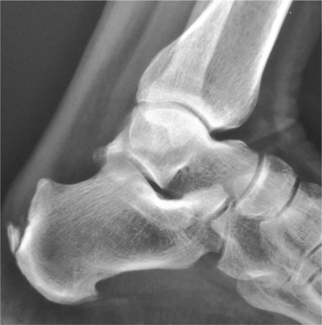
X-rayLateral radiograph of the calcaneus: posterosuperior bony prominence characteristic of Haglund deformity, preoperative.
Source : Natarajan et al., Malaysian Orthopaedic Journal, 2015, figure 1 · CC BY
In this chapter: the inventory of radiographic measurements proposed, what two independent case-control studies found when applying them, why good reproducibility does not make good diagnostic value, and what the radiograph really contributes.
The literature has produced an unusual number of indices to quantify the Haglund deformity. The paper by Nischal et al. published in Skeletal Radiology in 2023 lists them: Fowler-Philip angle, Ruch pitch, Chauveaux-Liet angle, calcaneal pitch angle, parallel pitch lines and X/Y ratio12. And the authors add straight away, in their introduction, that most of these indices lack specificity and have variable intra- and inter-observer reproducibility.
A French and Irish surgical team goes further. In their series of fifty Zadek osteotomies published in Foot and Ankle Surgery in 2022, Tourné et al. write that they measured only the X/Y ratio and the calcaneal inclination angle, because the radiographic measurements classically described in Haglund syndrome are not reliable13. When the surgeons who operate on the deformity give up the measurements that define it, the clinician who receives the report may allow themselves the same caution.
The most important result in this chapter was published in 2012 and has never been contradicted since. Kang, Thordarson and Charlton compared, in Foot & Ankle International, 48 heels from patients with insertional tendinopathy with 50 control heels without tendinopathy, on weight-bearing lateral radiographs10. They measured the height and the peak angle of the deformity, the Böhler angle, the Fowler-Philip angle and the parallel pitch lines sign.
No difference reaches statistical significance. The mean height of the deformity is 9.6 mm in the patients against 9.0 mm in the controls; the peak angle is 105 degrees in both groups ; the Böhler angle and the Fowler-Philip angle are comparable. And the result that overturns intuition: the parallel pitch lines sign is positive in 60 % of the controls against 41.7 % of the painful patients. The sign is therefore more frequent in people who hurt nowhere.
Eleven years later, Lee, Giro and Crymes took the question up again with a new measurement system, on 55 feet matched for age and sex with 55 controls11. Their system shows excellent intra- and inter-observer reproducibility. And it too finds no difference: a deformity angle of 6.0 degrees in both groups, a height of 3.3 mm against 3.2 mm.
Painful heels against control heels: what radiographs really measure
Five measurements, two independent studies, no significant difference
Each panel is scaled to its own unit: bar heights are only comparable within a panel. Sources: Kang S, Thordarson DB, Charlton TP. Foot Ankle Int 2012;33(6):487-491 (PMID 22735321), 48 painful heels against 50 controls. Lee W, Giro ME, Crymes C. Foot Ankle Int 2023;44(8):719-726 (PMID 37218117), 55 feet against 55 controls matched for age and sex. None of the differences shown reaches the threshold of statistical significance in the original study.
The same study by Lee et al. answers this, and that is its main contribution. The multivariate logistic regression analysis identifies three independent factors, none of which is the size of the deformity11 :
The confidence interval of the odds ratio for calcification is very wide, which reflects a modest sample and requires the point estimate to be read as an order of magnitude and not as a measure of precision. But its lower bound remains at 11.23: the association is solid in its direction, even if its magnitude is poorly estimated. Kang et al. moreover observed an insertional calcification in 73 % of their patients with pain10.
The radiograph does not say whether the heel hurts. It says whether there is calcium in the tendon, and that is useful.
A figure this article does not use
The indexed abstract of Lee et al. reports, for the calcaneal pitch angle, the comparison “5.2 against 23.1 degrees” while describing the diseased group as having the higher angle. The two numbers are incompatible with the sentence that introduces them, and an order of magnitude of 5.2 degrees is incompatible with the usual definition of this angle. This value is therefore not used here : only the odds ratio of 6.32, consistent with the rest of the analysis, is cited. Flagging an inconsistency in a source rather than copying it out is part of the verification work.
A word is needed on the BRINK angle, proposed by Nischal et al. in 2023 as a new, more reliable measurement12. The study compares 20 radiographs of ankles carrying a deformity with 100 normal radiographs. The mean angle is 20.04 degrees (standard deviation 4.88) in the normal cohort and 25.1 degrees (standard deviation 3.3) in the Haglund cohort, with a p below 0.0001 and excellent reproducibility, kappa at 0.8.
Two remarks are called for, and they do not diminish the merit of the work. First, a difference in means of five degrees with standard deviations of 4.88 and 3.3 means that the two distributions overlap widely : a threshold placed between the two means would misclassify a large share of the subjects in each group. A significant difference between two means is not an ability to decide about an individual, and that is exactly the confusion this kind of measurement encourages.
Second, the Haglund cohort was assembled on the presence of the deformity. The study therefore establishes that the angle does measure the shape of the bone, reproducibly, which is useful for planning an osteotomy. It does not establish, and does not seek to establish, that it predicts pain. The authors say so themselves in concluding that their angle can support the surgical decision.
| Measure | What it quantifies | What the literature says about it | Reasonable use |
|---|---|---|---|
| Fowler-Philip angle | Inclination of the posterior surface of the calcaneus | Comparable between painful heels and controls10 | Descriptive only |
| Parallel pitch lines (Pavlov) | Projection of the posterosuperior border | Positive in 60 % of controls against 41.7 % of patients10 | No longer to be used as a diagnostic argument |
| Height and peak angle | Size of the prominence | 9.6 against 9.0 mm and 105 against 105 degrees, not significant10 | Descriptive only |
| Haglund angle and height (Lee) | Size of the prominence, standardised method | Excellent reproducibility, but 6.0 against 6.0 degrees11 | Reliable to measure, without diagnostic value |
| BRINK angle | Shape of the posterosuperior angle | Kappa 0.8, widely overlapping distributions12 | Surgical planning |
| Calcaneal pitch angle | Overall inclination of the calcaneus | Independent factor, OR 6.3211 | Worth recording, with its margin of uncertainty |
| X/Y ratio (Tourné) | Length of the calcaneus more than size of the bump | Correlated with the result of the Zadek osteotomy13 | Surgical decision and planning |
| Intratendinous calcification | Calcium deposit in the tendon | Independent factor, OR 55.67 (CI 11.23 to 275.91)11 ; present in 73 %10 | This is what to look at |
A practical nuance brought by Tourné et al. deserves to be known: the problem is not always a big bump, it is sometimes a calcaneus that is too long13. The authors suggest that the failure of some posterior debridement series is explained by resecting a prominence without correcting the overall geometry of the bone. That is also what explains the rationale for the osteotomy, which is revisited in the surgical chapter.
Key points
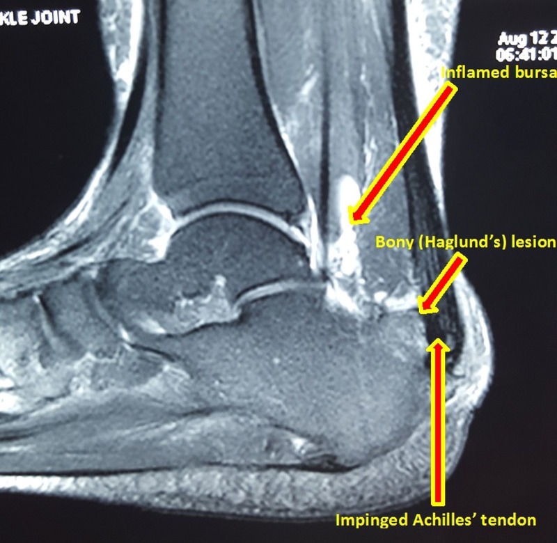
MRISagittal MRI of the ankle: posterosuperior bony prominence of the calcaneus (Haglund deformity), retrocalcaneal bursitis and Achilles tendon impingement, with arrows annotating each feature.
Source : Vaishya et al., Cureus, 2016, figure 3 · CC BY
In this chapter: what ultrasound and MRI really contribute, the major limit of the reasoning “it hurts so it is inflamed”, and the rule that avoids unnecessary investigations.
The diagnosis of posterior heel pain is clinical. Imaging is requested only if its answer changes management. In practice, three situations justify it: doubt about a red flag, failure of well-conducted conservative treatment, and preparation for a surgical procedure.
The pictorial review in Radiographics from 2024 provides the complete imaging map of the heel and recalls that the differential diagnosis of heel pain essentially covers abnormalities of the Achilles tendon, the calcaneus and the plantar fascia1. It is the reference to keep at hand when a report is difficult to interpret.
Ultrasound shows the retrocalcaneal bursa, the thickening of the insertion, calcifications and neovascularisation on Doppler well. It is available, inexpensive and dynamic. The case reported by Lotliker et al. in 2024 moreover underlines its value, both diagnostic and interventional, in ankle disorders35.
But a Danish rheumatology study brings a useful correction to the use made of it. Felbo et al. examined with ultrasound, using the OMERACT enthesitis score, 27 patients with peripheral spondyloarthritis whose heel was painful on palpation29. Result: ultrasound signs of inflammatory enthesitis were found in only 48 % of them, and Doppler activity in 19 % only. In 26 %, ultrasound identified another inflammatory condition, tendinitis, arthritis or bursitis.
In other words: in a population where enthesitis is the expected lesion, a heel that is painful on palpation is an inflamed enthesis only one time in two. Tenderness at a point is not a tissue diagnosis, and ultrasound serves precisely to avoid settling for it. That is an argument for imaging, not against it, but an argument that assumes a willingness to be contradicted.
MRI remains the most complete examination for characterising tendon degeneration, bone oedema of the calcaneus, effusion in the bursa and the state of Kager's fat pad. It is justified mainly before a surgical decision : that is how Tourné et al. used it in their series, to assess the state of the distal end of the tendon before osteotomy13.
The corollary is worth telling the patient: finding MRI abnormalities on a painful heel surprises no one, and finding them on a painless heel does not either. It is the same reasoning as for the radiograph, with higher resolution and therefore a higher risk of over-interpretation.
Key points
In this chapter: the mechanism that makes the shoe a cause and not a detail, what a heel lift really does, the true level of evidence for these measures, and how to prescribe them in practice.
The reasoning is simple and it is mechanical. The posterosuperior prominence of the calcaneus is covered by skin, by a superficial bursa and sometimes by the edge of the tendon. The heel counter of a shoe, that rigid piece which wraps the back of the heel, presses exactly there. The higher and stiffer it is, the higher it presses, that is to say on the summit of the prominence.
That impingement is not settled by an exercise, for the same reason that a blister is not rehabilitated: as long as the source of the friction stays in place, the tissue keeps receiving the stress that irritates it. The synthesis by the American Academy of Orthopaedic Surgeons devoted to the management of insertional tendinopathy indeed places footwear modification and activity modification at the head of the conservative options, ahead of physiotherapy, injections and shockwaves23.
The level of evidence, stated honestly
Footwear modification is recommended by review articles and by clinical consensus, but it has been the subject of no randomised controlled trial in this indication. It rests on coherent mechanical reasoning and on experience, not on a demonstration of efficacy. That does not mean it should not be done: it is cheap, riskless and immediately reversible. It means it must be presented to the patient for what it is, and that its failure must not be held against them as if it were an established treatment.
The useful instruction is not “change your shoes”, which means nothing, but a series of precise checks.
A heel lift raises the calcaneus and puts the ankle into slight plantarflexion. It therefore reduces the dorsiflexion reached during the step, and with it the compression of the tendon against the posterosuperior angle. It is the same mechanism as the one described by the cadaveric work of Malagelada et al., which showed the rise in pressure in terminal dorsiflexion4.
Two limits must be set out. First, a heel lift does not change the impingement against the heel counter: it can even worsen it by raising the heel inside the shoe, and so moving the counter's point of pressure upwards. In a patient whose pain is first of all a footwear impingement, the heel lift is the wrong tool.
Second, the neighbouring article on Achilles tendinopathy recalls it for the midportion of the tendon: relief is transient and the heel lift must be withdrawn gradually. The same principle holds here. A heel lift is not a treatment, it is a temporary unloading tool that makes exercise possible, and exercise is the treatment.
Key points
In this chapter: the study that tipped practice by showing that full range failed, the protocol in detail, what does and does not transfer from the midportion of the tendon, and the pain-monitoring model that allows running to continue.
It is the paper by Jonsson, Alfredson, Sunding, Fahlström and Cook, published in the British Journal of Sports Medicine in 2008, and its detail must be known14. The authors' starting point: painful eccentric training, in which the patient loads the tendon through to full dorsiflexion, gives good results in tendinosis of the midportion of the tendon, but only 32 % good clinical results in insertional tendinopathy.
They therefore tested a new model, without loading into dorsiflexion. Twenty-seven patients, twelve men and fifteen women of mean age 53, for 34 painful tendons for a mean of 26 months. Protocol: 3 sets of 15 repetitions, twice a day, seven days a week, for twelve weeks.
At follow-up, at a mean of four months, 18 patients out of 27, that is 67 %, corresponding to 23 tendons out of 34, were satisfied and had returned to their previous activity. Their pain on exertion on a visual analogue scale fell from 69.9 to 21.0 (p < 0.001). The nine dissatisfied patients, that is 11 tendons, still saw their pain fall from 77.5 to 58.1 (p < 0.01).
What this study does not establish
It is a pilot study without a control group, on 27 patients, with short follow-up. It does not demonstrate the superiority of the limited-range protocol: it shows an encouraging result in a group where the classic protocol gave 32 %. The comparison between the two figures sets two different populations side by side, not two arms of the same trial. The systematic review by Wiegerinck et al. goes the same way in concluding that full-range eccentric work shows low patient satisfaction compared with exercises at floor level15, but no randomised trial has since directly compared the two ranges. That is, to date, the main gap in the literature on this subject.
At the midportion of the tendon you seek range; at the insertion you seek load, and you give up range.
The reason is the one from the first chapter: at the insertion, the tendon undergoes a compression against the bone that grows with dorsiflexion4. Dropping the heel below the edge of a step, the emblematic movement of Alfredson's protocol for the midportion of the tendon, amounts to deliberately crushing the injured zone at every repetition. What is the treatment in one case is the insult in the other.
Phase 1, weeks 1 to 2: establish the load without provoking
Phase 2, weeks 3 to 8: concentric and eccentric loading at floor level
Phase 3, weeks 9 to 12 and beyond: heavy loading and return to activity
Load progression at the insertion, and what distinguishes it from the midportion of the tendon
The dorsiflexion range stays capped as long as compression is painful
Volumes and frequency taken from the protocol published by Jonsson P et al., Br J Sports Med 2008 (PMID 18184750). The division into three phases, the phase 3 loads and the criteria for moving on are an editorial proposal based on this protocol and on the progressive loading principles of Beyer R et al., Am J Sports Med 2015 (PMID 26018970); they have not been evaluated as such in a trial.
Two findings obtained on the midportion of the tendon illuminate practice at the insertion, provided the extrapolation is declared.
The randomised trial by Beyer et al. published in the American Journal of Sports Medicine in 2015 compared, in 58 patients with tendinopathy of the midportion of the tendon for more than three months, classic eccentric work with a protocol of heavy slow resistance over twelve weeks20. Both groups improve significantly, and maintain it at 52 weeks, with no difference between them. On the other hand, compliance was 92 % in the heavy loading group against 78 % in the eccentric group (p < 0.005), and satisfaction at 12 weeks 100 % against 80 % (p = 0.052). What transfers here is therefore not a superiority in efficacy, it is an adherence argument: heavy loading three times a week is better followed than twice-daily eccentric work.
The 2007 trial by Silbernagel et al., also on Achilles tendinopathy, tested the pain-monitoring model : 38 patients divided between a group allowed to continue running and jumping under pain monitoring and a group put on active rest for six weeks21. No difference between the groups, and both improve markedly, the VISA-A-S rising from 57 to 85 and from 57 to 91 at twelve months. Conclusion: no harmful effect from maintaining activity under pain monitoring.
This result is valuable in the consultation, because it means a runner need not be stopped. It must however be said that this study did not deal specifically with insertional forms, and that a patient whose pain is woken by impingement against the shoe or by dorsiflexion first needs that stress removed, before running volume is discussed.
The network meta-analysis by Ko et al., published in BMC Musculoskeletal Disorders in 2023, is the most complete synthesis of non-surgical treatments specifically devoted to insertional tendinopathy16. It brings together 9 randomised trials and 464 participants. The ranking puts in first place the combination of eccentric exercise and soft tissue therapy, with a SUCRA value of 84.8 and a mean rank of 1.9.
The authors nevertheless conclude, and it must be quoted in full, that the overall confidence in the non-surgical treatments from all the included trials was very low and that no recommendation of the best treatment can be made from this review. Nine trials, 464 patients: that is the whole randomised evidence base on a condition that fills thousands of sessions a year.
The gap that must be known
The reference clinical practice guideline for the Achilles tendon, published by the Academy of Orthopaedic Physical Therapy in JOSPT in December 2024, is entitled “Midportion Achilles Tendinopathy Revision” and explicitly covers the prevalence, the risk factors, the diagnosis, the imaging and the physiotherapy interventions for tendinopathy of the midportion of the tendon22. The insertional form, and a fortiori Haglund syndrome, do not fall within its scope. There is therefore to date no clinical practice guideline covering the presentation dealt with in this article. Applying the midportion guideline to a painful insertion is not following the recommendation, it is extending it beyond its declared scope.
Key points
In this chapter: the reversal of evidence on shockwaves between 2013 and 2025, the safety signal of corticosteroid injections, and a table of modalities by level of evidence.
It is a textbook case of a reversal of evidence, and it deserves to be told in order.
2013. The systematic review by Wiegerinck et al. concludes that shockwaves appear effective in patients with insertional tendinopathy that is not calcified, and that they are superior to no treatment as well as to an eccentric programme15.
2021. The systematic review by Zhi et al., covering 23 studies, concludes that the available evidence favours shockwaves or their combination with eccentric work17.
In that same year, 2021, a double-blind randomised placebo-controlled trial appears in the Journal of Bone and Joint Surgery. Mansur et al. included 119 patients with insertional tendinopathy, divided between eccentric work plus radial shockwaves and eccentric work plus sham shockwaves, three sessions every two weeks and three months of exercise18. Result on the primary outcome, the VISA-A at 24 weeks: 63.2 in the treated group against 62.3 in the placebo group, p = 0.876. No difference on any secondary outcome. The authors' conclusion is unambiguous: shockwaves do not potentiate the effects of eccentric strengthening.
Two secondary results deserve to be known because they run in opposite directions: the shockwave group showed a higher failure rate, 38.3 % against 11.5 % (p = 0.002), but a lower recurrence rate, 17.0 % against 34.6 % (p = 0.047). No complication was reported in either group.
2025. The randomised trial by Alsulaimani, Perraton, Vallance, Powers and Malliaras appears in Clinical Rehabilitation19. Seventy-six people, symptoms of more than three months, three sessions of radial shockwaves or placebo added to education and exercise identical in both arms. At twelve weeks, the between-group difference on the VISA-A is 4.6 points (95 % CI: −2.5 to 11.6), that is compatible with the absence of an effect. No difference at six weeks either, nor on any secondary outcome. No serious adverse event.
Two level I trials, four years apart, two independent teams, two continents: added to well-conducted exercise, shockwaves bring nothing measurable.
The reasonable reading is not that shockwaves “do not work”, but that they add nothing to a properly conducted exercise programme. It is a nuance that matters: the favourable reviews of 2013 and 2021 often compared shockwaves with weak comparators. When the comparator becomes well-conducted exercise in both arms, the difference disappears.
The anatomical reasoning of the first chapter finds its clinical consequence here. Turmo-Garuz et al. reported in Musculoskeletal Surgery in 2014 the case of three patients who presented with a combined rupture of the Achilles tendon and the gastrocnemius-soleus complex six months after a corticosteroid injection for retrocalcaneal bursitis24. To understand the mechanism, they injected a coloured contrast into the bursa of three cadaveric specimens: a connection between the bursa and the anterior and distal fibres of the tendon was found in every case.
This work joins that of Pękala et al. described above, which documents the same communication on twenty specimens and identifies the anteroinferior portion of the tendon as a zone of weakness5.
What these data establish, and what they do not
Three case reports and two cadaveric series do not demonstrate a causal relationship, and the authors themselves put forward a hypothesis. It is therefore not a matter of asserting that injection ruptures tendons, but of noting that there exists a plausible anatomical mechanism, documented twice independently, and compatible reported cases. Against a transient symptomatic benefit not demonstrated in the long term, that is enough to weigh in the balance, particularly in athletes and in patients about to resume heavy loading. The decision belongs to the prescriber; the physiotherapist's role is to know this signal and to be able to set it out.
Modalities and level of evidence in posterior heel pain from impingement
An editorial appraisal applying the GRADE principles, on the sources cited in this article
Built from Jonsson 2008 (PMID 18184750), Wiegerinck 2013 (PMID 23052113), Ko 2023 (PMID 36750789), Zhi 2021 (PMID 33785026), Mansur 2021 (PMID 34029235), Alsulaimani 2025 (PMID 39704142), Chen 2022 (PMID 35286285), Pękala 2017 (PMID 28765268) and Turmo-Garuz 2014 (PMID 24222527). The levels shown are an editorial appraisal applying the GRADE principles, and not a published GRADE assessment.
Key points
In this chapter: the threshold for referral, the three families of techniques and their compared results, the paradox of resecting the bump, and the physiotherapist's role after the operation.
Surgery is considered after failure of well-conducted conservative treatment, which assumes that this treatment did include a footwear modification, a progressive loading programme with limited range sustained for several months, and the correction of contributing factors. The review by the American Academy of Orthopaedic Surgeons sets out this sequence23, as does the meta-analysis by Yuen et al., which recalls that surgery is indicated in symptomatic patients after failure of a period of conservative treatment including analgesics, physiotherapy and modification of activity and footwear26.
The exact duration is fixed by no data. Six months is the interval most often used in practice; it is not an established finding but a custom.
Calcaneoplasty, open or endoscopic. It consists of resecting the posterosuperior prominence, excising the bursa and debriding the diseased tendon. The systematic review by Yuen et al. analysed 20 papers graded by level of evidence and concludes that both approaches are effective, with an advantage for endoscopy on operating time, complication rate and cosmetic result26. The systematic review by Alessio-Mazzola et al., published in Knee Surgery, Sports Traumatology, Arthroscopy in 2021, goes the same way and concludes that endoscopic calcaneoplasty gives better functional results, a lower complication rate and a shorter recovery than open procedures27.
The Zadek osteotomy. It is a dorsal closing-wedge osteotomy of the calcaneus, which does not resect the bump but changes the geometry of the bone so as to move the posterosuperior angle away from the tendon. The meta-analysis by Poutoglidou et al., published in Foot and Ankle Surgery in 2023, brings together 10 studies and 232 patients : functional scores and pain levels improve significantly (p < 0.00001), and the 22 complications reported were all considered minor, mainly superficial wound infections and sural nerve paraesthesia25. The authors conclude that the technique is safe and effective, while stressing that no well-designed randomised trial compares it with the other surgical treatments.
The series by Tourné et al. supplies the explanatory side: of 50 patients followed for a mean of 7 years, 40, that is 80 %, have an excellent result, and this result corresponds to the pre- and postoperative change in the geometric measurements, in particular the X/Y ratio13. That is what supports the idea that the target is not always the bump, but the general shape of the bone.
Associated tendon procedures : debridement and reattachment of the tendon, flexor hallucis longus transfer when tendon loss is substantial, gastrocnemius lengthening23. These procedures are met above all in advanced forms and in ruptures, as the clinical cases in the next chapter illustrate.
The two case-control studies from the radiographic chapter each draw, on their own, a surgical conclusion that deserves to be known by rehabilitation professionals. Kang et al. write that they believe it is possible that resection of the Haglund deformity is not necessary in the surgical treatment of insertional tendinopathy10. Lee et al., eleven years later, conclude that since the real size of the deformity, reliably measured, is not associated with insertional tendinopathy, a systematic resection of the deformity could be unnecessary11.
Two independent teams, two methodologies, one and the same conclusion: the most visible structure is not necessarily the one to remove. The question remains open and has not been settled by a trial, but it explains why approaches that change the geometry rather than shave the projection have gained ground.
The physiotherapist takes over. The principles do not change: initial unloading according to the surgical instruction, progressive return to loading, and particular caution with dorsiflexion, all the more so if the tendon has been detached and reattached. The timings vary considerably with the technique and with whether a tendon procedure was combined: they are obtained from the operating surgeon, never from a generic protocol. The review by Alessio-Mazzola et al. recalls that recovery is shorter after the endoscopic approach27, which is useful information to give the patient before their decision.
Key points
In this chapter: four published and verified case reports, chosen to cover four profiles that every practice meets, and what each teaches in practice.
A woman aged 69 years presents with difficulty moving the left leg and a deformity that appeared after a fall nine months earlier. The crucial point of the case: the pain had begun three months before the accident. Examination finds a seven-centimetre gap and a positive Thompson test. The ankle radiograph shows a Haglund deformity of the left calcaneus. The diagnosis retained is a chronic complete rupture of the Achilles tendon, treated by flexor hallucis longus transfer and resection of the deformity. One week after the operation, walking and the shape of the limb had improved.
What this case teaches: the deformity can act as a chronic wear mechanism and set the stage for rupture. The reported chronology, pain before trauma, is exactly the one the history must look for. And a rupture can go nine months unnoticed in an older person.
Usman MA, Murtaza B, Winangun PAN, Kennedy D. Medicina (Kaunas) 2022;58(9):1216. PMID 36143893
A professional handball player aged 39 years presents with a re-rupture of the Achilles tendon on a Haglund deformity that is large and painful. Treatment combined tendon debridement and reattachment with an anchor system. The authors recall in their introduction that the Haglund deformity frequently leads to retrocalcaneal bursitis as well as to thickening and inflammation of the tendon, a combination they call Haglund syndrome.
What this case teaches: the elite athlete carrying a symptomatic deformity is not in the same situation as the asymptomatic carrier. The distinction between shape and syndrome, set out in the first chapter, takes on its full prognostic value here.
Madi S, Hillrichs B. J Foot Ankle Surg 2022;61(2):410-413. PMID 34961680
A teacher aged 60 years has had left heel pain for three years and can neither stand nor walk for more than fifteen minutes. The diagnosis retained is Haglund's disease. The pain medicine team proposed ultrasound-guided injections targeting the superficial branches of the sural nerve, with lasting relief reported. The authors stress that the aetiology of Haglund's disease remains poorly understood, and cite among the probable causes a tight Achilles tendon, a cavus foot and a hereditary component.
What this case teaches: the most frequent profile in practice is not the runner, it is the adult in their fifties whose pain is long-standing and whose functional impact is major. The impact is assessed in minutes of standing, not in kilometres of running. The technique described remains, however, an isolated case, reported by a pain medicine team, and carries the weight neither of a recommendation nor of proof of efficacy.
Lotliker SD, Verma S, Bhalla A, Shah M, Mekewar S, Sharma A. J Orthop Case Rep 2024;14(2):12-17. PMID 38420222
A man aged 36 years presents with progressive pain and swelling of the left Achilles tendon insertion. He had surgery for an Achilles tendon lengthening in childhood. Imaging shows large multifocal calcium deposits in the tendon. Surgery combined a complete debridement of the calcified lesions, a resection of the Haglund deformity and a flexor hallucis longus transfer. Histology finds myxoid degeneration, neovascularisation and mature ossified tissue. At three months, pain, function and range were markedly improved.
What this case teaches: calcific insertional tendinopathy usually affects older subjects, and its appearance in a young adult should prompt a search for a local history36. It is a reminder that the old surgical history, often forgotten by the patient because it dates from childhood, is part of the assessment.
Li Z, Wei W, Yao Z, Zhao F. BMC Musculoskelet Disord 2025;26(1):1078. PMID 41316219
What the four cases say together
In this chapter: the consultation in four steps, the sentences that help the patient understand their imaging, and the criteria that should lead to referral.
One. Locate. Have the heel uncovered, inspect, then palpate in order: midportion of the tendon first, then the medial and lateral pinch above the upper border of the calcaneus, then the insertion with the tendon relaxed, then the posterosuperior bump. Note what exactly reproduces the patient's pain. Test passive dorsiflexion, then forced plantarflexion. This step takes three minutes and it determines all the rest.
Two. Have the shoes brought in. This is what distinguishes the management of posterior heel pain from the management of an ordinary tendinopathy. Look for the mark of the heel counter, the shiny patch, the chronological correlation with the purchase of a pair.
Three. Ask about the background. Age, body mass index, diabetes, dyslipidaemia, thyroid disorder, current medication. These elements do not change the rehabilitation, but they change the prognosis given and may justify a letter to the GP9.
Four. Set the baseline. VISA-A in the validated French version32, a numerical rating scale on the most bothersome movement, and the tolerated standing time. Three figures, written down, to be repeated at six weeks.
This is probably the most frequent situation, and the most poorly handled. The patient arrives with a report mentioning a “Haglund exostosis” and has understood that they have a bony outgrowth that will have to be removed.
Three useful ways of putting it
Key points
No, but the two often go together, and that is why this article deals with them on a single page. The Haglund's disease refers to the posterosuperior bony prominence of the calcaneus. The retrocalcaneal bursitis refers to inflammation of the bursa located between that zone and the tendon. The prominence can provoke the bursitis by impingement, but one can occur without the other. When the deformity, the bursitis and the insertional tendinosis come together, we speak of Haglund syndrome1.
Not routinely. It is useful for looking for an insertional calcification, which is the only radiographic feature robustly associated with insertional tendinopathy11, and for ruling out a bone lesion in case of doubt. It is not useful for measuring the size of the bump, since that size does not differ between painful heels and healthy heels1011.
Because dorsiflexion increases the compression of the tendon against the posterosuperior angle of the calcaneus, which has been measured directly on cadaveric specimens4. The full-range eccentric protocol, which gives good results at the midportion of the tendon, obtained only 32 % good results at the insertion; the same work performed without loading into dorsiflexion obtained 67 %14.
The published protocol lasts twelve weeks, and its assessment took place at a mean of four months14. A horizon of three to four months must therefore be announced, with the warning that the mean duration of symptoms before consultation in that same series was 26 months: patients often arrive late. It is more honest to announce a long timescale and a gradual improvement than a cure in six sessions.
Added to a well-conducted exercise programme, two independent randomised trials, in 2021 and in 2025, found no additional benefit1819. The earlier systematic reviews were favourable to them, but their comparators were often weaker. If access is easy and the patient wants it, the risk appears nil; one simply must not expect from them what exercise delivers.
It is a medical decision, but the safety signal must be known. Two independent cadaveric studies have shown that the retrocalcaneal bursa communicates with the anterior fibres of the tendon524, and three cases of combined rupture of the tendon and the gastrocnemius-soleus complex six months after injection have been reported24. This is not a demonstrated causality, but a plausible mechanism documented twice.
No. An isolated, asymptomatic Haglund deformity calls for no treatment: it is present in subjects with no pain at all, in proportions equivalent to those in patients1011. Reasonable advice is limited to the choice of shoes, to prevent the heel counter from setting up an impingement.
Not necessarily. The trial by Silbernagel et al. showed that maintaining running and jumping under a pain-monitoring model caused no harmful effect compared with six weeks of active rest21. The priority nevertheless remains to remove the impingement stress first, the shoe included; continuing to run in the shoe that hurts makes no sense.
Tendinopathy of the midportion of the tendon lies 2 to 7 cm above the insertion, undergoes mainly tensile stress, and is treated through full range, step drops included. Posterior heel pain from impingement lies at the insertion and above it, also undergoes compression against the bone, and is treated with capped range. The detail of the former is covered in the article on Achilles tendinopathy.
Pain at the back of the heel before the age of 15 suggests first of all calcaneal apophysitis, or Sever's disease, and not a Haglund syndrome. The systematic review by Nieto-Gil et al. brings together 11 studies and 1,265 participants of mean age 10.72 years, and identifies the limitation of ankle dorsiflexion as the most studied intrinsic factor30.
On the same subject
Every reference was verified individually in PubMed: identifier resolved, journal, year, volume, pagination and author list checked, abstract read to make sure the source does establish what is attributed to it.
Methodological note
The 36 references in this article were searched through the NCBI E-utilities API, crossing Haglund's disease, retrocalcaneal bursitis and insertional Achilles tendinopathy with anatomy, epidemiology, imaging, radiographic measurements, exercise, shockwaves, injections and surgery. Every identifier was resolved and every abstract read before citation.
Main limitation: the randomised evidence base specific to the insertional form amounts to nine trials and 464 patients, whose overall confidence was judged very low by the most recent network meta-analysis. No clinical practice guideline covers the presentation dealt with here, the reference in the field bearing explicitly on the midportion of the tendon. The levels of evidence in the modalities chart are an editorial appraisal applying the GRADE principles, and not a published GRADE assessment. The three-phase protocol takes up the volumes published by Jonsson et al. but its division and its criteria for moving on are an editorial proposal not evaluated as such.
Two figures were set aside during the work. The first is the calcaneal pitch angle comparison reported in the indexed abstract of Lee et al., whose two values contradict the sentence that introduces them; only the corresponding odds ratio is cited. The second concerns the fetal lengths in the work of Shaw et al. on the enthesis organ, whose published sequence contains an inconsistency in the series: only the composition of the enthesis organ, which does not depend on these values, is used here. Article written on 15 August 2026.
Behind this article
An author who explains, a reviewer who checks.
How we write and check our content

Physiotherapist · co-founder of Physio Learning
Marked for life by his first four-hour lecture without a single image, he took a master’s in instructional design so that it would never happen to anyone again. He hunts down publication bias and unreadable slides with the same intransigence.

Head of scientific content
Physiotherapist specialising in neuro-musculoskeletal practice and holder of a master’s in public health. He checks the methodological rigour of every article: primary sources, levels of evidence, no exceptions.