
















On-siteDPCFIFPL

Pelvic health rehabilitation
The basics of postnatal abdominal and pelvic floor rehabilitation: helping patients take charge
Julie Cantournet
Go further
Coccydynia (tailbone pain): dynamic sitting-standing X-ray thresholds, telling it from sacroiliac or low back pain, manual treatment, injections.
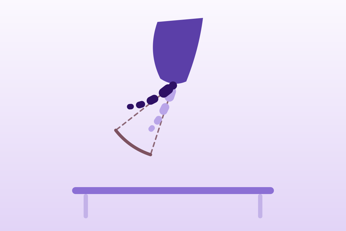
Coccyx pain relieved by standing suggests common coccydynia, pain at rest or at night does not, and calls for another cause to be sought.
1-3%of spinal pain is coccydynia
What follows covers each of these points in detail, with sources. It is there if you need it.
This topic is taught in a course: The basics of postnatal abdominal and pelvic floor rehabilitation: helping patients take charge, on-site with Julie Cantournet.
1 / 12
11 article chapters · 58 min in total
Understanding◔ 5 min
Coccydynia is pain in the coccyx, triggered by sitting and the sit-to-stand transition.
Mobility◔ 5 min
The coccygeal norm simply is not settled in the current literature.
Differentials◔ 5 min
Coccydynia is neither low back pain nor sacroiliac pain: three signs tell these apart.
Imaging◔ 4 min
Dynamic imaging does not predict response to treatment, even though the pathophysiological information is real.
Red flags◔ 4 min
Common coccydynia is a diagnosis of partial exclusion: serious causes are sought before treating.
Treatment◔ 14 min
The physiotherapist has a real place in coccydynia, but on weak to moderate evidence.
Postpartum◔ 4 min
The coccyx is mobilised during childbirth, and this physiological mobility can become injurious.
Interventions◔ 5 min
Local injection is the first-line interventional option, the only one with a formal positive recommendation.
Case studies◔ 4 min
Three cases out of four were treated as mechanical coccydynia for one to three years before the right diagnosis.
In practice◔ 4 min
Having the patient point to the pain with one finger rules out most confusions between coccydynia, sacroiliac pain and low back pain.
Questions◔ 4 min
The course is longer than usually said: 51% remain unfavourable at three years.
Train on this
Pelvic health rehabilitation
Julie Cantournet
Next comes practice: the course that teaches this topic, with Julie Cantournet.
Our courses on this topic may be covered by DPC or FIFPL, the French continuing-education funding schemes, depending on your profession and situation.
A rare presentation (1 to 3% of consultations for spinal pain) but one where the physiotherapist has a genuine therapeutic role, and where the gap between what the literature establishes and what is commonly said about it is particularly wide. This synthesis sets out the radiological thresholds, the differential diagnosis with sacroiliac pain and low lumbar pain, and the exact level of evidence for each modality, manual treatment included.
In brief
Coccydynia in three figures
Relative frequency, sex ratio and three-year outcome under conservative treatment.
Sources: Carayannopoulos 2026, Am J Med 139(4):405-412 (PMID 41241121); Benditz & Thoma 2025, Dtsch Arztebl Int 122(23):638-644 (PMID 40991348); Charrière 2021, Eur Spine J 30(10):3009-3018 (PMID 34216237). The figure of 51% aggregates residual pain above 3/10 and the coccygectomies performed during follow-up.
A pain rarely seen in practice, very largely female, whose true prevalence is unknown and whose aetiology remains uncertain in one patient out of three. This chapter sets out what is solidly established, and names what is not.
Coccydynia denotes pain localised to the coccyx, triggered or worsened by sitting and by the transition from sitting to standing. The definition stops there, and that is already informative: there is neither a consensus diagnostic criterion nor a reference test. Diagnosis remains clinical, based on the topography of the pain, its mechanical behaviour and reproduction of the symptom on direct palpation of the coccyx.12
Epidemiologically, the most useful datum is a relative frequency: coccydynia accounts for 1 to 3% of consultations for spinal pain.1 That is little, and it is enough to explain why most physiotherapists see only a few cases a year. Prevalence in the general population, for its part, is quite simply unknown: the 2025 German narrative review states this bluntly, and the only robust demographic datum available is the sex ratio, of about five women to one man.2
Four less well known markers
What the clinical series establish beyond the sex ratio.
Sources: Woon & Stringer 2012, Clin Anat 25(2):158-167 (PMID 21739475); Maigne 2000, Spine 25(23):3072-3079 (PMID 11145819); Maigne 2012, Eur J Phys Rehabil Med 48(3):387-392 (PMID 22820826); Güler & Ünal 2024, Agri 36(1):22-28 (PMID 38239122). The 7.3% and the 78% come from specialist hospital series: they describe a recruited population, not the general population.
The literature classifies coccydynia into three broad groups, to which is added a category that must be handled separately.
Post-traumatic. A backward fall onto the buttocks is the principal mechanism. One often ignored datum nevertheless qualifies the causal link: in 208 consecutive patients investigated under the same protocol, trauma altered the type of lesion only if it dated from less than a month before the pain appeared, the instability rate then rising from 55.6% to 77.1%.4 A long-standing trauma reported by the patient is therefore not automatically the cause of their current pain; it is often a memory that the history brings up because it goes looking for it.
Postpartum. It deserves a chapter of its own, further on. Note here that it accounts for 7.3% of coccydynia cases in women seen in specialist practice, and that it is distinguished by a coccygeal dislocation rate of 43.9% against 17.0% in other patients.13
Idiopathic. This is the category that recent literature puts at about a third of cases.14 The reference anatomical systematic review concludes in the same way: aetiopathogenesis remains uncertain in a third of affected patients.12 These forms are associated with repeated mechanical overload, prolonged sitting, weight changes, but association does not establish causation.
And the fourth, which is not an aetiology of common coccydynia: symptomatic causes, whether tumoural, infectious, inflammatory or referred visceral. They do not sit alongside the first three, they come before them in the reasoning. The chapter on red flags is devoted to them.
This is the most counter-intuitive and the most operational result in the literature. In 208 consecutive patients, body mass index did not merely alter the risk of coccydynia: it determined which lesion the patient developed. The proposed mechanism is simple and verifiable in practice: BMI conditions the way a person sits, and therefore the load the coccyx bears when seated.4
Type of coccygeal lesion according to body mass index
208 consecutive patients, same dynamic radiographic protocol. The four profiles are mutually exclusive and sum to 100% in each column.
Source: Maigne JY, Doursounian L, Chatellier G. Causes and mechanisms of common coccydynia: role of body mass index and coccygeal trauma. Spine 2000;25(23):3072-3079 (PMID 11145819), p < 0.0001 for the distribution. Single-centre study, consecutive series from specialist practice: the BMI distribution is that of referred patients, not that of the French population.
Two practical consequences follow. First, in a thin patient the dynamic examination has almost a one in two chance of being normal: imaging without abnormality therefore does not refute the diagnosis, it is the expected result. Second, the coccygeal spicule, that bony outgrowth on the dorsal surface of the last segment, is a lesion of thin patients, not to be looked for first in an obese patient. It is found in 22.3% of the 244 coccydynia patients in a recent Chinese series, but also in 23% of 112 CT scans of subjects without known coccygeal pathology.1516 In other words: a spicule on an image is never enough to explain a pain.
A morphological abnormality of the coccyx is a diagnosis only if it explains the pain of the patient sitting in front of you. Otherwise it is an anatomical variant that happened to be there.
Key points
It is hard to call a movement abnormal when the normal is poorly known. The coccyx long remained the least studied segment of the spine, and the two largest series of asymptomatic subjects do not agree with each other. This chapter sets out what is known, and the extent of what is not.
The coccyx is the terminal segment of the spine. It most often has four segments, found in 76% of 112 CT scans of subjects without coccygeal pathology, but their number varies from three to five between individuals, and the other large CT series, covering 500 asymptomatic subjects, most often finds three segments (50.8%).1617 This divergence on a point as elementary as the number of bones gives the measure of the problem: the coccygeal norm is not settled.
It articulates with the sacrum through the sacrococcygeal junction, a joint intermediate between a symphysis and a diarthrosis, and with its own segments through the intercoccygeal junctions. These junctions frequently fuse, and all the more so as one descends towards the tip: sacrococcygeal fusion is present in 57% of asymptomatic subjects in the New Zealand series.16 Around it insert the sacrotuberous ligament, the sacrospinous ligament, the gluteus maximus, and above all the coccygeus muscle and the iliococcygeal fibres of the levator ani, and it is this last point that gives the pelvic floor physiotherapist a direct therapeutic hold.
The question is not cosmetic: it probably explains part of the 5:1 sex ratio. The CT series of 112 adults finds a female coccyx that is significantly shorter (mean curved length 4.0 ± 0.8 cm against 4.4 ± 0.8 cm in men, p < 0.01), straighter, and more often retroverted: nine of the twelve coccyges with a retroverted tip in the series belonged to women.16 A shorter, straighter coccyx is a coccyx less protected when sitting and more exposed during childbirth.
Direct comparison between patients and healthy subjects points the same way, with one important caveat. In women with coccydynia, the coccyx is found to be more curved forwards, sacrococcygeal fusion less frequent (27% against 58%) and the coccygeal spicule more frequent (44% against 19%).18 The caveat: when the authors combine all the significant parameters in a logistic regression model, sensitivity peaks at 72 % and specificity at 71% in women. A model that is wrong one time in three is not a diagnostic test, it is a population association.
The Postacchini and Massobrio classification, published in 1983 from 120 asymptomatic subjects, remains the universal reading grid: type I slightly curved forwards, type II marked anterior curvature, type III sharp anterior angulation, type IV subluxed.19 The authors noted that 69% of their 51 operated patients were type II, III or IV, against 68% type I in asymptomatic subjects: the association looked strong.
Thirty years later, a series of 500 asymptomatic subjects investigated on multidetector CT recounted the numbers. The result is unsettling.
| Morphological type | Asymptomatic Postacchini 1983 (n = 120) |
Asymptomatic Przybylski 2013 (n = 500) |
Coccydynia patients Feng 2025 (n = 244) |
|---|---|---|---|
| Type I (slight anterior curvature) | 68 % | 16.2 % | 12.0 % |
| Type II (marked anterior curvature) | — | 40.0 % | 33.1 % |
| Type III (sharp anterior angulation) | — | 32.4 % | 28.1 % |
| Type IV (subluxation) | — | 11.4 % | 21.5 % |
| Type V (described later) | not described | not assessed | 5.4 % |
Postacchini 1983 (PMID 6226668) details in its abstract only the proportion of type I in asymptomatic subjects and the combined II+III+IV proportion in patients; Przybylski 2013 (PMID 23700101); Feng 2025 (PMID 41122360). The three series differ in imaging modality, radiography against CT, and in geographical origin.
Type I, which Postacchini described as the configuration of two healthy subjects out of three, concerns only 16.2% of the 500 asymptomatic subjects in the Polish series, and the authors themselves note that their results "differ from other results in the literature". In other words, the "normal" distribution of coccygeal types is not the same depending on which series you read. One direct consequence for practice: telling a patient that their coccyx is "of an at-risk type" is not reliable clinical information, and saying it can install a lasting structural belief where the prognosis depends mostly on something else.
The whole of dynamic radiographic reasoning rests on two threshold values established in 1996 in 91 patients and 47 healthy control subjects:
Two clarifications matter. The first: the precision of the angular measurement has been quantified at ± 2.6°, with intraobserver and interobserver variation of 15.3% and 12.5%.20 A coccyx measured at 26° of flexion is therefore a coccyx that cannot be said to exceed the threshold. The second: in the 1996 series, dislocation or hypermobility was found in only 48.4 % of coccydynia patients.3 Half of patients in pain therefore have normal coccygeal mobility, and that is a fact, not a failed examination.
On the origin of the pain itself, the strongest argument remains coccygeal discography: in the original series of 51 patients and 51 controls, the provocation-anaesthesia combination was positive in 15 of the 21 patients with normal mobility who were tested, leading the authors to estimate that common coccygeal pain arises from the coccygeal disc in about 70 % of cases.20 This is a figure to handle with caution, since discography is an invasive test and the series is old and single-centre, but it provides a pathophysiological hypothesis consistent with what MRI would show twenty years later.
The nuance that changes what you do
A coccyx that is stiff is not a healthy coccyx. In the manual treatment series it is in fact the profile with the worst prognosis: patients with an immobile coccyx obtained the poorest results of the three manual techniques compared, whereas patients with normal mobility obtained the best.21 The intuition that one should mobilise what is stiff runs up against the data here.
Key points
This is the practical crux of the presentation. The three pictures share a body region, a patient vocabulary ("my lower back hurts") and, for two of them, worsening on sitting. Three elements separate them reliably, and a fourth settles matters when doubt persists.
Coccydynia is not low lumbar pain, and it is not sacroiliac pain. This distinction, which looks obvious on paper, is regularly missed: the 2026 review in the American Journal of Medicine notes that coccygeal pain is frequently confused with lumbosacral, pelvic or gastrointestinal pathology, which delays management.1 The clinical case of an intrapartum coccygeal fracture missed for eighteen months, examined further on, illustrates exactly this mechanism.22
1. Topography. Coccydynia is midline : the pain sits below the sacrum and above the anus, and the patient points to it with one finger on the midline.23 Sacroiliac pain is lateralised, below L5, classically pointed to over the posterior superior iliac spine. Non-specific low back pain is more diffuse and extends above the lumbosacral junction.
2. Mechanics. Coccydynia is triggered or worsened by sitting, and above all by the transition from sitting to standing. This sign has particular value: in the 1996 radiographic series, pain on rising was one of the clinical features distinguishing the "dislocation" group from the "normal" group, and in the controlled trial of manipulation, pain on the sit-to-stand transition was among the predictors of a good result.321 Neither non-specific low back pain nor sacroiliac pain has this sitting signature.
3. Palpation. Direct palpation of the coccyx, external and then intrarectal if needed, reproduces the patient's complaint focally and exquisitely. It is the most discriminating element of the physical examination, and pain medicine recommendations set it as a mandatory step of the assessment.11
And the fourth, which settles matters by exclusion. The recommendation from the World Institute of Pain states a rule that is used far too rarely: in the absence of pain provocation by prolonged sitting and by manual examination of the coccyx, a neurological cause must be sought, first among them a lumbar disc herniation.11 Coccygeal pain that is reproduced neither on sitting nor on palpation is probably not coccydynia.
Decision tree: low back pain, three orientations
A three-stage clinical pathway. Red flags are sought first, before any mechanical hypothesis.
Built from: Patijn 2010, Pain Pract 10(6):554-559 (PMID 20825565), for the step 3 rule and referred visceral causes; Foye 2017, Phys Med Rehabil Clin N Am 28(3):539-549 (PMID 28676363) for midline topography; Staartjes 2026, Neurospine 23(2):365-379 (PMID 42097750) for the assessment sequence. This tree is an aid to reasoning, not a prospectively validated algorithm: none exists to date for this presentation.
| Feature | Coccydynia | Sacroiliac pain | Non-specific low back pain |
|---|---|---|---|
| Site pointed to | Midline, below the sacrum, above the anus | Lateral, below L5, over the posterior superior iliac spine | Diffuse, low lumbar, often bilateral |
| Aggravating position | Sitting, and above all the sit-to-stand transition | Single-leg loading, turning over in bed, prolonged standing | Flexion, prolonged standing, variable |
| Relieving position | Standing, side lying, sitting in anteversion | Unloading the affected side | Variable, often lying down |
| Direct palpation | Exquisite pain over the coccyx, reproduces the complaint | Tenderness of the sacroiliac sulcus, not very specific | Paravertebral guarding, not focal |
| Specific tests | Intrarectal mobility; no published validated test | Clusters of provocation tests | No attribution test |
| First-line imaging | Lateral radiograph, dynamic sitting/standing if persistent | None first line | None first line |
| Classic pitfall | Postpartum fracture missed and wrongly reassured | Attributing discogenic or coccygeal pain to the SIJ | Attributing midline coccygeal pain to the spine |
For the sacroiliac column, see our dedicated synthesis: mechanical sacroiliac pain and how long sacroiliac pain lasts. For the lumbar column: non-specific acute and chronic low back pain and sciatica from disc herniation.
Three pitfalls deserve to be named explicitly, because they keep coming back.
The "piriformis syndrome" pitfall. Deep buttock pain reproduced in the sitting position readily points towards the piriformis. But coccydynia is midline, and piriformis syndrome gives lateralised buttock pain with posterior thigh radiation. The two can coexist, and piriformis stretching is in fact among the interventions studied in coccydynia24 and that is one more reason not to settle for the first diagnosis found. See piriformis syndrome.
The anorectal pitfall. Proctalgia fugax, levator ani syndrome and pudendal neuralgia share the region and part of the vocabulary. The review in the Cleveland Clinic places them alongside coccydynia among the chronic anal pains and gives their distinguishing signs.25 Proctalgia fugax is brief, paroxysmal and nocturnal; levator syndrome gives a dull, higher pain, reproduced by posterior traction on the levator and not by bony palpation of the coccyx; pudendal neuralgia follows a nerve territory and worsens on sitting but spares lying down, with an anterior perineal topography.
The inflammatory pitfall. Coccydynia may reveal axial spondyloarthritis. A small Turkish series finds coccydynia in 13 of 42 patients followed for axial spondyloarthritis, with significantly more impaired functional scores.26 The sample is too small to derive a prevalence from it, but the signal is enough to justify asking about morning stiffness, waking in the second half of the night and improvement with exercise, three features that do not belong to mechanical coccydynia. See ankylosing spondylitis.
Key points
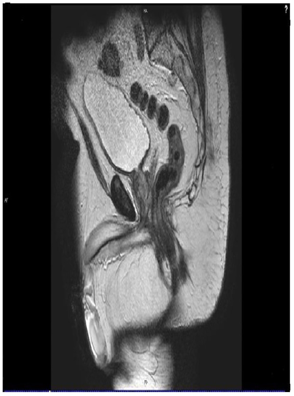
MRISagittal T2-weighted pelvic MRI: anteversion of the coccyx with rectal impingement, an identified cause of coccydynia and pain on defecation.
Source : Salar et al., Journal of Medical Case Reports, 2012, figure 1 · CC BY
It is the emblematic investigation for this presentation, and the most overrated. It provides real pathophysiological information and an argument for surgical discussion. It does not predict response to treatment, and a study of 184 operated patients demonstrates this in the most direct way there is.
The principle is elegant. A lateral film is taken standing, then a second in the sitting position that triggers the pain and not in a comfortable sitting position. Superimposing the two films measures the displacement of the coccyx under load, something no static imaging can show. It is today the first-line imaging recommended by the 2026 review in the American Journal of Medicine, and the first radiological step according to the 2024 German radiological update.114
Its descriptive performance is good: applied to 208 consecutive patients, the protocol identifies a causal lesion in 69.2 % of cases.4 It also allows patients to be sorted into four mobility groups, dislocation, hypermobility, immobility and normal mobility, and this classification has prognostic value for manual treatment, as we shall see.
Coccygeal MRI was for a long time supported by no data at all. The series of 172 patients with severe chronic coccydynia filled that gap, and its main finding has great internal coherence: the type of visible abnormality depends on the mechanical behaviour of the coccyx.27
What MRI shows according to coccygeal mobility
172 patients with severe chronic coccydynia, MRI and dynamic radiography in each.
Source: Maigne JY, Pigeau I, Roger B. Magnetic resonance imaging findings in the painful adult coccyx. Eur Spine J 2012;21(10):2097-2104 (PMID 22354690, full text PMC3463700). Single-centre series of severe coccydynia referred to a specialist centre: the frequency of abnormalities there is probably higher than in primary care. No asymptomatic control group was imaged.
The practical reading is direct: a coccyx that moves too much hurts through its disc, a coccyx that no longer moves hurts through its tip and the soft tissues around it. This matches the historical discography datum, a discogenic origin in about 70% of cases20 and it provides a different target depending on the profile: in one case the mobile segment, in the other the pericoccygeal tissues and the levator ani.
The 2026 recommendations frame the use: plain anteroposterior and lateral radiographs mainly to exclude serious pathology, dynamic sitting-standing radiographs to be considered when mechanical pain is suspected and symptoms persist, cross-sectional imaging reserved for trauma, red flags, suspected tumour or infection, or inconclusive baseline imaging.28
Three converging results should temper enthusiasm.
It does not reliably discriminate patients from healthy subjects. The most complete published morphological model, built by combining all the parameters that differed significantly between 107 patients and 112 controls, reaches 72% sensitivity and 71% specificity in women, 52% and 92% in men.18 These values are incompatible with individual diagnostic use.
It does not predict response to treatment. This is the clearest demonstration. Of 184 patients operated on with coccygectomy and reviewed after 37 months on average, 33 had normal preoperative imaging and 138 abnormal imaging. The surgical failure rate was 24% in the normal imaging group against 32% in the abnormal imaging group and that difference is neither clinically nor statistically significant, and it runs the opposite way to intuition.29 The authors conclude that surgery should not be refused on the sole ground that the images are normal.
Some of its associations rest on almost nothing. The increase in intercoccygeal angle in idiopathic coccydynia is regularly cited. It comes from a study comparing 106 control subjects with ten patients.30 Ten. The result is statistically significant (p = 0.002) and clinically uninterpretable; it appears here because it circulates, not because it concludes anything.
Dynamic imaging explains the pain of one patient in two to three. It does not say which one will recover, nor with which treatment. Mistaking it for a prognostic test is the commonest error in this presentation.
Key points
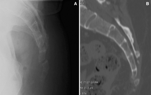
X-rayLateral X-ray and sagittal CT reconstruction of the coccyx showing a fracture, in a patient with coccydynia that developed after a fall six months earlier.
Source : Patel et al., Current Reviews in Musculoskeletal Medicine, 2008, figure 1 · CC BY-NC
Common coccydynia is a diagnosis of partial exclusion. Symptomatic causes are rare but all documented by real published cases, and they share one trait: the patient was treated for months or years for mechanical coccydynia before the diagnosis was made.
The mechanism of the delay is always the same. The patient has coccyx pain, coccyx pain is commonplace, it is treated as such, and time passes. Three years for the glomus tumour, two years for the dermoid cyst, eighteen months for the postpartum fracture: these are not exceptional wandering times in the published cases, they are the usual ones.
Red flags in coccygeal pain
Faced with any of these features, cross-sectional imaging, MRI first line, does not wait.28
Sacrococcygeal chordoma. This is the main fear, and it is rare: the reference epidemiological systematic review puts the incidence of chordoma, all locations combined, between 0.18 and 0.84 cases per million population per year, with a clear male predominance and a diagnosis made on average in the late fifties. Depending on the series, the sacrococcygeal location accounts for 29% to 45% of cases.31 Its presentation is insidious; a recent case describes a woman of 73 whose progressive low back pain with bilateral thigh radiation progressed to cauda equina syndrome with S3-S5 saddle hypoaesthesia before MRI revealed the lesion.32
Benign tumours. Two published cases deserve to be known for their teaching value. A precoccygeal dermoid cyst in a man of 20, treated for two years as refractory coccydynia before an MRI showed the cystic lesion; excision with coccygectomy gave an excellent result.33 And a glomus tumour in a woman of 57, with more than three years of coccygeal pain, sharp pain at the slightest pressure and, a characteristic sign, worsened by exposure to cold ; excision of the 2 cm lesion, without resection of the coccyx, left her asymptomatic at ten months.34
Infections. Isolated coccygeal tuberculosis is exceptional but documented: a man of 35 with three months of coccygeal and buttock pain, in whom CT and MRI showed bone destruction with a mass extending anteriorly and posteriorly, histology concluding tuberculous granulomatosis.35 Pilonidal disease, for its part, is common and is recognised clinically by the midline cutaneous opening of the natal cleft; it appears explicitly in the radiological differential.36
Referred pain of visceral origin. The recommendation from the World Institute of Pain mentions them expressly: disorders of the rectum, the sigmoid colon and the urogenital system can refer pain into the coccygeal region.11 In women, endometriosis is part of this differential, in particular with involvement of the rectovaginal septum or the uterosacral ligaments. See endometriosis and the physiotherapist's role.
Inflammatory rheumatic disease. Besides the axial spondyloarthritis already mentioned, a deposit of calcium crystals can cause acute coccydynia, described in a published observation.37 The picture is one of sudden, intense pain, without trauma, in a patient with no mechanical factor.
What the physiotherapist can do with this list
None of these conditions falls within their therapeutic remit, and that is not the point. The point is the delay. A patient referred for coccydynia who is not progressing after six to eight weeks of well conducted treatment, or in whom even one of the red flags above is present at the initial assessment, should leave with a letter for their doctor, not with a further block of sessions. In the published cases, it is months of ineffective treatment that separate the patient from their diagnosis.
Key points
This is the chapter where the temptation to embellish is strongest, because it is the one that bears directly on our practice. The honest result is this: the physiotherapist has a real and documented place in coccydynia, and that place rests on weak to moderate evidence, never strong. Here is which evidence, and what it says exactly.
Let us start with the point that structures everything else: there is no randomised controlled trial of physiotherapy in coccydynia with more than sixty patients. The 2025 Spanish systematic review gathered nine of them, totalling 532 participants; the Indian review of the same year gathered ten, totalling 515 adults.387 That is the whole of the randomised material available for this presentation, all interventions combined. Any categorical claim about the comparative efficacy of two techniques is, arithmetically, impossible there.
A single randomised controlled trial has evaluated intrarectal coccygeal manipulation. It deserves to be reported in detail, because it is almost always cited wrongly, in one direction or the other.
One hundred and two patients with chronic coccydynia, randomised into two groups of fifty-one. The experimental group received three sessions of coccygeal manipulation guided by the dynamic radiographs ; the control group received low-power external physical therapy. Assessment at one month and at six months.6
At one month, the median change in scores is more favourable in the manipulation group: −34.7% against −19.1% on the visual analogue scale (p = 0.09, borderline), −36.0% against −7.7% on the McGill (p = 0.03), −20.0% against +20.0 % on the Paris functional questionnaire (p = 0.02) and −33.8% against −15.7% on the Dallas (p = 0.02). Three criteria out of four reach significance.
But the criterion that matters to the patient is the rate of good results, and here it is: 36% against 20% at one month (p = 0.075), and 22% against 12% at six months (p = 0.18). Neither reaches significance. The authors conclude in one sentence: "we found a mild efficacy of intrarectal manipulation in chronic coccydynia".
This result is not nothing. A doubling of the rate of good results, maintained at six months, in a condition where placebo achieves 12%, is clinically interesting, but it is measured on a sample that does not allow it to be asserted. The same author had in fact calculated, in his earlier pilot study, that it would take 190 patients to settle the question with 80% power.21 That trial has never been carried out.
The 2001 pilot study is clinically richer than the 2006 trial, because it compares three manual techniques and cross-tabulates the results with the mobility profile. Patients were randomised between massage of the levator ani, joint mobilisation and gentle stretching of the levator, at a rate of three to four sessions, with independent assessment at 7 days, 30 days, 6 months and 2 years.21
The rate of satisfactory results is 25.7% at six months and 24.3% at two years and it is remarkably stable, which suggests that what is gained is gained durably. Three lessons follow:
The caveat, set out by the authors themselves: none of these results reaches significance, because of the low overall success rate. They are orientations for reasoning, not decision rules.
One important point for reading these data: the 2025 systematic review concludes that, among all the physiotherapy interventions studied, only stretching or massage of the levator ani shows maintenance of the analgesic benefit in the long term.7 The convergence between the 2001 pilot and the 2025 synthesis is the best argument manual therapy has in this presentation.
The target is not the bone, it is the muscle that inserts on it. Massage and stretching of the levator ani do better than joint mobilisation, and they are the only techniques whose effect holds in the long term in systematic review.
Two randomised trials and three systematic reviews converge: ESWT is today the best supported physiotherapy intervention in coccydynia. The 2026 recommendations state it explicitly: physiotherapy is first line, "with extracorporeal shockwave therapy having the most robust support".28
The first trial compared ESWT (20 patients) with physical modalities (21 patients) over four weeks: a significant fall in pain in both groups, significantly more marked under ESWT, with no significant difference on the Oswestry disability index, and greater satisfaction under ESWT.8 The second compared three weekly sessions of radial shockwave therapy with a "blind" corticosteroid injection in 34 patients: the time × group interaction is significant in favour of ESWT over six months of follow-up (p = 0.043).9
Forty-one patients, then thirty-four. It has to be said every time the word "effective" is uttered.
The methodologically best constructed trial in the whole corpus is, curiously, on kinesiotaping. Sixty obese patients with coccydynia were randomised double-blind between active kinesiotaping plus an exercise programme and sham kinesiotaping plus the same programme, over three weeks with follow-up at four weeks. The differences are significant in favour of active taping on pain, mobility on the modified Schober test and disability (p < 0.001).39 It is a genuine placebo-controlled trial, which is rare here, but a single trial, in a selected population, with four weeks of follow-up. The 2025 review notes moreover that kinesiotaping improves the perception of pain but has limited impact on disability.38
A less expected line deserves mention: stretching of the piriformis and the iliopsoas. Forty-eight patients divided into three groups: stretching alone, stretching plus thoracic mobilisation of hypomobile segments, and a conventional group combining a cushion, sitz baths and phonophoresis. The two stretching groups significantly improved pressure pain threshold and pain-free sitting time, with the benefit maintained after treatment stopped; the conventional group did not improve significantly.24 This detail is instructive: the cushion, often presented as the first thing to do, served here as the comparator and produced nothing.
This is the intervention closest to our pelvic floor rehabilitation offering, and the one to speak about with the greatest caution. A review of records covering 124 consecutive patients referred to a pelvic floor rehabilitation clinic analysed 93 patients who actually followed the programme, of whom 79 completed it, with a mean of nine sessions. The main intervention was pelvic floor physiotherapy aimed at muscle relaxation rather than strengthening.40
The figures are impressive: mean pain from 5.08 to 1.91 (p < 0.001), maximum pain from 8.81 to 4.75 (p < 0.001), overall subjective improvement of 71.9%. The subgroup of 17 patients with pain after coccygectomy improved from 6.64 to 3.27.
And here is why they should not be quoted without their context: this is a retrospective review of records, with no control group, of evidence level III, in which 19% of patients also received baclofen, 17% coccygeus trigger point injections and 8% a ganglion impar block. The effect measured is not that of physiotherapy alone. The result remains the best available signal in favour of pelvic floor work in this presentation: it does not constitute proof of efficacy.
Two series you need to know how to read
Two publications report spectacular effects of osteopathic manual treatment: a series of 50 patients going from a VAS of 6.5 to 1.2 after three sessions41 and a practice improvement project covering 16 patients, of whom 10 were analysed and only 5 had follow-up data.42 These effects are reproduced by no controlled trial. In the first study, patients were first treated for three months with physiotherapy and medication, with no improvement in pain (p = 0.065), then referred to the osteopaths: this sequential design, with no randomisation and no simultaneous comparator, does not allow the effect of treatment to be separated from regression to the mean and from expectation. A gap of this size between uncontrolled studies and randomised trials is in itself information about the nature of the effect measured.
A five-year British prospective trial of 120 patients, published in 1991, is still regularly invoked to disqualify physiotherapy in this presentation. Its conclusion is indeed unequivocal: "physiotherapy was of little help", whereas 60% of patients responded to local injection and the combination of manipulation and injection cured about 85% of them.60
Two clarifications change the reach of that sentence. The first is chronological: the "physiotherapy" evaluated in 1991 in a British orthopaedic department meant passive physical agents, shortwave and ultrasound, and not what physiotherapy management of coccydynia covers today, that is to say manual work targeted on the levator ani, adaptation of seating and education. The study does not refute an intervention that did not exist in that form when it was conducted. The second is that this same study established a point rarely borrowed from it: the authors found no element of neurosis in their patients and concluded that the condition is real and disabling, a result which, at the time, went against the dominant discourse on this presentation.
It remains that 85% cure by combined manipulation and injection is a figure that no controlled study has reproduced since. Thirty-five years later, the best available randomised trial obtains 22% good results at six months. The gap is not trivial: it measures what moving from an open design to a controlled design takes away from a result.
The table below summarises the level of evidence for each modality. One methodological clarification is needed: no formal and complete GRADE assessment has been published for the whole of these interventions. The only one that exists, applied to seven conservative studies in 2013, concluded that evidence ran from moderate to very weak and that recommendations ran from weakly in favour to weakly against, that is to say the impossibility of recommending anything at all.43 The levels given here aggregate that assessment, the graded levels published in 202244, the 2025 systematic reviews and the specific 2026 GRADE assessment of the ganglion impar block.
| Modality | Level of evidence | What the data show | Basis |
|---|---|---|---|
| Education, seating relief, simple analgesics | Weak | First line by expert consensus; no trial isolating these measures | Staartjes 2026 |
| Extracorporeal shockwave therapy | Moderate | Superior to physical modalities and to blind injection over 6 months; effect sustained to 6 months in some trials | 2 RCTs (n = 41; n = 34), 3 systematic reviews |
| Stretching / massage of the levator ani | Weak | 25.7% satisfactory results at 6 months, 24.3% at 2 years; the only modality with long-term maintenance in systematic review | Randomised pilot (about n = 35 per arm), SR 2025 |
| Intrarectal coccygeal manipulation | Weak | Good results 22% vs 12% at 6 months, not significant; "mild" efficacy according to the authors; better in the recent phase | 1 RCT (n = 102) |
| Kinesiotaping combined with exercise | Weak | Superior to placebo taping on pain, mobility and disability at 3 and 7 weeks; limited impact on disability in synthesis | 1 double-blind placebo-controlled RCT (n = 60) |
| Stretching of the piriformis and iliopsoas | Very weak | Improvement in pain threshold and sitting time, maintained at 1 month; unblinded | 1 three-arm randomised trial (n = 48) |
| Pelvic floor physiotherapy | Very weak | Mean pain 5.08 → 1.91 in 9 sessions, but retrospective series with no control, with co-interventions in a third of patients | 1 review of records (n = 124), level III |
| Offloading cushion, seating adaptation | Very weak | No dedicated trial; "mixed" results in review; ineffective as comparator in the only trial that tested it as such | Narrative reviews |
| Local corticosteroid and anaesthetic injection | Weak | Recommended in the severe chronic phase with grade 2 C+; prefer prednisolone acetate to cortivazol | WIP 2010 recommendation |
| Ganglion impar block | Very weak | Pain reduction in the short, medium and long term, with no serious adverse effect, but "very low" GRADE certainty on every outcome | 2026 meta-analysis (11 studies, n = 391) |
| Radiofrequency | Very weak | 55.5% mean relief in 12 patients; advised only under study conditions by the WIP recommendation | Case series (n = 12), WIP recommendation |
| Coccygectomy | Weak | Mean fall of 5 points maintained beyond 36 months; 8% complications, 3% revisions, but 18 of the 21 studies are retrospective, and a formal recommendation advises against it | Meta-analysis (n = 826), level III |
Scroll the table horizontally on a small screen. Detailed references in the bibliography.
Modalities ranked by certainty of evidence
Stacked horizontal cards. Width represents certainty, not the size of the effect: a modality can be very effective in some patients and still sit at the "very weak" level.
The authors' synthesis based on: Howard 2013, J Man Manip Ther (PMID 24421634, the only published GRADE assessment of the conservative field); White 2022, PM R 14(9):1143-1154 (PMID 34333873, level grading); Blanco-Diaz 2025, BMC Musculoskelet Disord 26(1):514 (PMID 40420056); Sidiq 2025, Arch Physiother 15:77-89 (PMID 40308532); Jevotovsky 2026, Reg Anesth Pain Med 51(5):512-523 (PMID 40081927, "very low" GRADE); Staartjes 2026, Neurospine (PMID 42097750); Patijn 2010, Pain Pract (PMID 20825565). This classification is an editorial reading of heterogeneous sources, not a formal GRADE assessment carried out de novo.
A 2022 systematic review, the largest published with 1,980 patients across 64 studies, proposes an attractive ranking of modalities by mean fall on the visual analogue scale: radiofrequency 5.11 cm, shockwave therapy 5.06, coccygectomy 4.86, injection 4.22, ganglion block 2.98, stretching and manipulation 2.19, usual conservative care 1.69.45
This ranking is regularly taken up as it stands, and it does not mean what it is made to say. These are aggregated within-group changes, not comparisons. Radiofrequency and coccygectomy are offered to refractory patients, very painful at the outset, therefore with a great deal to lose on the scale, after everything else has failed, whereas "usual conservative care" includes less severe patients. The ranking measures the indication at least as much as the efficacy. Of the 64 included studies, only five are randomised trials; 45 are retrospective.
The most useful datum in this whole file is probably prognostic, and it is darker than the usual message.
Pain trajectory under conservative treatment over three years
115 adults with chronic coccydynia present for 18 months on average. Coccygeal injection first line, manual therapy or coccygectomy second line.
Source: Charrière S, Maigne JY, Couzi E, Lefèvre-Colau MM, Rannou F, Nguyen C. Conservative treatment for chronic coccydynia: a 36-month prospective observational study of 115 patients. Eur Spine J 2021;30(10):3009-3018 (PMID 34216237). The values at 6 and 36 months are reconstructed from the baseline intensity (6.5 ± 2.0) and the mean changes reported. Observational single-centre study in a specialist centre, with no comparison arm: it describes an outcome, it does not evaluate a treatment.
Three messages follow for the consultation.
Time matters, and it is the only proven lever. Each extra month of symptoms before management increases the odds of an unfavourable course by 4%. That justifies treating early, and not letting a patient "wait for it to pass" for a year.
You should announce a partial and slow improvement, not a cure. A mean fall of 2.8 points out of 10 over three years, with a one in two chance of staying above 3/10, is an honest trajectory. Promising otherwise exposes the patient to a disappointment that feeds chronicity, and recent reviews do underline the risk of pain chronicity in this presentation.2
The psychological impact is not anecdotal. In a series of 60 patients with coccydynia, 78.3% had depressive symptoms, 81.6% anxiety and 76.7% were poor sleepers.46 The sample is small and the cross-sectional design does not allow conclusions about the direction of the relationship, but the order of magnitude is an invitation not to treat a coccyx alone.
Key points
Yes, and on three counts: its lesion mechanism, its radiographic profile and its risk of being missed. It is also the situation where the pelvic floor rehabilitation offering and coccygeal management meet most naturally, since the patient is already being followed, access to the pelvic floor is already established, and the reason for treatment is already legitimate.
The coccyx is mobilised during childbirth: its retropulsion widens the anteroposterior diameter of the pelvic outlet at the moment of expulsion. This physiological mobility becomes injurious when it exceeds the tolerance of the sacrococcygeal junctions, and the mechanism is all the more constrained when labour is long or instrumented.
The only dedicated series compares 57 women with postpartum coccydynia with 192 women with coccydynia of other origin, all investigated by dynamic radiography. Its results outline a distinct entity.13
Postpartum coccydynia: circumstances and lesions
57 women with postpartum coccydynia, compared with 192 women with coccydynia of other cause.
Source: Maigne JY, Rusakiewicz F, Diouf M. Postpartum coccydynia: a case series study of 57 women. Eur J Phys Rehabil Med 2012;48(3):387-392 (PMID 22820826). Series from a specialist hospital clinic: instrumented deliveries are probably over-represented there compared with the general obstetric population. A sample of 57 patients, so the percentages should be read as orders of magnitude.
The classification of coccygeal fractures established in 104 consecutive patients identifies three mechanisms, and one of them is specifically obstetric. Fractures by extension (38 cases out of 104) are obstetric, sit in the coccyx low down, and their characteristic feature is a progressive separation of the fragments over time.47 The authors specify that, unlike flexion fractures which generally heal spontaneously, obstetric fractures and compression fractures are mostly unstable.
This detail has a direct clinical consequence: in a patient whose postpartum pain does not settle, a reassuring initial radiograph does not exclude a lesion that will reveal itself later, as the fragments separate. The case published in 2025 of a 31-year-old primipara illustrates this exactly: severe pain after vaginal delivery, reassured at the postpartum consultation as "inflammation and coccydynia", then eighteen months later an MRI showing a transverse fracture of the second coccygeal segment.22
The sentence not to say in the postpartum period
"It's normal, it will pass." Coccygeal pain that appears the first time the patient sits after delivery, that persists beyond six to eight weeks and that prevents sitting is not an ordinary consequence of childbirth: it is a dislocation in 44% of cases and a fracture in 5% in the only series available. It justifies a clinical examination of the coccyx and, if it persists, imaging. The most recent published case cost a patient eighteen months of professional and personal life for having been reassured too quickly.22
This is where the link with pelvic floor rehabilitation makes full sense. The postpartum patient is already directed towards a pelvic floor assessment; examination of the coccyx and of the levator ani insertion fits in with no additional step, and management can start early, and delay is the only modifiable prognostic factor identified.5
A recent randomised trial evaluated this population specifically: sixty women aged 25 to 35 with postpartum coccydynia for at least six weeks, divided into three groups: photobiomodulation combined with pelvic floor exercises, photobiomodulation alone, or placebo photobiomodulation combined with exercises. All groups improved; the group combining photobiomodulation and pelvic floor exercises did significantly better than the other two on pain, lumbar mobility and disability (p < 0.001).48
Two readings overlap. The first, cautious one: sixty patients, a single centre, and a modality, photobiomodulation, that is not standard in France. The second, more interesting for us: in the two arms that included pelvic floor exercises, the results were better than in the arm that did not. This is an indirect argument in favour of pelvic floor work in this indication, consistent with the pelvic floor rehabilitation series already cited.40
On the content of that work, the principle emerging from the data as a whole is this: you are seeking relaxation of the levator ani, not its strengthening. The largest pelvic floor rehabilitation series explicitly describes an intervention "aimed at muscular relaxation of the pelvic floor"40, and the randomised pilot study finds that excessive levator tone on examination is a predictor of a good response to manual treatment.21 The perineal bracing reflex, useful elsewhere, is counterproductive here.
For the general framework of postpartum pelvic floor management, see our syntheses on stress urinary incontinence and on diastasis recti, which share with this presentation the population, the timeline and part of the examination.
Key points
The physiotherapist does not set these indications, but does support patients who are considering them, fear them, or are recovering from them. This chapter sets out what each option really brings, what it costs, and a major contradiction between recommendations that is better known in advance than discovered in front of a patient.
This is the first-line interventional option, and the only one with a formal positive recommendation. The evidence-based pain medicine recommendation gives manual treatment and local injection of anaesthetic and corticosteroid into the painful segment the grade 2 C+, in patients in the chronic phase with severe pain.11 In the French prospective cohort, coccygeal injection was the systematic first-line treatment.5
One technical precaution deserves to be known: four cases of intercoccygeal disc calcification after cortivazol injection have been published, two of which probably caused additional pain. The author recommends preferring prednisolone acetate for coccygeal intradiscal injections.49
The ganglion impar is the terminal sympathetic ganglion, lying in front of the sacrococcygeal junction. Blocking it has become the most studied intervention of the last ten years in this presentation. The 2026 meta-analysis is the reference: seventeen studies describing 625 patients, of which eleven studies and 391 patients in the quantitative meta-analysis, with a mean baseline pain of 7.93 out of 10.50
The standardised mean differences are −2.73 (95% CI −3.45 to −2.01) up to three months, −2.13 (−2.82 to −1.45) between three and six months, and −1.86 (−2.58 to −1.15) beyond six months. No serious adverse effect was reported. And yet the authors conclude explicitly that the GRADE assessment indicates "very low" certainty for all outcomes. It is a textbook case: large effects, consistent over time, on a corpus whose methodological quality does not allow any conclusion.
An earlier systematic review covering 189 patients split between ganglion impar block (104) and radiofrequency (85) reports failure rates of the same order for the two techniques, 13.9% and 14.1%, and complication rates of 2.9% and 0%.51 A randomised trial of 56 patients has moreover shown that an ultrasound-guided coccygeal nerve block does as well as a fluoroscopy-guided ganglion impar block at twelve weeks, with neither superior to the other.52
The data are thin. The most cited series covers twelve patients, with mean relief of 55.5% but a standard deviation of 30.3% and a range from 0 to 100%; two neuritides occurred, resolving spontaneously.53 The recommendation from the World Institute of Pain classifies radiofrequency, like the ganglion impar block and intradiscal injection, at grade (0) : to be reserved for study conditions.11
And one published case is a reminder of the anatomy: the proximity of the rectum to the sacrococcygeal region exposes the patient to rectal perforation by thermal spread, a complication described after coccygeal radiofrequency ablation whose management required a surgical and gastroenterological team.54
This is the last resort, and the subject on which the literature contradicts itself most plainly.
What the meta-analysis says. Twenty-one studies, 826 patients of whom 75% were women, 720 total coccygectomies and 106 partial ones. The aetiology was traumatic in 56% of cases and idiopathic in 33%. The pooled mean difference on pain is 5.03 points at 6-12 months, 5.02 beyond 12 months and 5.41 beyond 36 months, crossing the minimal clinically important difference threshold at each time point; the Oswestry index improves by 23.49 points. Pooled complications reach 8 % (95% CI 5-12%), essentially surgical site infections and wound dehiscence, and revisions 3%.10 The caveat is a large one: 18 of the 21 studies are retrospective.
What the recommendation says. The same procedure is explicitly advised against by the pain medicine recommendation, "because of moderate long-term results and the risk of major complications".11 It dates from 2010 and predates the meta-analysis; it has not been formally revised.
What the recent series say. In 184 patients operated on and reviewed, the success rate, meaning a patient fully recovered or clearly improved, is 71 %, and 89% of those operated on would have consented to the procedure again had they known the result. The postoperative infection rate fell from 10% to 2% by extending antibiotic prophylaxis from 24 to 48 hours (p = 0.018).55 A Parisian team reports a comparable trajectory: nine infections requiring revision in 61 operated patients in a first series56, then an infection rate that fell to 0% in 80 patients after a protocol combining skin glue, double 48-hour antibiotic prophylaxis, preoperative enema and two-layer closure.57
The overall reading is therefore: a procedure whose analgesic efficacy is real and durable, whose main complication is infectious, and whose complication rate has fallen a great deal with standardisation of the protocol but which rests on a literature that is almost exclusively retrospective, and which a formal, unrevised recommendation still advises against.
The claim that "it works better on traumatic forms" rests on sixteen patients
You very often read that coccygectomy gives better results in post-traumatic coccydynia than in idiopathic forms. The source is a 2005 study reporting 88% good or excellent results in the traumatic group against 38% in the idiopathic group, with a mean follow-up of 7.3 years. Its numbers: eight patients per group.58 Seven patients out of eight against three out of eight. A single patient tipping one way or the other shifts the result by more than ten points. This study is regularly cited as an argument for surgical selection; it cannot carry that weight.
The indication criteria adopted by the 2026 synthesis are more useful than a success rate: chronic pain limiting function, documented failure of conservative treatment and interventional treatment, and above all agreement between clinical examination, imaging and diagnostic blocks.28 It is this agreement, and not the isolated radiological abnormality, that grounds the indication.
Key points
Four published observations, all verifiable by their PubMed identifier. Two come from a physiotherapy journal and describe two opposite outcomes in two patients with an almost identical picture. The other two are missed diagnoses, and they say why.
Published in the Journal of Orthopaedic & Sports Physical Therapy, this double observation is the most instructive in the corpus for a physiotherapist, precisely because it reports a failure.59
The shared picture. Two women aged 26 and 31, persistent coccygeal pain, increased by prolonged sitting and intensified on the sit-to-stand transition. In both, the intrarectal mobility examination concludes there is hypomobility of the sacrococcygeal junction and reproduces the symptoms. In both, examination of the lumbar spine neither reproduces nor relieves the pain, a remarkable methodological point, and exactly the differential reasoning set out above. Both receive manual therapy of the sacrococcygeal junction over three sessions.
What separates them. The first has a traumatic onset and was referred to physiotherapy as soon as symptoms appeared. The second has a non-traumatic onset, on prolonged sitting; she first received a cortisone injection, and was referred to physiotherapy only when the symptoms returned, a year later.
The outcomes. The patient with a traumatic onset and early management achieves near complete resolution. The patient with a non-traumatic onset, managed late, achieves only temporary relief ; she will go on to need further investigation and surgical excision.
These two trajectories match point by point the predictors identified in the randomised trial: traumatic aetiology and short symptom duration herald a good response to manual treatment.6 And they match the single prognostic factor from the prospective cohort: the duration of symptoms before management.5 Three independent sources, one and the same message.
The best predictor of response to manual therapy is not the technique used. It is the delay between the onset of the pain and the first session.
A 31-year-old primipara, a dental hygienist and therefore working seated, presents with severe low back pain after a vaginal delivery. At the postpartum consultation she is reassured: inflammation and coccydynia. Conservative management for this presumed coccydynia brings no relief. The pain persists and seriously affects her daily life and her working activity.22
A year and a half later, an orthopaedic surgeon takes the history again, examines her, and requests an MRI: a transverse fracture of the second coccygeal segment. The patient refuses the coccygectomy offered and finally achieves control of her pain with shockwave therapy and caudal blocks performed abroad.
What is missing from this story is not a technology: it is a clinical examination of the coccyx in the postpartum period. The authors themselves note that the true incidence of intrapartum coccygeal fractures is unknown, because of under-reporting and diagnostic error, the symptoms being taken for ordinary postpartum discomfort.
A woman of 57 presents with pain in the coccygeal region present for more than three years. She describes sharp pain as soon as pressure is applied to the coccyx, which looks like ordinary coccydynia, but also pain worsened by exposure to cold, which belongs to no mechanical coccydynia.34
MRI shows a 2 cm coccygeal lesion with sharp margins. Excision, without resection of the coccyx, allows a histological diagnosis of glomus tumour. Ten months later, the patient has no coccygeal pain and no difficulty sitting.
The useful sign is there: pain triggered by a non-mechanical stimulus. It appears in the red flag box of this article for that reason.
A man of 20 presents with typical coccydynia symptoms, refractory to conservative treatment for two years. It is the impact on his daily activities that finally prompts an MRI: a well circumscribed precoccygeal cystic lesion. Excision with coccygectomy confirms a benign dermoid cyst and makes the symptoms disappear.33
The authors state the lesson in their conclusion: an MRI must be performed in persistent coccygeal pain. The patient's profile, a young man with no trauma, no mechanical factor and no improvement, ticked several boxes which, taken together, fell outside the frame of common coccydynia.
What these four cases have in common
What this file concretely changes in a session, an assessment and a letter to the doctor. Nothing here is an official recommendation: it is a practical translation of the data set out above, with their uncertainties acknowledged.
| Profile | What the data suggest | What you tell the patient |
|---|---|---|
| Recent post-traumatic (less than 3 months) |
Best response profile for manual treatment. Levator work, seating adaptation, progressive return to activity. | Favourable prognosis, but time matters: start now. |
| Postpartum | Integrate into the pelvic floor assessment. Levator relaxation, no strengthening. Imaging if it persists beyond 6 to 8 weeks. | This is not an ordinary consequence of childbirth, and treatment exists. |
| Chronic with a mobile coccyx | Massage and stretching of the levator in preference to joint mobilisation. Shockwave therapy if available. | Probable but partial improvement, over several months. |
| Chronic with an immobile coccyx | The profile with the worst prognosis under manual treatment. Do not persist; refer for a medical opinion for interventional discussion. | Manual therapy is less effective in your case: here are the other options. |
| Long-standing idiopathic, no mechanical factor | Question the diagnosis again. Look for an inflammatory rhythm, an anorectal differential, a visceral cause. Address the psychological impact and sleep. | We are going to check the origin of this pain again first. |
To say. That the prognosis is one of gradual and partial improvement, over months rather than weeks. That early management is the only documented lever. That sitting will have to be adapted, and that this adaptation is an active strategy, not a surrender. That coccyx pain is recognised, that it is not "in the head": the British prospective series of 120 patients said so as long ago as 1991, its authors finding no element of neurosis in their patients and describing the condition as real and disabling.60
Not to say. That the coccyx is "out of place" and needs "putting back": manipulation obtains 22% good results at six months against 12% under control treatment, and the vocabulary of repositioning installs a structural belief that the data do not support. That the shape of the coccyx on the radiograph explains everything: two series of asymptomatic subjects give 68% and 16% for the same morphological type. That a few sessions will be enough: that is false in half of cases at three years, and saying it prepares a disappointment.
On the cushion
The offloading cushion, U-shaped or wedge-shaped and open at the back, is almost always the first piece of advice given, and it is one of the least supported interventions in this whole file. No trial has evaluated it in isolation; the German narrative review classes it among the measures with "mixed" results2 ; and in the only trial that used it as a comparator, the arm combining cushion, sitz baths and phonophoresis did not improve significantly, unlike the two stretching arms.24 That does not mean it should be banned: it costs little, carries no risk, and relieves some patients. It means it must not constitute the treatment, and that a patient who has received only a cushion has not been treated.
Longer than is usually said. In the only long prospective cohort available, 115 patients followed for 36 months under conservative treatment, pain intensity fell on average by 1.5 points out of 10 at six months and by 2.8 points at three years, but 51% of participants still had an unfavourable course at the end of the three years, defined as pain above 3/10 or a coccygectomy performed.5 Recent forms do markedly better than long-standing ones: symptom duration at the time of management was the only factor independently associated with an unfavourable course.
It has a real place, at a modest level of evidence. The only randomised controlled trial of intrarectal coccygeal manipulation shows 22% good results against 12% at six months, a non-significant difference that its authors describe as "mild efficacy".6 A 2025 systematic review covering ten studies and 515 adults concludes that there are short-term improvements across all interventions, and finds long-term maintenance only for stretching or massage of the levator ani.7 Extracorporeal shockwave therapy is the intervention best supported by randomised trials, none of which exceeds 60 patients.
By three elements. Topography: coccydynia is midline, below the sacrum and above the anus, whereas sacroiliac pain is lateral, below L5. Mechanics: coccydynia is triggered by sitting and above all by the sit-to-stand transition, which is not the sacroiliac signature. Palpation: coccydynia gives exquisite focal pain on direct palpation of the coccyx, which reproduces the complaint. And a fallback rule: in the absence of provocation by prolonged sitting and by manual examination of the coccyx, look for a neurological cause, lumbar disc herniation first of all.11 Our dedicated synthesis: mechanical sacroiliac pain.
Useful, not decisive. It compares a standing lateral view with a lateral view in the painful sitting position; hypermobility is defined as flexion greater than 25° and dislocation as displacement greater than 25%.3 The protocol identifies a lesion in about 69% of patients.4 But in 184 operated patients, surgical failure ran at 24% with normal imaging against 32% with abnormal imaging: the image does not sort the responders.29 The 2026 recommendations reserve it for persistent mechanical pain, plain radiography serving first of all to rule out serious pathology.28
Four families. Tumours, chordoma first of all: 29 to 45% of chordomas are sacrococcygeal, for an overall incidence of 0.18 to 0.84 cases per million per year31 but also benign tumours, dermoid cyst or glomus tumour. Infections, including isolated coccygeal tuberculosis.35 Inflammatory disease, since coccydynia may reveal axial spondyloarthritis. And referred pain of rectal, sigmoid or urogenital origin.11 The warning signals: non-mechanical or night pain, pain that does not ease on standing, deterioration in general condition, a palpable mass, saddle hypoaesthesia, sphincter disturbance.
Coccygectomy is a last resort, after documented failure of conservative and interventional treatment, and where clinical findings, imaging and diagnostic blocks agree.28 The reference meta-analysis pools 826 patients and finds a mean fall of 5.03 pain points at 6-12 months, maintained beyond 36 months, for 8% complications and 3% revisions.10 Two important caveats: 18 of the 21 included studies are retrospective, and a formal pain medicine recommendation still advises against the procedure because of moderate long-term results and the risk of major complications.11 The literature is not unanimous, and it is honest to say so to the patient.
No study has evaluated return to sport in this presentation, and that is a gap, not an oversight on our part. The available reasoning is mechanical: activities that load the coccyx in the sitting position are the ones that cause problems, cycling and rowing first of all, with the pain medicine recommendation explicitly citing cycling among the provoking circumstances.11 Weight-bearing standing activities, walking and running are not affected. Adapting the saddle, reducing continuous sitting time and breaking outings into shorter sessions are reasonable measures, presented as such and not as evidence-based recommendations.
Yes, and it is in fact a frequent situation. About a third of cases are classified as idiopathic.14 The reference anatomical systematic review concludes for its part that aetiopathogenesis remains uncertain in a third of patients.12 In the thin patient in particular, the dynamic examination is normal in 48.1% of cases: the absence of an abnormality does not refute the diagnosis.4
Sixty references, each verified individually in PubMed: identifier confirmed, then the abstract read to check what each source actually establishes. The PMID links open the PubMed record, the DOIs the publisher's page.
How this article was built
Systematic search in PubMed on the terms coccydynia, coccygodynia, coccyx and their combinations, supplemented by targeted queries on anatomy, dynamic imaging, physiotherapy, interventional procedures, surgery, the postpartum period and differential diagnoses. Each identifier was confirmed through the NCBI programmatic interface, then the abstract of each retained article was read to check the match between the datum cited and what the source establishes.
Three limitations are acknowledged and flagged in the text: the over-representation of retrospective surgical series, often old; the small sample sizes (no randomised physiotherapy trial exceeds 60 patients in this presentation); and the absence of a formal GRADE assessment covering all the modalities, the grading table presented here being an editorial reading of heterogeneous sources. No French or European good practice recommendation dedicated to coccydynia exists to date.
Disclaimer. This article is intended for healthcare professionals. It does not replace a clinical examination or individual medical advice, and must not be used for self-diagnosis. Any persistent coccygeal pain warrants a medical consultation.
Behind this article
An author who explains, a reviewer who checks.
How we write and check our content

Physiotherapist · co-founder of Physio Learning
Marked for life by his first four-hour lecture without a single image, he took a master’s in instructional design so that it would never happen to anyone again. He hunts down publication bias and unreadable slides with the same intransigence.

Head of scientific content
Physiotherapist specialising in neuro-musculoskeletal practice and holder of a master’s in public health. He checks the methodological rigour of every article: primary sources, levels of evidence, no exceptions.
